Innovative Intraoperative Titanium Mesh Preparation for Safer Implantation
Abstract
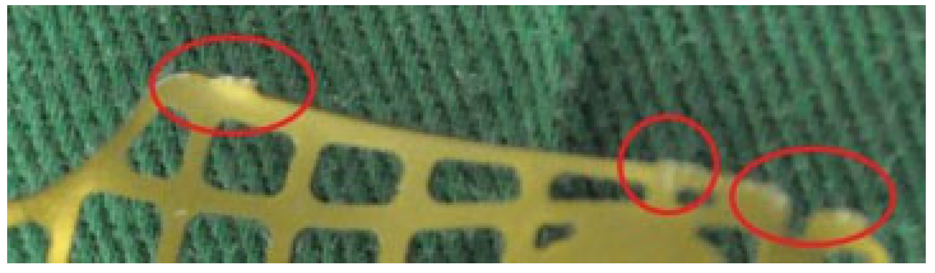
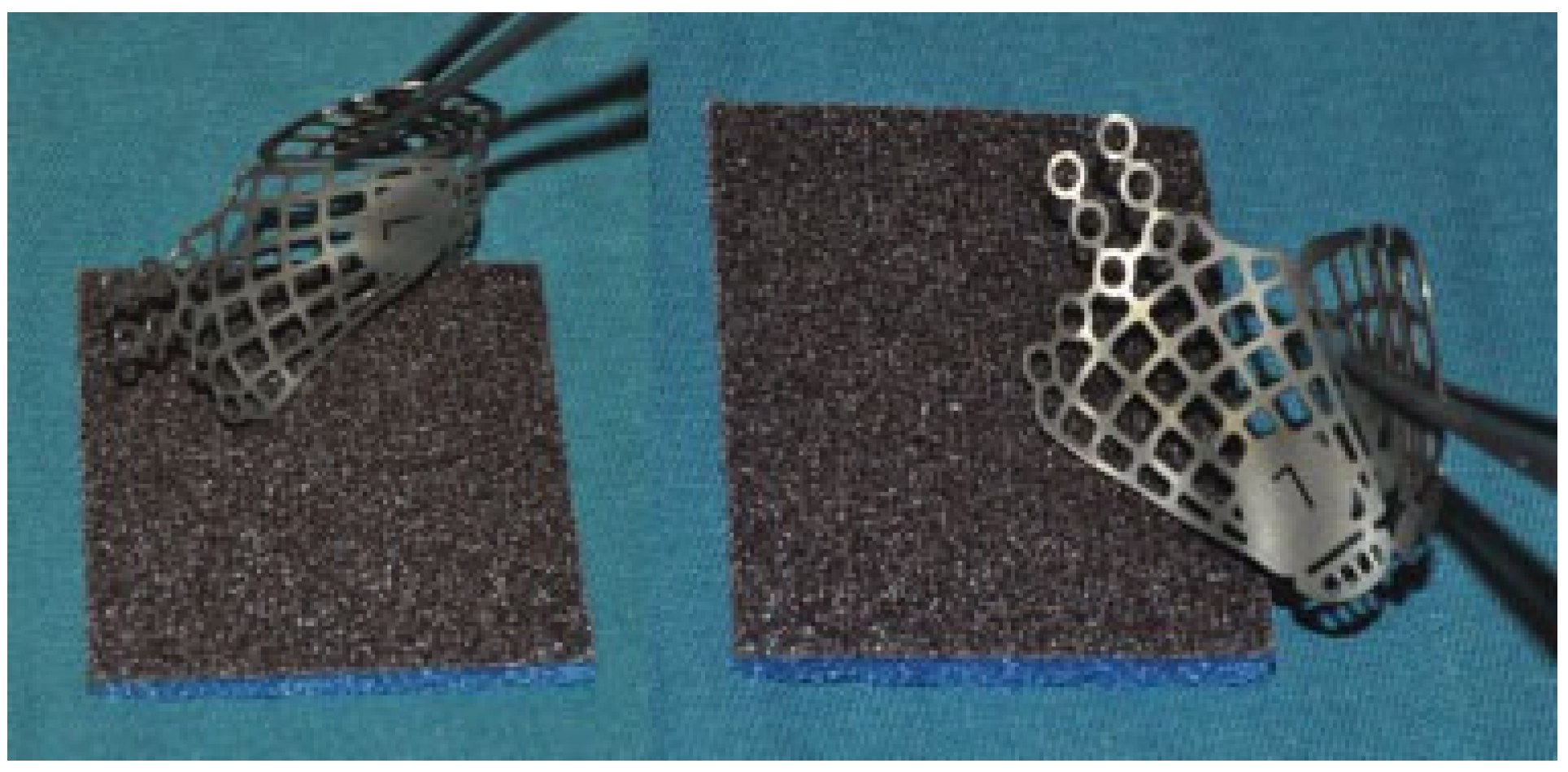
:- Dear Sir,
References
- AO Foundation Website. Midface Orbital Floor Fracture—Orbital Reconstruction. Available online: http://www2.aofoundation.org/wps/portal/surgerymobile?showPage=redfix&bone=CMF&segment=Midface&classification=92-Orbit,%20Orbital%20floor%20fracture&treatment=operative&method=Open%20treatment&implantstype=Orbital%20reconstruction&approach=&redfix_url (accessed on 12 March 2009).
- Janecka, I.P. New reconstructive technologies in skull base surgery: role of titanium mesh and porous polyethylene. Arch Otolaryngol Head Neck Surg 2000, 126, 396–401. [Google Scholar] [CrossRef]
- Merbs, S.L.; Iliff, N.T.; Grant, M.P.; Garibaldi, D.C. Use of MEDPOR TITAN Implants in orbital reconstruction. [E-Abstract] Invest Ophthalmol Vis Sci 2005, 46, 4210–4568. [Google Scholar]
- Zanetti, D.; Nassif, N. Transmastoid repair of minor skull base defects with flexible hydroxyapatite sheets. Skull Base 2003, 13, 1–11. [Google Scholar] [CrossRef]

© 2013 by the author. The Author(s) 2013.
Share and Cite
Nallathamby, V.; Lee, H.; Lim, J.; Ong, W.C.; Lim, T.C. Innovative Intraoperative Titanium Mesh Preparation for Safer Implantation. Craniomaxillofac. Trauma Reconstr. 2013, 6, 65-66. https://doi.org/10.1055/s-0032-1329544
Nallathamby V, Lee H, Lim J, Ong WC, Lim TC. Innovative Intraoperative Titanium Mesh Preparation for Safer Implantation. Craniomaxillofacial Trauma & Reconstruction. 2013; 6(1):65-66. https://doi.org/10.1055/s-0032-1329544
Chicago/Turabian StyleNallathamby, Vigneswaran, Hanjing Lee, Jane Lim, Wei Chen Ong, and Thiam Chye Lim. 2013. "Innovative Intraoperative Titanium Mesh Preparation for Safer Implantation" Craniomaxillofacial Trauma & Reconstruction 6, no. 1: 65-66. https://doi.org/10.1055/s-0032-1329544
APA StyleNallathamby, V., Lee, H., Lim, J., Ong, W. C., & Lim, T. C. (2013). Innovative Intraoperative Titanium Mesh Preparation for Safer Implantation. Craniomaxillofacial Trauma & Reconstruction, 6(1), 65-66. https://doi.org/10.1055/s-0032-1329544