Next Article in Journal
Frey's Syndrome Consequent to an Unusual Pattern of Temporomandibular Joint Dislocation: Case Report with Review of Its Incidence and Etiology
Previous Article in Journal
Investigation of Severe Craniomaxillofacial Battle Injuries Sustained by U.S. Service Members: A Case Series
 
 
Craniomaxillofacial Trauma & Reconstruction is published by MDPI from Volume 18 Issue 1 (2025). Previous articles were published by another publisher in Open Access under a CC-BY (or CC-BY-NC-ND) licence, and they are hosted by MDPI on mdpi.com as a courtesy and upon agreement with Sage.
Short Note

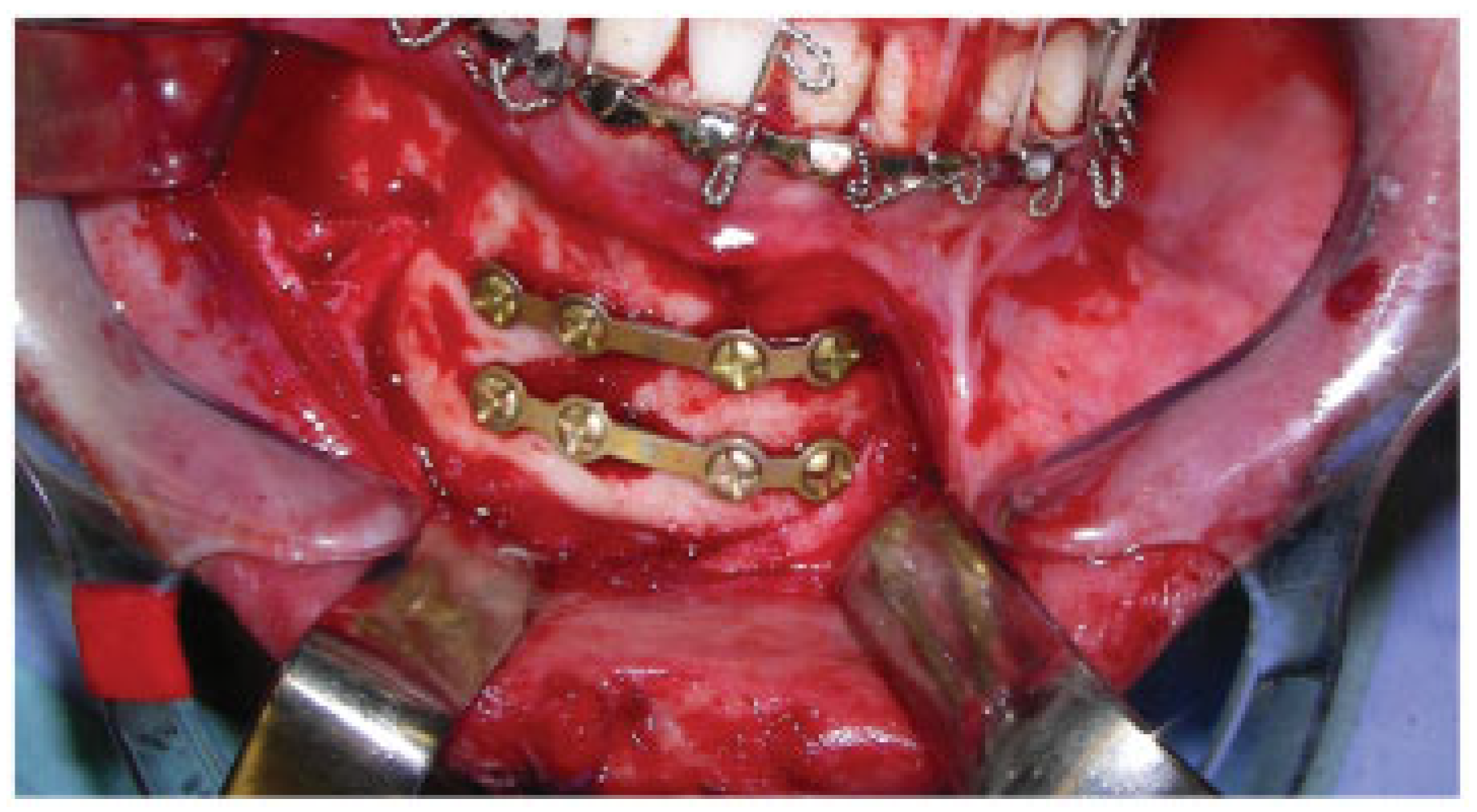
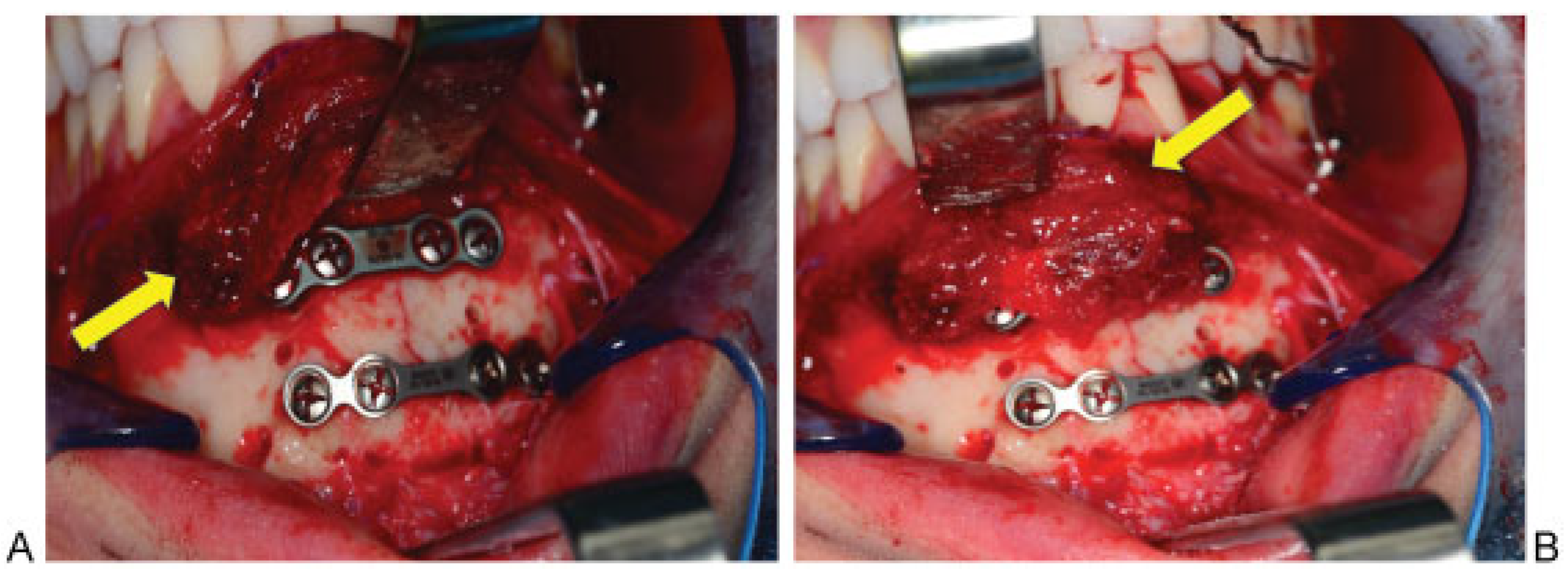
Soft Tissue Coverage for Mandibular Fractures Using Two Miniplates

by
Ajul Shah
,
Anup Patel
and
Derek Steinbacher
*
Department of Plastic Surgery, Yale, 330 Cedar Street, BB 3rd Floor, New Haven, CT 06520, USA
*
Author to whom correspondence should be addressed.
Craniomaxillofac. Trauma Reconstr. 2012, 5(4), 253-254; https://doi.org/10.1055/s-0032-1329543
Submission received: 25 November 2011 / Revised: 12 February 2012 / Accepted: 12 February 2012 / Published: 23 October 2012

Abstract

:
Recent reports have raised the concern that the two miniplate fixation technique for mandibular symphysis and body fractures may lead to greater complications than previously thought. However, it is possible that the surgical exposure and methods of soft tissue closure may be a major contributor to plate exposure. In this article, we detail a technique for vascularized tissue coverage of the hardware used to repair these mandibular fractures. We believe that this soft tissue coverage is crucial for minimizing complications associated with plate fixation.

Vascularized tissue coverage of fixation is critical to uneventful fracture healing. Numerous studies have validated the principle of robust soft tissue coverage over metal plates being necessary to prevent exposure or extrusion [1,2]. Furthermore, a durable soft tissue covering is often placed to salvage exposed hardware and may obviate plate removal [3]. The purpose of this article is to review the concept of soft tissue coverage in relation to mandibular fracture hardware placement and potential complications, as recently described by Ellis [4].
We commend the author of the recent report entitled “A Study of 2 Bone Plating Methods for Fractures of the Mandibular Symphysis/Body” for continued contributions relating to mandibular fracture treatment [4]. This systematic, retrospective review comparing outcomes using two miniplates versus one stronger plate for fractures of the mandibular symphysis/body seemed to suggest that the two miniplate technique resulted in a greater frequency of wound dehiscence, plate exposure, and the need for plate removal, despite equivalency relating to osseous healing and occlusal results. Considering patients in both groups have similar demographics, these complications may be attributed to a foreign body in close approximation to the incisional closure.
However, the surgical exposure, as depicted in the intraoperative photographs in this report. Figure 1B,C, Figure 2A and Figure 4A, demonstrates only a thin mucosal flap with minimal muscle, left near the mucogingival line, to participate in closure and plate coverage [4]. If the images included in this analysis are representative of all the vestibular approaches performed in the series, this may be a major contributor to the finding of superior plate exposure. In our own experience, we have performed a similar dissection, undermining the superior flap near the mucogingival junction (Figure 1). We would not want to draw conclusions on a fixation method when it may very well be a soft tissue coverage, and prior to abandoning the two miniplate technique, this should be investigated more thoroughly.
We have recently altered our technique and suggest that conserving a thicker musculomucosal flap during the dissection will optimize tissue closure and may minimize plate exposure (Figure 2A,B and Figure 3) [5,6]. For symphyseal or parasymphyseal fractures, the mucosal incision is performed 1 cm from the mucogingival line, and the dissection is continued in a perpendicular fashion through the retracted muscle for several millimeters before reorienting toward the bone. This leaves a thicker cuff of mentalis muscle along the superior aspect of the deep flap, which allows robust coverage of the superior plate. Following plate fixation, the previously divided mentalis muscle is reattached using heavy braided, absorbable sutures to preserve function and prevent lower lip and chin ptosis. Posterior to the mental foramen (i.e., for mandibular body fractures), the same methodology is used except that the buccinator is used for muscular coverage. For further support, a chin dressing is applied, composed to two strips of thermoplastic tape: one horizontal piece is placed on the anterior surface of the chin where the mentalis has been reattached, and one piece is placed submentally with the lateral portions being placed in an upward fashion to cover the lateral wings of the first piece of tape (Kaban LB, personal communication, 2007).
Soft tissue coverage is crucial for minimizing complications associated with hardware fixation. The technique of two plate fixation for mandibular body and symphyseal fractures has been embraced by maxillofacial practitioners and is a common treatment modality for these types of fractures. The biomechanical rationale for this treatment approach has been elucidated by several sources [7]. The recent report by Ellis calls into question the complication rate experienced by such a technique. However, we would caution that prior to abandoning this technique, we should investigate the role of soft tissue coverage over the superior plate. In our last five consecutive mandibular fractures treated by the technique stated previously, leaving robust musculomucosal coverage over the superior plate has obviated complications related to dehiscence and infection during at least a 1-year follow-up period. Attention should be paid, long-term, to a myriad of factors, including the role of mucosal flap coverage of plate fixation, relating to the successful treatment of mandibular fractures.

References

  1. Cordeiro, P.G.; Hidalgo, D.A. Soft tissue coverage of mandibular reconstruction plates. Head Neck 1994, 6, 112–115. [Google Scholar] [CrossRef] [PubMed]
  2. Klotch, D.W.; Prein, J. Mandibular reconstruction using AO plates. Am. J. Surg. 1987, 154, 384–388. [Google Scholar] [CrossRef] [PubMed]
  3. Viol, A.; Pradka, S.P.; Baumeister, S.P.; Wang, D.; Moyer, K.E.; Zura, R.D.; Olson, S.A.; Zenn, M.R.; Levin, S.L.; Erdmann, D. Soft-tissue defects and exposed hardware: a review of indications for soft-tissue reconstruction and hardware preservation. Plast. Reconstr. Surg. 2009, 123, 1256–1263. [Google Scholar] [CrossRef] [PubMed]
  4. Ellis, E., III. A study of 2 bone plating methods for fractures of the mandibular symphysis/body. J. Oral Maxillofac. Surg. 2011, 69, 1978–1987. [Google Scholar] [CrossRef] [PubMed]
  5. McCormick, S.U.; Stern, J.C. Split mentalis flap for reconstruction of the anterior mandible. J. Oral Maxillofac. Surg. 1996, 54, 1031–1033. [Google Scholar] [CrossRef] [PubMed]
  6. Hochberg, J.; Ardenghy, M.; Yuen, J.; Gonzalez-Cruz, R.; Miura, Y.; Conrado, R.M.; Pait, T.G. Muscle and musculocutaneous flap coverage of exposed spinal fusion devices. Plast. Reconstr. Surg. 1998, 102, 385–389. [Google Scholar] [CrossRef] [PubMed]
  7. Cienfuegos, R.; Cornelius, C.P.; Ellis, E.; Kushner, G. Mandible–Body: Simple–ORIF. In: Kushner G, ed. AO Foundation, AO Surgery Reference. Available online: www.aofoundation.
Figure 1. Two miniplate plate fixation of mandibular parasymphyseal fracture without soft tissue coverage.
Cmtr 05 00033 g001
Figure 2. (A) Elevating the musculomucosal flap for coverage of the plate. (B) The musculomucosal flap shown covering the superior plate of the two miniplates.
Cmtr 05 00033 g002
Figure 3. Mentalis muscle reapproximation to provide soft tissue coverage for miniplates.
Cmtr 05 00033 g003

