The authors present the sad case of a patient born with an incomplete Tessier number 5 oro-ocular cleft, who underwent 25 operations between infancy and the age of 21. He presented to their institution 20 years later, seeking further treatment.
Whatever work was done originally, we can surmise from the chronology, was before Paul Tessier had even presented his classification of facial clefts, centered around the orbital cavity like time zones [1]. The most commonly encountered oro-ocular clefts are the number 3 and number 4, which begin either through or slightly lateral to the philtral column, and then pass through the medial portion of the lower eyelid, either without a bony septation between the nasal cavity and the maxillary sinus (the more difficult number 3), or with a bony septation (number 4). The number 5 cleft is much rarer, accounting for only 0.3% of a series of 345 atypical facial clefts 2 (versus 4.4% for number 3 and 3% for number 4) [2].
The principles of correcting any of these three types of oro-ocular clefts, however, remain the same. There is a coloboma of the eyelid, which must be corrected. There is a shortness in the distance between the lip and the lower eyelid compared with the normal contralateral side (if there is one), and this shortness must be corrected at the primary operation. A rotation advancement procedure will provide lengthening of the upper lip (and the scar should be placed along the desired philtral column, discarding some tissue lateral to that if necessary); the distance from the eyelid to the lip must be lengthened by either transposition flaps or Z-plasties (which of course are convergent transposition flaps).
The next very important principle is to restore the missing element of the orbit—the orbital floor—with a bone graft as soon as possible. Initially, I did this several months after the primary operation, but now I do it during the primary operation if possible, using a full-thickness calvarial graft (at 6 months of age, the donor site will quickly reossify by itself). The reason for this is to prevent the prolapse of the globe and lack of transverse development of the orbit, with the development of a true vertical orbital dystopia, which will require a subsequent transcranial procedure for correction. This should be done whether there is a functioning, sighted eye present or not. We do not know what had been done in the original 25 operations performed on the patient presented, nor whether he initially had a sighted eye or not. From the photo shown of the patient as an infant, I would wager that this was initially a sighted eye.
Pichler [3] issued the dictum, ‘‘First the bone, and then the soft tissue,’’ and the authors followed that advice. I think they were ill advised, and I would suggest (easy in the retrospectoscope) that it would have been better in this instance to provide more healthy and unscarred tissue in the upper lip to eyelid dimension before embarking on their transcranial orbital dystopia correction. When they proceeded in this fashion, predictably there was inadequate, scarred soft tissue and a severe ectropion, which eventually led to removal of the patient’s eye, provision of soft tissue by a free flap, and an alloplastic epiprosthesis, an acceptable outcome for an invasive cancer but not for an oroocular cleft. One final item that I might take issue with would be the use of an enucleation (was this not an exenteration, removing orbital contents and eyelids?) rather than an evisceration. Most ophthalmologists I work with no longer subscribe to the concept of sympathetic ophthalmia.
Of course, the authors cannot be held responsible for the score of operations performed before the patient arrived on their door step. They are courageous to present what was a series of complications and ending up with less of an epiprosthesis.
I will present two cases that illustrate the two principles I alluded to above.
Case 1 was a complete number 4 cleft showing how a calvarial bone graft at the primary operation has corrected the combination of hypoglobus due to the orbital floor defect and prevented development of a vertical orbital dystopia (Figure 9).
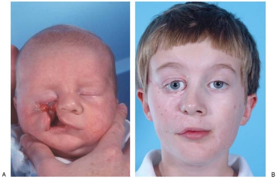
Figure 9.
(A,B) Complete number 4 cleft.
Case 2 was a complete number 5 cleft, asymmetric bilateral, showing how lengthening was obtained by a rotation advancement and z-plasties (Figure 10). (Note that both cases still have slight scleral show.)
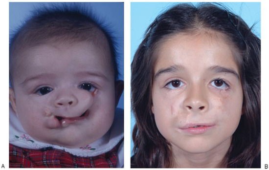
Figure 10.
(A,B) Complete number 5 cleft.
References
- Tessier, P. Anatomical classification facial, cranio-facial and latero-facial clefts. J. Maxillofac. Surg. 1976, 4, 69–92. [Google Scholar] [CrossRef] [PubMed]
- Kawamoto, H.K., Jr.; Patel, P.K. Atypical facial clefts. In Plastic Surgery; Bentz, J., Ed.; Appleton and Lange: Stamford, CT, USA, 1998; p. 199. [Google Scholar]
- Pichler, M.A. In facial reconstruction first the bone then the soft tissues. Personal communication, 1948. [Google Scholar]
Disclaimer/Publisher’s Note: The statements, opinions and data contained in all publications are solely those of the individual author(s) and contributor(s) and not of MDPI and/or the editor(s). MDPI and/or the editor(s) disclaim responsibility for any injury to people or property resulting from any ideas, methods, instructions or products referred to in the content. |
© 2011 by the author. The Author(s) 2011.