Next Article in Journal
Mandibular Osteoradionecrosis: Use of Sequential Fibula Free Flaps for a Remote Sequence of Complications
Previous Article in Journal
Cleft Lip Repair: Technical Refinements for the Wide Cleft
 
 
Craniomaxillofacial Trauma & Reconstruction is published by MDPI from Volume 18 Issue 1 (2025). Previous articles were published by another publisher in Open Access under a CC-BY (or CC-BY-NC-ND) licence, and they are hosted by MDPI on mdpi.com as a courtesy and upon agreement with Sage.
Article

Treatment of Pseudoaneurysm of Internal Maxillary Artery: A Case Report

by
Jimmy Charles Melo Barbalho
1,
Eliardo Silveira Santos
2,
José Maria Sampaio Menezes
3,
Fabricio Rocha Gonçalves
4 and
Otacilio Luiz Chagas
5,*
1
Private Practice, Currais Novos, RN, Brazil
2
Oral and Maxillofacial Surgery Department, General Hospital of Fortaleza, Fortaleza, CE, Brazil
3
Oral and Maxillofacial Surgery Residency Program of Batista Memorial Hospital, Fortaleza, CE, Brazil
4
Private Practice, Pelotas, RS, Brazil
5
Department of Oral and Maxillofacial Surgery and Maxillofacial Prosthodontics, Faculty of Dentistry, Federal University of Pelotas, Rua Gonçalves Chaves, 412 / 705 – Centro, Pelotas 96015-560, RS, Brazil
*
Author to whom correspondence should be addressed.
Craniomaxillofac. Trauma Reconstr. 2010, 3(2), 87-89; https://doi.org/10.1055/s-0030-1254378 (registering DOI)
Submission received: 26 August 2009 / Revised: 1 January 2010 / Accepted: 23 January 2010 / Published: 31 May 2010

Abstract

:
Pseudoaneurysms are rare lesions secondary to blunt or penetrating trauma, temporomandibular joint surgery, or orthognathic surgery. Nonsurgical interventions are the treatment of choice for pseudoaneurysms. In the case reported here, endovascular injection of acrylic glue was successful in the treatment of a pseudoaneurysm of the internal maxillary artery secondary to fracture of the zygomatic bone.

A pseudoaneurysm is an extravascular hematoma that communicates with the intravascular space. This rare lesion receives its name from the fact that its vascular wall is composed of fibrous connective tissue (pseudocapsule) that develops after rupture of the endothe-lium Blood leaks into the tissues through a hole during systole and is contained only by muscle fascia. Leaking continues until the hematoma applies enough pressure to counterbalance arterial pressure and control hemorrhage in the affected area [1,2,3]. This pulsating liquefied hematoma develops for 1 to 8 weeks and forms a new vascular wall (pseudocapsule) that may rupture and trigger severe hemorrhage or thromboembolism [1,2,3].
Pseudoaneurysms are usually secondary to blunt or penetrating trauma, temporomandibular joint surgery, or orthognathic surgery [2,3,4,5]. The main signs and symptoms are asymmetries, neurological deficits, and thromboembolism [6]. Arteriography is the standard procedure to identify pseudoaneurysms in the maxillofacial region. Other imaging studies, such as ultrasound, CT, and magnetic resonance, may also be used [1,2,3]. Nonsurgical interventions are the choice of treatment for this lesion because the large contralateral vascular network makes it difficult to achieve adequate hemostasis when surgical ligation is used [2]. This report describes the treatment of a pseudoaneurysm in the left maxillary sinus secondary to facial trauma. Treatment consisted of selective embolization of the internal maxillary artery with acrylic glue. Also, the importance of early diagnosis and medical care by a multidisciplinary team is discussed.

Case Report

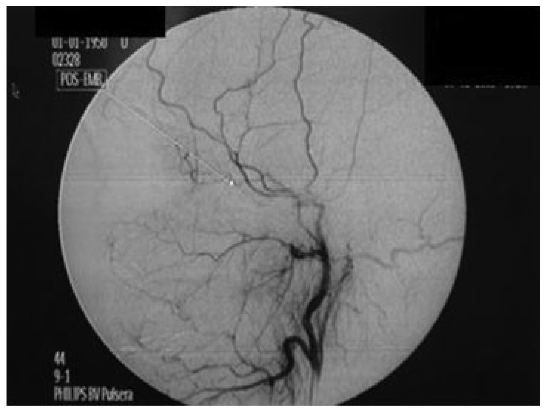
A 22-year-old white woman injured in a car accident in December 2004 was first seen in the Oral and Maxillofacial Surgery (OMS) Department of Batista Memorial Hospital and immediately referred to the General Hospital of Fortaleza (GHF), a level 1 trauma center. She presented edema in the left zygomatic-maxillary region, limited mouth opening, recurrent epistaxis from left nostril at intervals of ~15 days (three previous episodes), left eye divergent strabismus, nonreactive left eye pupil, intact consensual light reflex, normal eye movements, loss of vision, and proptosis in left eye. Physical examination and imaging studies revealed a comminuted fracture of the left zygomatic bone and probable vascular impairment. The patient was evaluated by the specialists in the GHF departments of Oral and Maxillofacial Surgery, Neurology, Ophthalmology, and Endovascular Neurosurgery, and the following studies were requested: arteriography, fundoscopy and magnetic resonance of the orbit, and maxillary sinuses. Results confirmed the diagnosis of pseudoaneurysm of the internal maxillary artery without rupture of the optical nerve. On the same day, after a new episode of epistaxis, the patient was hospitalized in the Emergency Service of GHF to undergo minimally invasive endovascular embolization of the internal maxillary artery. Under local anesthesia, the right femoral artery was punctured with the aid of digital fluoroscopy (Figure 1). Acrylic glue (Histoacryl [1]) was injected by the endovascular neurosurgery and interventional radiology team. Postoperative arteriography revealed instantaneous total regression of the lesion (Figure 2). No bleeding was observed and the edema was substantially reduced on the first day after the procedure. Fifteen days later, the patient returned to treat facial fractures at the OMS Department of HGF, where she has been followed up without any signs or symptoms of the lesion.

Discussion

Blunt trauma and fractures may sometimes be associated with pseudoaneurysm formation [2,3,7]. Rogers et al. [8]. described two cases of aneurysm of the maxillary artery secondary to trauma. One of their patients was injured in a car accident, had a Le Fort II fracture, and was successfully treated with selective embolization. In the case reported here, the patient was injured in a car accident and presented with comminuted fracture of the left zygomatic bone. She was also successfully treated with selective embolization. In 2005, two patients with pseudoaneurysm of the maxillary artery were referred to the interventional radiology service of the General Hospital in Fortaleza, and selective embolization was the treatment used.
Pseudoaneurysms of the intracavernous portion of the internal carotid artery are close to other cavernous structures and may be associated with lesion of the II, III, IV, V, or VI cranial nerve or the ophthalmic artery. Clinically, the classic symptoms of unilateral loss of vision, orbit fractures, and massive epistaxis may be considered pathognomonic for pseudoaneurysm of the internal carotid artery. In the case reported here, these three symptoms were unexpectedly associated with a pseudoaneurysm of the internal maxillary artery [9].
Fan and Mao [10] reported that pseudoaneurysms appear as abnormal high-density masses after contrast injection on magnetic resonance imaging. Results of careful anamnesis and clinical examination of our patient suggested the diagnosis of a pseudoaneurysm of the maxillary artery. CT of the facial sinuses and MRI and angiography studies of the internal maxillary artery were thus requested. Whenever a pseudoaneurysm is suspected, angiography is used to establish the diagnosis and prognosis [1,2,3,4]. The lumen of the lesion is usually opaque and fills up with contrast, but an initial angiography may fail to demonstrate the development of the lumen [9]. Angiographic findings of this case revealed that the maxillary sinus filled up after contrast injection. Selective embolization is a safe and effective method for the treatment of pseudoaneurysms that affect vessels which are difficult to reach by surgery. Also, it reduces the risk of bleeding and revascularization [1,2,3,6,8,10].
The internal maxillary artery is a hard-to-reach, complex anatomic structure. Therefore, our interventional radiology team chose selective embolization, thus reducing the chances of postoperative bleeding and revascularization. Different materials, such as absorbable gelatin sponge, polyvinyl alcohol particles, acrylic glues, and metal coils, may be used for embolization [2,3,5,11]. A recent study with 16 patients reported on the use of cyanoacrylate glue for embolization in a variety of anatomic sites. Embolization was successful in most patients even when previous coil or particulate embolization had failed [11]. Jordan et al. [12] have reported some desired Histoacryl properties, such as good arrangement between penetration and permanence. Even though the adherence to the vascular walls, fast polymerization, and the adding of radiopacifying agent (lipiodol) improve the capacity of manipulating cyanoacrylate, they also interfere deeply in Histoacryl polymerization, turning the embolization control more critical. Besides, exothermic polymerization and toxic products make Histoacryl prone to cause inflammatory reactions [13].
Polyvinyl alcohol is considered the easiest embolic agent to handle [14], and it also shows interesting properties like its capacity of vascular occlusion higher than that of metal coils [12]. Although polyvinyl has been used in several embolizations, it does not have the same acceptance as Histoacryl, mainly due to revascularization procedures, which seem to be more frequent with polyvinyl alcohol [15]. The material available at Brazilian public health service to endovascular treatment is the acrylic glue (Histoacryl), and that is the reason it has been chosen.
Selcuk et al. [16] reported a case of pseudoaneurysm of an ethmoidal branch of the ophthalmic artery caused by removal of a nasograstric tube and treated with embolization with 25% NBCA-histoacryl. The case reported here confirmed the effectiveness of acrylic glue in the treatment of a pseudoaneurysm of the internal maxillary artery, and clinical and imaging studies confirmed the total regression of the lesion at follow-up.
Complications of embolization, such as stroke or blindness, may result from the reflux of embolization material outside the intended area of embolization [11]. Immediate and late follow-up examinations of our patient did not show any clinical signs or symptoms of complications of maxillary artery embolization.

Conclusions

Pseudoaneurysms may sometimes develop after maxillofacial trauma. They should be treated by a multidisciplinary team of specialists in a tertiary hospital prepared to provide a full range of medical care services. Early diagnosis and treatment of a pseudoaneurysm is essential to minimize morbidity and mortality.

References

  1. Zachariades, N.; Skoura, C.; Mezitis, M.; Marouan, S. Pseudoaneurysm after a routine transbuccal approach for bone screw placement. J Oral Maxillofac Surg 2000, 58, 671–673. [Google Scholar] [PubMed]
  2. Krishnan, D.G.; Marashi, A.; Malik, A. Pseudoaneurysm of internal maxillary artery secondary to gunshot wound managed by endovascular technique. J Oral Maxillofac Surg 2004, 62, 500–502. [Google Scholar] [PubMed]
  3. Zachariades, N.; Rallis, G.; Papademetriou, G.; Papakosta, V.; Spanomichos, G.; Souelem, M. Embolization for the treatment of pseudoaneurysm and transection of facial vessels. Oral Surg Oral Med Oral Pathol Oral Radiol Endod 2001, 92, 491–494. [Google Scholar] [CrossRef] [PubMed]
  4. Procopio, O.; Fusetti, S.; Liessi, G.; Ferronato, G. False aneurysm of the sphenopalatine artery after a Le Fort I osteotomy: Report of 2 cases. J Oral Maxillofac Surg 2003, 61, 520–524, discussion 524–525. [Google Scholar] [CrossRef] [PubMed]
  5. Chen, Y.L.; Chou, A.S.B.; Wong, H.F.; Wu, S.H.; Wan, Y.L. Pseudoaneurysm of the internal maxillary artery. Available online: http://www.eurorad.org/case.php?id=1627 (accessed on 30 November 2005).
  6. D’Orta, J.A.; Shatney, C.H. Post-traumatic pseudoaneurysm of the internal maxillary artery. J Trauma 1982, 22, 161–164. [Google Scholar] [CrossRef] [PubMed]
  7. Schwartz, H.C.; Kendrick, R.W.; Pogorel, B.S. False aneurysm of the maxillary artery. An unusual complication of closed facial trauma. Arch Otolaryngol 1983, 109, 616–618. [Google Scholar] [PubMed]
  8. Rogers, S.N.; Patel, M.; Beirne, J.C.; Nixon, T.E. Traumatic aneurysm of the maxillary artery: The role of interventional radiology. A report of two cases. Int J Oral Maxillofac Surg 1995, 24, 336–339. [Google Scholar] [CrossRef] [PubMed]
  9. De Sál, P.M.; Machado, M.C.; Da Silva, D.A.; Brito, L.L.; El Hassan, S. Delayed epistaxis secondary to intracavernous pseudoaneurysm of internal carotid artery. Rev Bras Otorrinolaringol 2003, 69, 715–718. [Google Scholar]
  10. Fan, X.; Mao, Q. Life-threatening oral haemorrhage of a pseudoaneurysm after raising of a fractured zygoma. Br J Oral Maxillofac Surg 2002, 40, 508–509. [Google Scholar] [CrossRef] [PubMed]
  11. Kish, J.W.; Katz, M.D.; Marx, M.V.; Harrell, D.S.; Hanks, S.E. N-butyl cyanoacrylate embolization for control of acute arterial hemorrhage. J Vasc Interv Radiol 2004, 15, 689–695. [Google Scholar] [CrossRef] [PubMed]
  12. Jordan, O.; Doelker, E.; Rüfenacht, D.A. Biomaterials used in injectable implants (liquid embolics) for percutaneous filling of vascular spaces. Cardiovasc Intervent Radiol 2005, 28, 561–569. [Google Scholar] [PubMed]
  13. Vinters, H.V.; Galil, K.A.; Lundie, M.J.; Kaufmann, J.C. The histotoxicity of cyanoacrylates. A selective review. Neuroradiology 1985, 27, 279–291. [Google Scholar] [PubMed]
  14. Hirohata, M.; Abe, T.; Morimitsu, H.; Fujimura, N.; Shigemori, M.; Norbash, A.M. Preoperative selective internal carotid artery dural branch embolisation for petroclival meningiomas. Neuroradiology 2003, 45, 656–660. [Google Scholar] [CrossRef] [PubMed]
  15. Nakstad, P.H.; Bakke, S.J.; Hald, J.K. Embolization of intracranial arteriovenous malformations and fistulas with polyvinyl alcohol particles and platinum fibre coils. Neuroradiology 1992, 34, 348–351. [Google Scholar] [PubMed]
  16. Selcuk, H.; Soylu, N.; Albayram, S.; et al. Endovascular treatment of persistent epistaxis due to pseudoaneurysm formation of the ophthalmic artery secondary to nasogastric tube. Cardiovasc Intervent Radiol 2005, 28, 242–245. [Google Scholar] [PubMed]
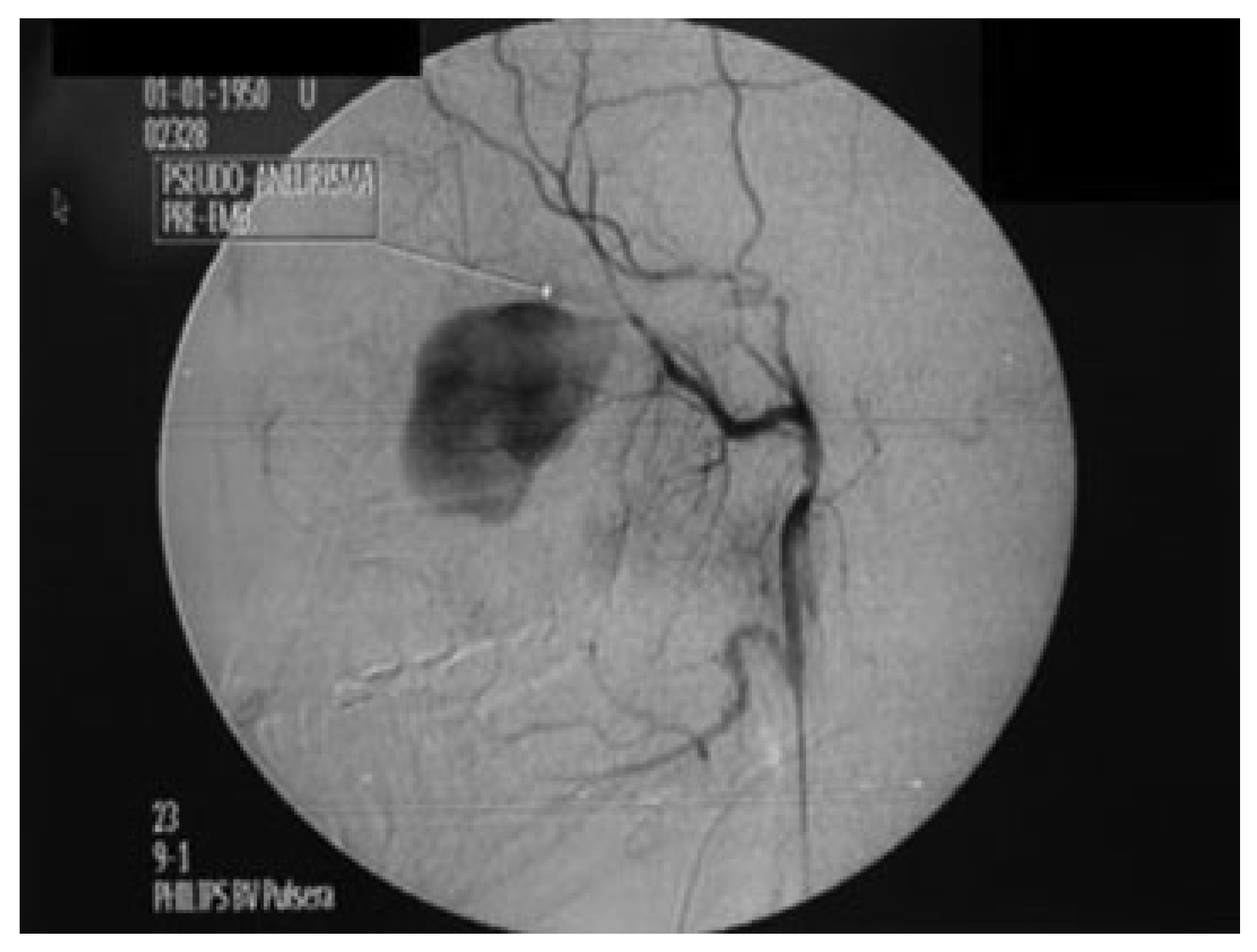
Figure 1. Initial arteriogram using digital fluoroscopy showed pseudoaneurysm of internal maxillary artery (arrow).
Cmtr 03 00009 g001
Figure 2. Control arteriogram using digital fluoroscopy after embolization of internal maxillary artery (arrow).
Cmtr 03 00009 g002