Next Article in Journal
Frontal Sinus Fractures and Traumatic Brain Injury: Predictors of Mortality in Surgical Management
Previous Article in Journal
Comparison of Strengths of Mandibular Angle Fractures Following Different Plate Designs: A Human Cadaver Study
 
 
Craniomaxillofacial Trauma & Reconstruction is published by MDPI from Volume 18 Issue 1 (2025). Previous articles were published by another publisher in Open Access under a CC-BY (or CC-BY-NC-ND) licence, and they are hosted by MDPI on mdpi.com as a courtesy and upon agreement with Sage.
Article

The Versatility of the Supraclavicular Flap for Head and Neck Reconstruction

by
Zubiate Illarramendi Imanol
1,*,
Ferrari Leonardo
2,
Cariati Paolo
1,
Monsalve Fernando
1 and
Martínez Lara Ildefonso
1
1
Department of Oral and Maxillofacial Surgery, Hospital Universitario Virgen de Las Nieves, Av Juan Pablo II S/N, 18014 Granada, Spain
2
Department of Oral and Maxillofacial Surgery, Universidad de Granada, Granada, Spain
*
Author to whom correspondence should be addressed.
Craniomaxillofac. Trauma Reconstr. 2024, 17(4), 306-313; https://doi.org/10.1177/19433875241226535
Submission received: 1 November 2023 / Revised: 1 December 2023 / Accepted: 1 January 2024 / Published: 6 January 2024

Abstract

Study Design: Descriptive study of 17 patients with primary or secondary reconstruction using a supraclavicular flap. Objective: Describe the different options for primary and secondary reconstruction using the supraclavicular flap, as well as the complications that may arise. Methods: The present study analyzed the outcomes of 17 patients who underwent reconstruction using the supraclavicular artery island flap at the Maxillofacial Department of Hospital Universitario Virgen de las Nieves (Granada, Spain) from November 2017 to June 2023. Results: The SCAIF served as the primary reconstructive option in 9 cases, covering skin defects in the parotid region (4 patients), cervical area (1 patient), partial tongue defects (3 patients), and cheek defects (1 patient). Additionally, the flap was used as a secondary reconstructive option in 8 patients affected by osteoradionecrosis (ORN) with bone and plate exposure. Complications included a single case of shoulder joint septic arthritis and 3 patients who experienced cervical fistula and neck infection. Conclusions: The supraclavicular flap represents a highly effective option for oncological reconstruction within the head and neck region. This flap is particularly useful in patients with significant clinical concerns and contraindications, both local and systemic, for microsurgical free flap procedures due to its reliability, versatility, and safety.

Introduction

The supraclavicular artery island flap (SCAIF) is a fasciocutaneous pedicled flap that has seen a recent rise in popularity as an alternative for head and neck reconstruction. Notably, malignant head and neck tumors typically affect older patients with numerous comorbidities. Consequently, given the fragility of these patients, microsurgical flaps may not always be a feasible option (despite being the preferred surgical alternative). In this regard, the SCAIF offers a solution that provides reliability, versatility, and safety [1].
The supraclavicular flap was first described by Lamberty in 1979 [2]. However, the technique was improved and popularized by Pallua and Machens in 1997 [3]. The flap is supplied by the supraclavicular artery, a branch of the transverse cervical artery. The venous drainage originates from comitant veins that run alongside the artery and empty into the transverse cervical or external jugular veins [4]. The flap is easy and quick to harvest in addition to being hairless, thin, and pliable. Hence, it has been proposed as a valid alternative to free flaps in patients with high surgical risk and contraindications for microsurgical reconstruction [5]. It offers an alternative for covering skin defects of the parotid region and neck and coverage of exposed plates due to osteoradionecrosis, partial tongue defect reconstruction, and oncological recurrences in irradiated patients [6].
The present study aimed to determine the potential indications and complications of this flap.

Material and Methods
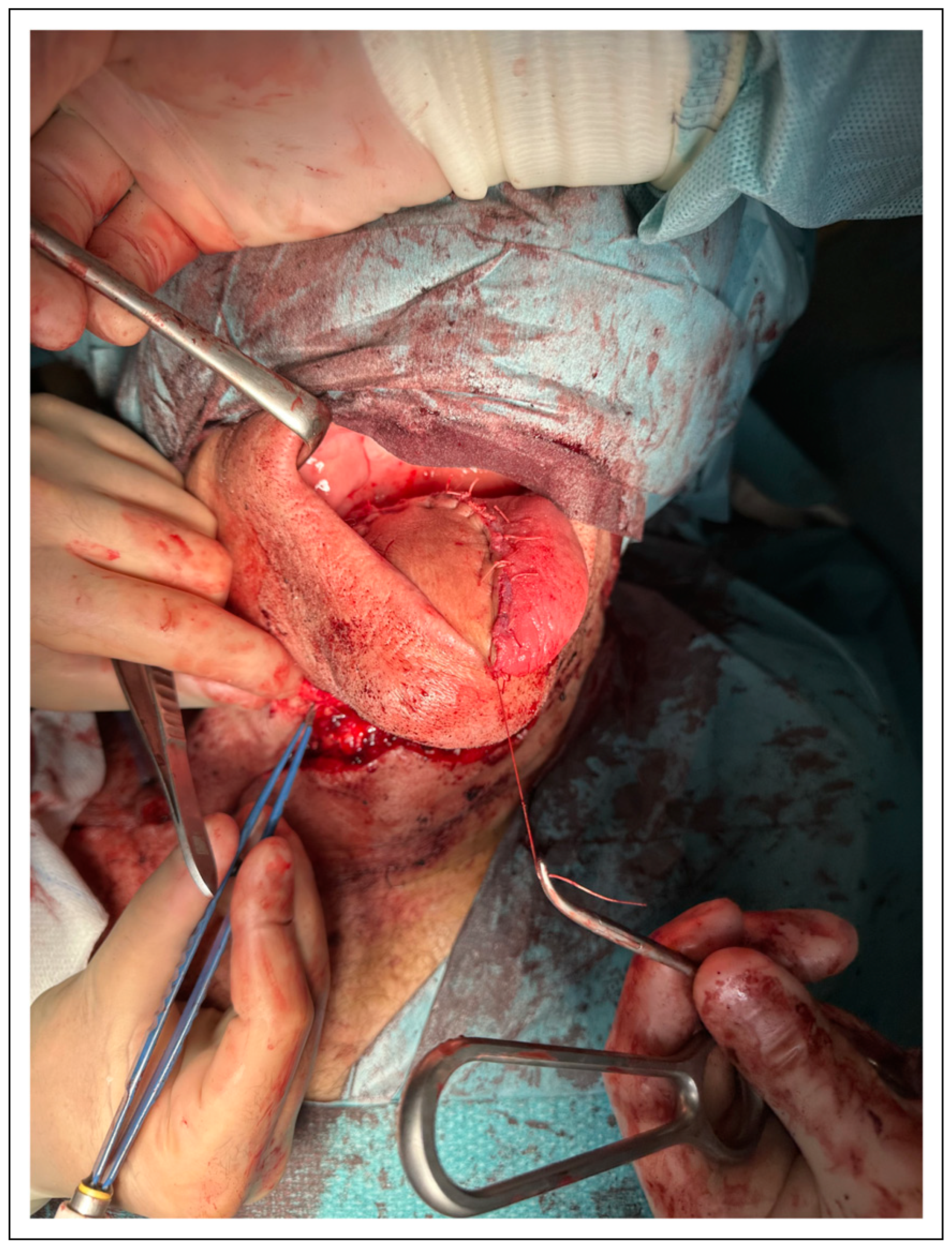
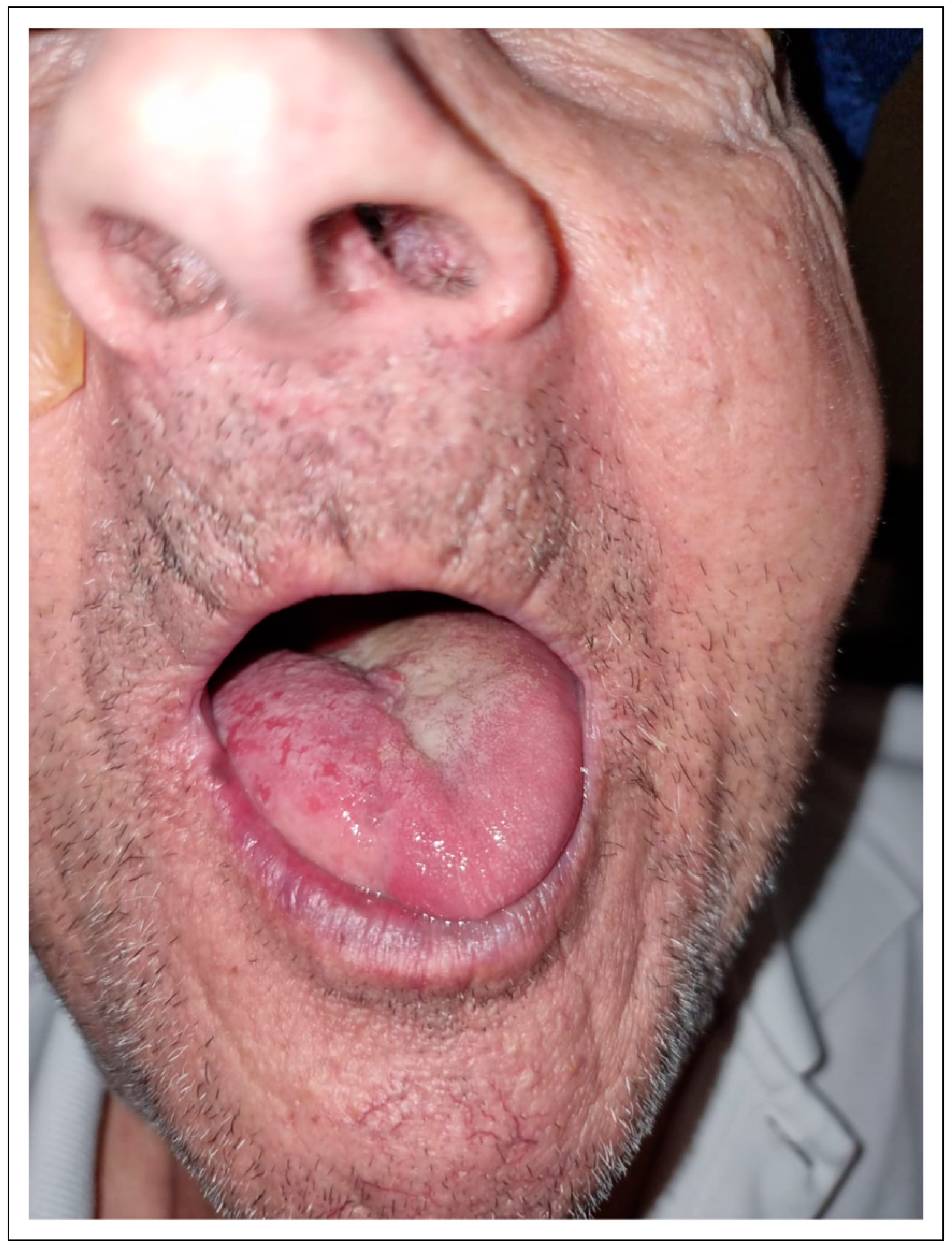
The study sample consisted of patients who underwent reconstruction with a supraclavicular artery island flap at the Maxillofacial Department of the Hospital Universitario Virgen de las Nieves (Granada, Spain) from November 2017 to June 2023. All patients signed the informed consent and this study was accepted by the ethical committee of the Virgen de las Nieves University Hospital. All patients were head and neck oncological patients, and the SCAIF was used for performing the primary oncological reconstruction or attempting to address complications arising from previous surgical procedures. This flap was chosen as the first reconstructive option in fragile patients with high surgical risk and as a secondary reconstructive option in patients who presented complications secondary to other surgical procedures or radiotherapy. The demographic data of the patients are displayed in Table 1.
The presence of the supraclavicular artery was confirmed using a pencil Doppler in all cases. Additionally, a cervical angio CT scan was also performed in patients with a depleted neck due to previous surgeries.
Kokot’s technique was used in all patients. The pedicle was located in a triangular region between the posterior edge of the sternocleidomastoid muscle, the external jugular vein, and the clavicle. The flap size was determined according to the location of the reconstruction site and the extension of the defect. The dissection of the flap began distally and proceeded proximally in a subfascial plane. The dissection continued until reaching the flap’s most proximal portion, where the supraclavicular artery arises from the transverse cervical artery.
After completion of the harvesting process, the flap was rotated to reach the reconstruction site. In many instances, the intermediate portion of the flap was deepithelialized, tunneling the flap beneath the cervical skin. For facial and cervical skin reconstruction, the flap could either be tunneled or simply rotated to cover the defect. After a simple rotation of the flap, the pedicle could be cut after a minimum of 3 weeks to optimize both aesthetic and functional outcomes.

Results

This study included 17 patients who underwent primary or secondary reconstruction with a pedicled supraclavicular flap. The flap was used as the first reconstructive option in 9 patients. In these cases, the flap was used to cover skin defects of the parotid region in patients affected by cutaneous squamous cell carcinoma (4 cases), cutaneous defects of the cervical area resulting from cervical metastasis of oral squamous cell carcinoma (OSCC) (1 case), partial tongue defects resulting from OSCC of the tongue (3 cases) (Figure 1, Figure 2 and Figure 3), and cheek defects arising from OSCC of the buccal mucosa (1 case).
The flap was used as a secondary reconstructive option in 8 patients affected by osteoradionecrosis (ORN) with bone and plate exposure (Figure 4). All these patients had previously undergone mandibular reconstruction with fibula-free flap.
Primary closure of the donor area was performed in 14 patients, while a skin graft was used in the other 3 patients.
No total flap necrosis was observed in our series. Only 1 patient experienced partial necrosis of the more distal portion of the flap that was used for covering a cutaneous defect of the parotid region. The area of the remaining defect after debridement of the distal necrosis was small, and a skin graft was used to cover this area.
The average intensive care unit (ICU) stay was 48 hours. No deaths were recorded during surgery or in the immediate postoperative period using this flap.

Complications

Donor Area

Only 1 significant complication was observed at the donor site. Specifically, in 1 patient, the continuity of the shoulder joint was compromised by a rupture of its articular capsule. In the postoperative period, the patient experienced septic arthritis with pain and fever. Joint lavage and local wound care were insufficient to resolve this complication, and it was necessary to cover the exposed joint area with a pedicled latissimus dorsi flap (Figure 5).
Suture dehiscence of the donor site was observed in 3 patients. However, local wound care was sufficient to manage this complication.

Recipient Area

Three patients who underwent intraoral reconstruction experienced cervical fistulas and neck infections. These complications occurred when the flap was used to address hemiglossectomy defects in 2 patients and a cheek defect in another patient. Thus, it is probable that the tunnel created at the level of the floor of the mouth to pass the flap was the primary cause of this complication. Fortunately, these issues were successfully managed through local wound care and systemic antibiotic treatment.
Moreover, 1 patient who underwent reconstruction of the parotid region presented a salivary fistula. This complication was managed with local wound care and the administration of botulinum toxin infiltration.
No other complications were observed in our series (Table 1).

Discussion

The results observed in these series are similar to others described in the current literature, demonstrating that the supraclavicular flap is a reliable and safe alternative for head and neck reconstruction. Moreover, this procedure offers great versatility and represents a feasible option for reconstructing several head and neck defects, particularly in fragile patients [7].
The pectoralis flap, submental flap, and pedicled latissimus dorsi flap are among the regional flaps that might be used for head and neck reconstruction, particularly in cases where major reconstructive surgeries such as microsurgical free flap procedures are contraindicated [8]. However, the supraclavicular flap offers several advantages over these alternative reconstructive tools. For instance, this method does not pose problems related to oncological safety secondary to the lymphatic drainage of the region, as occurs with the submental flap. Moreover, the supraclavicular flap is hairless, making it a preferred choice for oral reconstruction since the presence of excessive hair can result in aesthetic and functional concerns when utilizing the pectoralis flap, submental, and latissimus dorsi for oncological reconstruction in male patients [9]. Moreover, the scar at the donor site remains aesthetically acceptable when primary closure is achievable without affecting a highly visible and aesthetic area, as occurs with the pectoralis flap. In particular, the supraclavicular flap tends to be thin, and the increase in volume at the cervical level is less pronounced when it is tunneled. Notably, there was no need for a second surgery to cut the pedicle for aesthetic reasons in our patient cohort.
Another significant advantage of this flap is that it can be harvested rapidly, even in nonexpert hands [10]. This feature is particularly advantageous when dealing with fragile patients where swift and safe surgeries are paramount. ASA III/IV patients with >65 years old and with multiple pathologies such as systemic arterial hypertension, diabetes mellitus, chronic obstructive pulmonary disease, and cardiac pathologies such as a history of acute myocardial infarction or arrhythmias could benefit from this type of reconstruction because the surgical time and complications are reduced comparing with other local flaps. However, it is essential to emphasize the importance of accurate preoperative assessment of flap feasibility. The SCAIF is pedicled on the supraclavicular artery, a branch of the transverse cervical artery, and the pencil Doppler probe is a very useful tool for locating the artery both preoperatively and intraoperatively. Nevertheless, in our experience, accurately tracing the course of the artery preoperatively can sometimes be challenging. In cases where there is doubt about the presence and location of the supraclavicular artery, angio-CT can be a valuable resource for ensuring the safe harvesting of the flap. This is especially important for patients who have previously undergone neck surgeries [11].
Another notable advantage of this flap is its versatility. It can be effectively used to reconstruct defects of the oral cavity, pharynx, larynx, and skin defects in the facial and cervical regions. Importantly, the skin color of the flap typically provides a satisfactory match with the recipient area. Furthermore, there is a possibility to dissect at 360° the pedicle to have a real island flap thus achieving greater rotation of the flap. However, one of the disadvantages of this flap is that it is not possible to work on 2 fields at the same time [12].
In our series, the supraclavicular flap was highly effective for covering bone and plate exposure in the mandibular area secondary to radiotherapy. In particular, none of the patients who underwent reconstruction with the supraclavicular flap for this indication required any further procedures at follow-up. Additionally, the supraclavicular flap proved to be highly effective for tongue reconstruction. To illustrate, the flap was employed to reconstruct hemiglossectomy defects in 3 cases. Although 2 of these 3 patients presented local complications such as cervical fistulas and neck infections, they showed satisfactory outcomes in terms of swallowing and speech at the 12 month follow-up after reconstruction.
Another important characteristic of the supraclavicular flap is its reliability. In our series, no instances of flap loss were evidenced. Notably, the literature also reports a low incidence of flap loss, making this flap a suitable option for older patients. It remains important to emphasize that eventual flap loss does not preclude the possibility of other reconstructive options, including free tissue transfer [13].
However, like any reconstructive technique, the SCAIF is not devoid of complications and should only be indicated when appropriate. Complications such as closure dehiscence, partial skin necrosis of the donor area, septic arthritis of the shoulder, shoulder pain syndrome, orocervical fistula, hematoma, and seroma have been described [14]. For instance, our study noted a case of septic arthritis secondary to rupture of the shoulder joint capsule and 3 cases of orocervical fistula. Nonetheless, we firmly believe that this surgical technique must be considered an indispensable tool in the armamentarium of any head and neck reconstructive surgeon. Complications of other local flaps such as the pectoralis major myofasciocutaneous flap, the submental flap, and the latissimus dorsi flap are partial necrosis, infection, wound dehiscence, orocervical fistula, injury of the marginal branch of the facial nerve (submental flap), and inability to raise the shoulder (latissimus dorsi flap). These complications occur in 30–60% of cases [15].

Conclusions

The supraclavicular flap emerges as a highly effective option for oncological reconstruction of the head and neck area. This flap is of particular value for patients with significant medical problems and local and systemic contraindications to microsurgical free flap procedures due to its reliability, versatility, and safety. Nevertheless, further research is needed to evaluate this flap in comparison to other reconstructive techniques, considering patient comorbidities and the advantages offered by each method.

Author Contributions

This article was written by F.L., M.F. and Z.I.I., and was supervised and corrected by C.P. and M.L.I. All authors have read and agreed to the published version of the manuscript.

Funding

The author(s) received no financial support for the research, authorship, and/or publication of this article.

Institutional Review Board Statement

Ethically approved by the HUVN Ethical Committee.

Conflicts of Interest

The author(s) declared no potential conflicts of interest with respect to the research, authorship, and/or publication of this article.

References

  1. Nikolaidou, E.; Pantazi, G.; Sovatzidis, A.; et al. The supraclavicular artery island flap for pharynx reconstruction. J. Clin. Med. 2022, 11, 3126. [Google Scholar] [CrossRef] [PubMed]
  2. Lamberty, B.G. The supra-clavicular axial patterned flap. Br. J. Plast. Surg. 1979, 3, 207–212. [Google Scholar] [CrossRef] [PubMed]
  3. Pallua, N.; Machens, H.G.; Rennekampff, O.; et al. The fasciocutaneous supraclavicular artery island flap for releasing postburn mentosternal contractures. Plast. Reconstr. Surg. 1997, 7, 1878–1884, discussion 1885–1886. [Google Scholar] [CrossRef] [PubMed]
  4. Karabulut, B. Supraclavicular flap reconstruction in head and neck oncologic surgery. J. Craniofac Surg. 2020, 4, e372–e375. [Google Scholar] [CrossRef] [PubMed]
  5. Hamidian Jahormi, A.; Horen, S.R.; Miller, E.J.; et al. A comprehensive review on the supraclavicular flap for head and neck reconstruction. Ann. Plast. Surg. 2022, 6, e20–e32. [Google Scholar] [CrossRef] [PubMed]
  6. Zhang, S.; Chen, W.; Cao, G.; et al. Pedicled supraclavicular artery island flap versus free radial forearm flap for tongue reconstruction following hemiglossectomy. J. Craniofac Surg. 2015, 26, e527. [Google Scholar] [CrossRef] [PubMed]
  7. Alves, H.R.N.; de Faria, J.C.M.; Dos Santos, R.V.; et al. Supraclavicular flap as a salvage procedure in reconstruction of head and neck complex defects. J. Plast. Reconstr. Aesthetic Surg. 2019, 72, e9–e14. [Google Scholar] [CrossRef] [PubMed]
  8. Welz, C.; Canis, M.; Schwenk-Zieger, S.; et al. Oral cancer reconstruction using the supraclavicular artery island flap: Comparison to free radial forearm flap. J. Oral. Maxillofac. Surg. 2017, 75, 2261–2269. [Google Scholar] [CrossRef] [PubMed]
  9. Atallah, S.; Guth, A.; Chabolle, F.; et al. Supraclavicular artery island flap in head and neck reconstruction. Eur. Ann. Otorhinolaryngol Head. Neck Dis. 2015, 132, 291–294. [Google Scholar] [CrossRef] [PubMed]
  10. Li, Y.; Zhao, Z.; Wu, D.; et al. Clinical application of supraclavicular flap for head and neck reconstruction. Eur. Arch. Oto-Rhino-Laryngol. 2019, 276, 2319–2324. [Google Scholar] [CrossRef] [PubMed]
  11. Adams, A.S.; Wright, M.J.; Johnston, S.; et al. The use of multislice CT angiography preoperative study for supraclavicular artery island flap harvesting. Ann. Plast. Surg. 2012, 69, 312–315. [Google Scholar] [CrossRef] [PubMed]
  12. Lee, S.; Cho, H.M.; Kim, J.K.; et al. The supraclavicular artery island flap: A salvage option for head and neck reconstruction. Maxillofac. Plast. Reconstr. Surg. 2018, 40, 25. [Google Scholar] [CrossRef] [PubMed]
  13. Nthumba, P.M. The supraclavicular artery flap: A versatile flap for neck and orofacial reconstruction. J. OralMaxillofac Surg. 2012, 70, 1997–2004. [Google Scholar] [CrossRef] [PubMed]
  14. Chiu, E.S.; Liu, P.H.; Friedlander, P.L. Supraclavicular artery island flap for head and neck oncologic reconstruction: Indications, complications, and outcomes. Plast. Reconstr. Surg. 2009, 124, 115–123. [Google Scholar] [CrossRef] [PubMed]
  15. Shires, C.B.; Sebelik, M. The submental flap: Be wary. Clin. Case Rep. 2022, 1, e05260. [Google Scholar] [CrossRef] [PubMed]
Figure 1. Reconstruction of hemiglossectomy with supraclavicular flap, intraoperative picture.
Cmtr 17 00083 g001
Figure 2. Reconstruction of hemiglossectomy with supraclavicular flap, postoperative picture (7 days after surgery).
Cmtr 17 00083 g002
Figure 3. Reconstruction of hemiglossectomy with supraclavicular flap, postoperative picture (3 months after surgery).
Cmtr 17 00083 g003
Figure 4. Coverage of exposed osteosynthesis plate in lower jaw with supraclavicular flap.
Cmtr 17 00083 g004
Figure 5. Septic arthritis with active suppuration in the donor área.
Cmtr 17 00083 g005
Table 1. Descriptive Table With Number of Patients, Age, Sex, Type of Surgical Intervention, and Type of Complication.
AgeSexReconstruction (Primary/Secondary)Complication
Patient 165MalePrimary. Cutaneous reconstruction parotid regionSalivary fistula
Patient 270MaleSecondary. Plate exposureNo
Patient 374FemalePrimary. Tongue reconstructionCervical fistula and neck infection
Patient 462MaleSecondary. Bone exposureSuture dehiscence in donor site
Patient 580MalePrimary. Tongue reconstructionNo
Patient 681MaleSecondary. Plate exposureShoulder septic arthritis
Patient 762FemalePrimary. Cheek defect reconstructionCervical fistula and neck infection
Patient 863FemaleSecondary. Plate exposureNo
Patient 973MalePrimary. Tongue reconstructionCervical fistula and neck infection
Patient 1070MalePrimary. Cutaneous reconstruction parotid regionSuture dehiscence in donor site
Patient 1174MalePrimary. Cutaneous reconstruction parotid regionNo
Patient 1276MaleSecondary. Plate exposureNo
Patient 1374FemaleSecondary. Plate exposure
Patient 1480MalePrimary. Cutaneous reconstruction parotid regionNo
Patient 1575MalePrimary. Cervical cutaneous metastasisNo
Patient 1671FemaleSecondary. Bone exposureSuture dehiscence in donor site
Patient 1772MaleSecondary. Plate exposureNo

