Next Article in Journal
Orbital Trauma Epidemiologic Characteristics by Life Stage
Previous Article in Journal
Deoxycholic Acid for Submental Convexity: A MAUDE Database Analysis
 
 
Craniomaxillofacial Trauma & Reconstruction is published by MDPI from Volume 18 Issue 1 (2025). Previous articles were published by another publisher in Open Access under a CC-BY (or CC-BY-NC-ND) licence, and they are hosted by MDPI on mdpi.com as a courtesy and upon agreement with Sage.
Article

Repair of Inferior Alveolar Nerve in Orthognathic Surgery Simulator (RIANOS): A Novel, Open-Source, Combined 3D Printed, and Ex-Vivo Chicken Sciatic Nerve Training Model

by
Alfonso Navia
1,2,*,
Sebastian Tapia
3,
Maria Fernanda Rojas
1,
Francisco Rojas
3,
Alex Vargas
3,
Claudio Guerra
1,
Alvaro Cuadra
1,
Susana Searle
1,
Hernan Ramírez
2,3 and
Cristian Teuber
3
1
Experimental Surgery and Simulation Center, Section of Plastic and Reconstructive Surgery, Surgery Division, School of Medicine, Pontificia Universidad Católica de Chile, Santiago, Chile
2
Instituto de Seguridad del Trabajo, Santiago, Chile
3
Department of Surgical Oncology and Maxillofacial Surgery, Surgery Division, School of Medicine, Pontificia Universidad Católica de Chile, Santiago, Chile
*
Author to whom correspondence should be addressed.
Craniomaxillofac. Trauma Reconstr. 2024, 17(4), 43; https://doi.org/10.1177/19433875241236322
Submission received: 1 November 2023 / Revised: 1 December 2023 / Accepted: 1 January 2024 / Published: 14 March 2024

Abstract

:
Study Design: Face and content validation of a surgical simulation model. Objective: Accidental transection of the inferior alveolar nerve (IAN) during bilateral sagittal split osteotomies (BSSO) has a reported incidence of up to 7%, determining important sensory disturbances in patients. Proper repair demands the need of microsurgical anastomosis skills. No previous training models have been described to simulate this. Therefore, we present a validated simulation model for intraoral repair of transected IAN. Methods: A CT scan of an orthognathic surgery patient was modified and a 3D model of a mandible with BSSO was printed. Chicken thigh anatomy was reviewed, and 2.5 mm sciatic nerves were dissected and mounted in the model. In order to simulate intraoral work depth, it was put inside a dental phantom or medical glove box. The model was tested by a group of experts (n = 12), simulating a transected IAN repair inside the mouth with both loupes and a double visor surgical training microscope. A survey was conducted to assess Face and Content validity. Results: The model was named RIANOS after Repair of Inferior Alveolar Nerve in Orthognathic Surgery Simulator. The printing cost of each model was approximately US$3 and the design file is open-source and available for download. All experts “Strongly Agreed” that the model was useful for training inferior alveolar nerve microsurgical repair and would consider implementing it with their residents. Conclusions: We developed a low cost, reproducible, open-source simulator for IAN injury repair training during BSSO. Face and Content validity was achieved through evaluation by a group of experts.

Introduction

Orthognathic surgery combines bimaxillary osteotomy techniques to correct dentofacial malformations to a pre-planned esthetic and functional position. Mandibular osteotomies for prognathism corrections were first described by Berger, Angle, and Blair, but it was Obwegeser who first described the intraoral bilateral sagittal split osteotomy (BSSO) of the mandible.[1,2] The BSSO technique performs osteotomy lines with a reciprocating saw, often finishing the mandibular splitting with osteotomes and a mallet.[3] The whole procedure needs caution and understanding of the inferior alveolar nerve (IAN) position to avoid its transection as it is one of the critical steps. On average, the IAN canal is 4.9 mm from the buccal cortical margin and 17.4 mm from the alveolar crest[4] and 10.09 ± 3.69 mm overall from the inferior border of the mandible.[5] Injury to the IAN during orthognathic surgery is commonly caused by traction neuropraxia, with a frequency of 9–86%.[6,7] Most cases resolve without treatment in 6–8 months; however, those that involve an accidental nerve transection do not resolve, producing severe sensitive disturbances, and in some cases disabling trigeminal post-traumatic neuropathies. IAN transection during BSSO has an incidence of .1%–1.8% up to 4%–7% of cases according to different reported series[8,9] and its repair demands the need of microsurgical skills.[10] Microsurgical training among plastic and maxillofacial surgery residency programs varies worldwide;[11] however it’s a skill recommended to be trained, especially considering that due to the limited intraoral access, it is more difficult than vascular anastomosis in the neck or other areas with better exposure. Microsurgery requires a high level of manual dexterity usually practiced in simulators prior to the clinical setting.[12] Nerve repair training models have been published previously, including biological models like human cadaveric nerves,[13] rat femoral nerve,[14] and chicken leg nerves,[15,16] as well synthetic nerve models.[17] However, no previous training models have been described to simulate the specific scenario of IAN repair during orthognathic surgery, which represents a greater challenge due to intraoral depth and reduced working space. The aim of our study was to develop and validate a low cost, reproducible simulator for IAN injury repair training during BSSO.

Materials and Methods

Model Design

After signed consent, an orthognathic surgery patient’s CT scan was modified using Pro-Plan (Materialise, Leuven, Belgium) and a 3D model of a mandible with a BSSO was developed and printed. Chicken thighs were bought at a local store and sciatic nerves were dissected and mounted in the model. To simulate intraoral work depth, it was put inside either a dental phantom or medical glove box.

Evaluation: Face and Content Validity

The model was tested by a group of experts (n = 12), simulating an IAN repair inside the mouth with 2.5x loupes and a double training microscope with the help of an assistant. Microsurgery instruments and 8-0 Nylon sutures were used. Experts considered eligible were plastic or maxillofacial surgeons with experience in orthognathic surgery and/or microsurgery. A 5-level Likert survey was conducted to experts using Google Forms (Google Inc, California, US) to assess Face and Content validity. Survey results were analyzed in Microsoft Excel v.16.9 (Microsoft Corporation, Washington, US). Descriptive statistics were used.

Results

Model Development

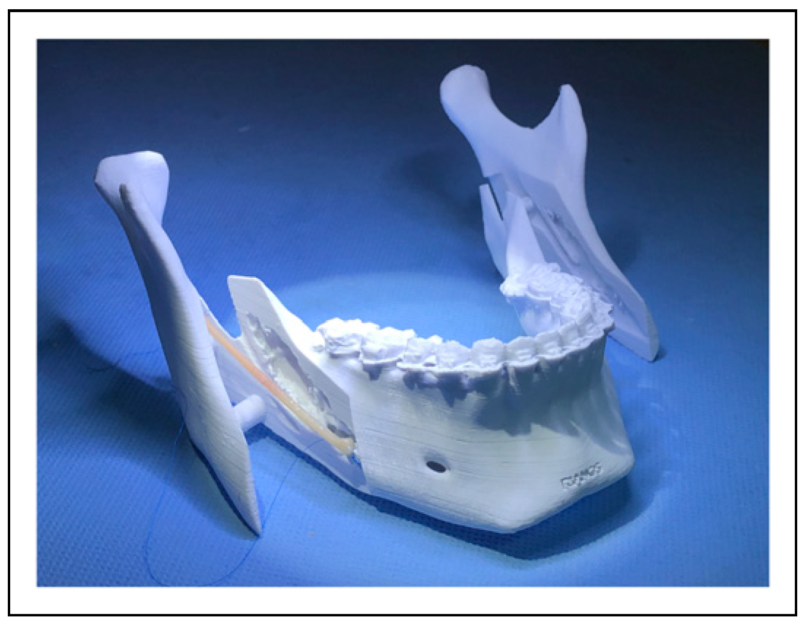
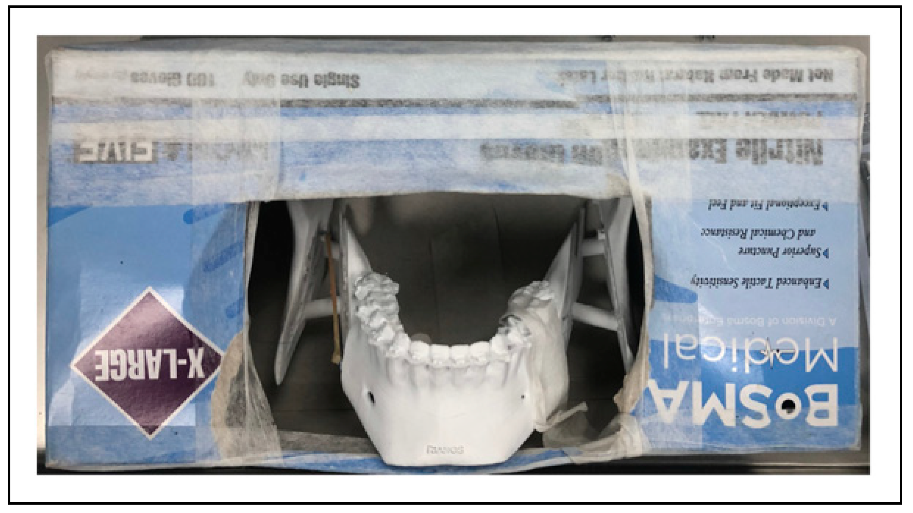
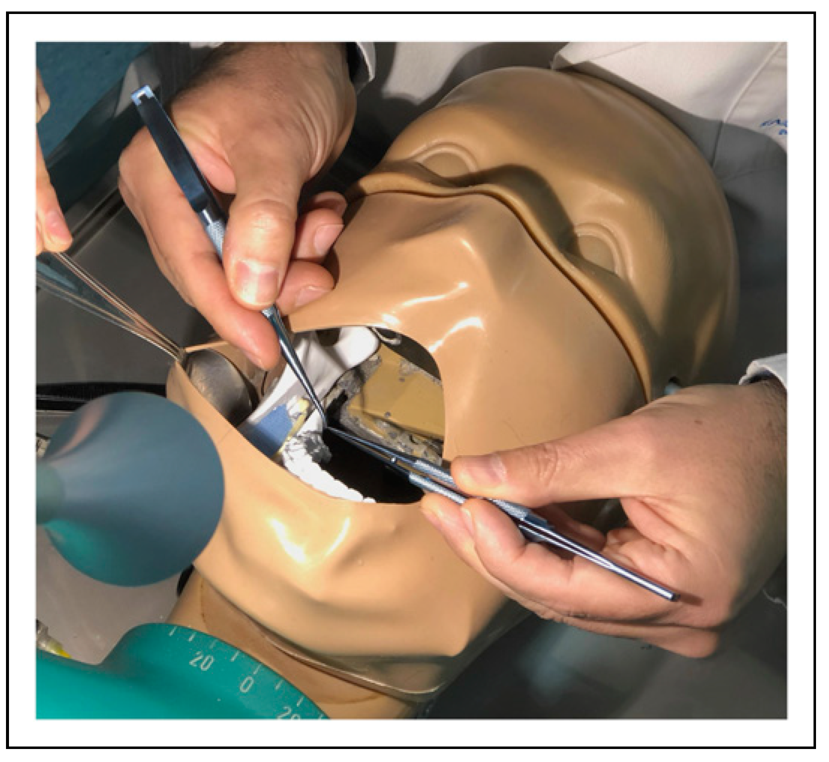
The 3D modeled mandible with BSSO file is open source and available for download at the National Institutes of Health 3D print exchange website (https://3dprint.nih.gov Model ID 3DPX-015091). Chicken thighs contain both the sciatic and femoral neurovascular bundles and had a cost of approximately US$1 per thigh (Figure 1). The model was named RIANOS after Repair of Inferior Alveolar Nerve in Orthognathic Surgery Simulator (Figure 2). Each 3D model was printed with a Ender 5 3D Printer of Fused Deposition Modeling (Creality, Shenzhen, China) using 100g of polylactic acid (PLA) which had a cost of approximately US$3. The RIANOS model was mounted and tested both inside a glove box, offering a low cost and easy to acquire setting (Figure 3), and a more realistic dental phantom (Figure 4). The instruments needed to allow nerve repair in this area of poor visualization and access were microsurgery forceps, microsurgery scissors, microsurgery needle holder, 90 Nylon sutures, and 2 Langenbeck retractors.

Face and Content Validation: Survey Results

All experts “Strongly Agreed” or “Agreed” that the chicken sciatic nerve and 3D printed mandible with BSSO properly simulated the real scenario. The dental phantom was preferred over the glove box to simulate intraoral depthness as well as the microscope over loupes. Regarding content validity, experts “Strongly Agreed” that the model was useful for training IAN repair, both for residents (100%) and experienced surgeons (91.7%). Survey results are shown in Table 1.

Discussion

Microsurgery simulation training has long demonstrated to be a fundamental tool for acquiring skills. Multiple models have been described; however, a minority of them refer to nerve repair.[18] Chicken sciatic nerves measure 2.5–3 mm in diameter, resembling the 2.2–2.4 mm diameter of the IAN.[19,20] Even though the use of chicken sciatic nerves is not new,[15,16] no previous study had combined their use with 3D printing in order to simulate a specific, more challenging scenario, as is the practice of intraoral microsurgical epineurorraphy. A study by Usón-Gargallo et al. described a methacrylate block where jaws of animals could be positioned to practice microsurgical suture techniques; however, their model did not simulate intraoral depthness and is less reproducible.[21] Supplemental Video S1 summarizes the complete mounting and testing process of RIANOS.
Experts’ evaluation of the model was positive. Even though the dental phantom was preferred, the medical glove box was also rated favorably (41.7% “Agreed” and 33.3% “Strongly Agreed”), being a more affordable option. The 3D printing material cost was US$3. Although this considers having a 3D printer available, the average printing cost of the model at different companies in our country ranged US$30–200 depending on the material used. Even though this could be considered expensive, it is completely reusable, and therefore, it is a one-time expense, being the chicken sciatic nerves the only variable cost (US$1).
RIANOS, however, has some pitfalls. The patient from whom the CT scan that was modeled had a small mandible, nevertheless, this did not affect training experience. Also, chicken thighs require refrigeration and need prior dissection mounting. Finally, RIANOS lacks construct validity and model-to-patient skill transferability, both subjects to be assessed in further studies.

Conclusions

We developed a novel, low cost, reproducible, open-source simulator for IAN injury repair training during BSSO. Face and Content validity was achieved through evaluation by experts. Further research must be aimed to assess RIANOS’ construct validity and clinical skill transferability.

Funding

The author(s) received no financial support for the research, authorship, and/or publication of this article.

Supplemental Material

Supplemental Material for this article is available online.

Acknowledgments

The authors would like to thank Andres Campolo and Camila Foncea for their contributions to this project.

Conflicts of Interest

The author(s) declared no potential conflicts of interest with respect to the research, authorship, and/or publication of this article.

References

  1. Steinhäuser, EW. Historical development of orthognathic surgery. J Craniomaxillofac Surg. 1996, 24, 195–204. [Google Scholar] [CrossRef] [PubMed]
  2. Hausamen J-E. The scientific development of maxillofacial surgery in the 20th century and an outlook into the future. Journal of Cranio-Maxillofacial Surgery. 2001, 29, 2–21. [Google Scholar] [CrossRef]
  3. Obwegeser HL Orthognathic surgery and a tale of how three procedures came to Be: a letter to the next generations of surgeons. Clinics in Plastic Surgery. 2007, 34, 331–355. [CrossRef] [PubMed]
  4. Levine MH, Goddard AL, Dodson TB. Inferior alveolar nerve canal position: a clinical and radiographic study. J Oral Maxillofac Surg. 2007, 65, 470–474. [Google Scholar] [CrossRef]
  5. Al-Siweedi SYA, Nambiar P, Shanmuhasuntharam P, Ngeow WC. Gaining surgical access for repositioning the inferior alveolar neurovascular bundle. The Scientific World Journal. 2014, 2014, 719243. [Google Scholar] [CrossRef]
  6. D’Agostino A, Trevisiol L, Gugole F, Bond´ı V, Nocini PF. Complications of orthognathic surgery: the inferior alveolar nerve. J Craniofac Surg. 2010, 21, 1189–1195. [Google Scholar] [CrossRef]
  7. Panula K, Finne K, Oikarinen K. Incidence of complications and problems related to orthognathic surgery: a review of 655 patients. J Oral Maxillofac Surg. 2001, 59, 1128–1136. [Google Scholar] [CrossRef]
  8. Choi B-K, Lee W, Lo L-J, Yang E-J. Is injury to the inferior alveolar nerve still common during orthognathic surgery? Manual twist technique for sagittal split ramus osteotomy. Br J Oral Maxillofac Surg. 2018, 56, 946–951. [Google Scholar] [CrossRef]
  9. Teltzrow T, Kramer F-J, Schulze A, Baethge C, Brachvogel P. Perioperative complications following sagittal split osteotomy of the mandible. J Craniomaxillofac Surg. 2005, 33, 307–313. [Google Scholar] [CrossRef]
  10. Tay ABG, Poon CY, Teh LY. Immediate repair of transected inferior alveolar nerves in sagittal split osteotomies. J Oral Maxillofac Surg. 2008, 66, 2476–2481. [Google Scholar] [CrossRef]
  11. Allouni A, Dunne J, Collin T, Saleh D. A survey of micro-surgery training among UK plastic surgery and maxillofacial surgery trainees. Microsurgery. 2014, 34, 419–420. [Google Scholar] [CrossRef]
  12. Rodriguez JR, Yañez R, Cifuentes I, Varas J, Dagnino B. Microsurgery workout: a novel simulation training curricu-lum based on nonliving models. Plast Reconstr Surg. 2016, 138, 739e–747e. [Google Scholar] [CrossRef]
  13. Odobescu A, Dawson D, Goodwin I, Harris PG, BouMerhi J, Danino MA. High-fidelity microsurgical simulation: the Thiel cadaveric nerve model and evaluation instrument. Plast Surg (Oakv). 2019, 27, 289–296. [Google Scholar] [CrossRef]
  14. Safi A-F, Safi S, Timmer M, Goldbrunner R, Kauke M. Epineurial nerve coaptation: a biological nonliving training model using gradually thawed cryopreserved sciatic nerves. J Craniofac Surg. 2018, 29, e708–e712. [Google Scholar] [CrossRef]
  15. Jeong H-S, Moon M-S, Kim H-S, Lee H-K, Yi S-Y. Microsurgical training with fresh chicken legs. Ann Plast Surg. 2013, 70, 57–61. [Google Scholar] [CrossRef]
  16. Couceiro J, Castro R, Tien H, Ozyurekoglu T. Step by step: microsurgical training method combining two nonliving animal models. J Vis Exp 2015, e52625. [CrossRef]
  17. Gul BU, Yanilmaz DK, Arslan D, Bayramicli M, Akbulut O. Silicone-based simulation models for peripheral nerve microsurgery. J Plast Reconstr Aesthetic Surg. 2019, 72, 477–483. [Google Scholar] [CrossRef]
  18. Javid P, Aydın A, Mohanna P-N, Dasgupta P, Ahmed K. Current status of simulation and training models in microsurgery: a systematic review. Microsurgery. 2019, 39, 655–668. [Google Scholar] [CrossRef]
  19. Svane TJ, Wolford LM, Milam SB, Bass RK. Fascicular characteristics of the human inferior alveolar nerve. J Oral Maxillofac Surg. 1986, 44, 431–434. [Google Scholar] [CrossRef]
  20. Ikeda K, Ho KC, Nowicki BH, Haughton VM. Multiplanar MR and anatomic study of the mandibular canal. AJNR Am J Neuroradiol. 1996, 17, 579–584. [Google Scholar]
  21. Gargallo JU, Rubio EA, Pe´rez JLG, Lagares DT, Va´zquez CC. New dental simulator for microsurgical training preliminary overview. Med Oral Patol Oral Cir Bucal. 2007, 12, E582–E584. [Google Scholar]
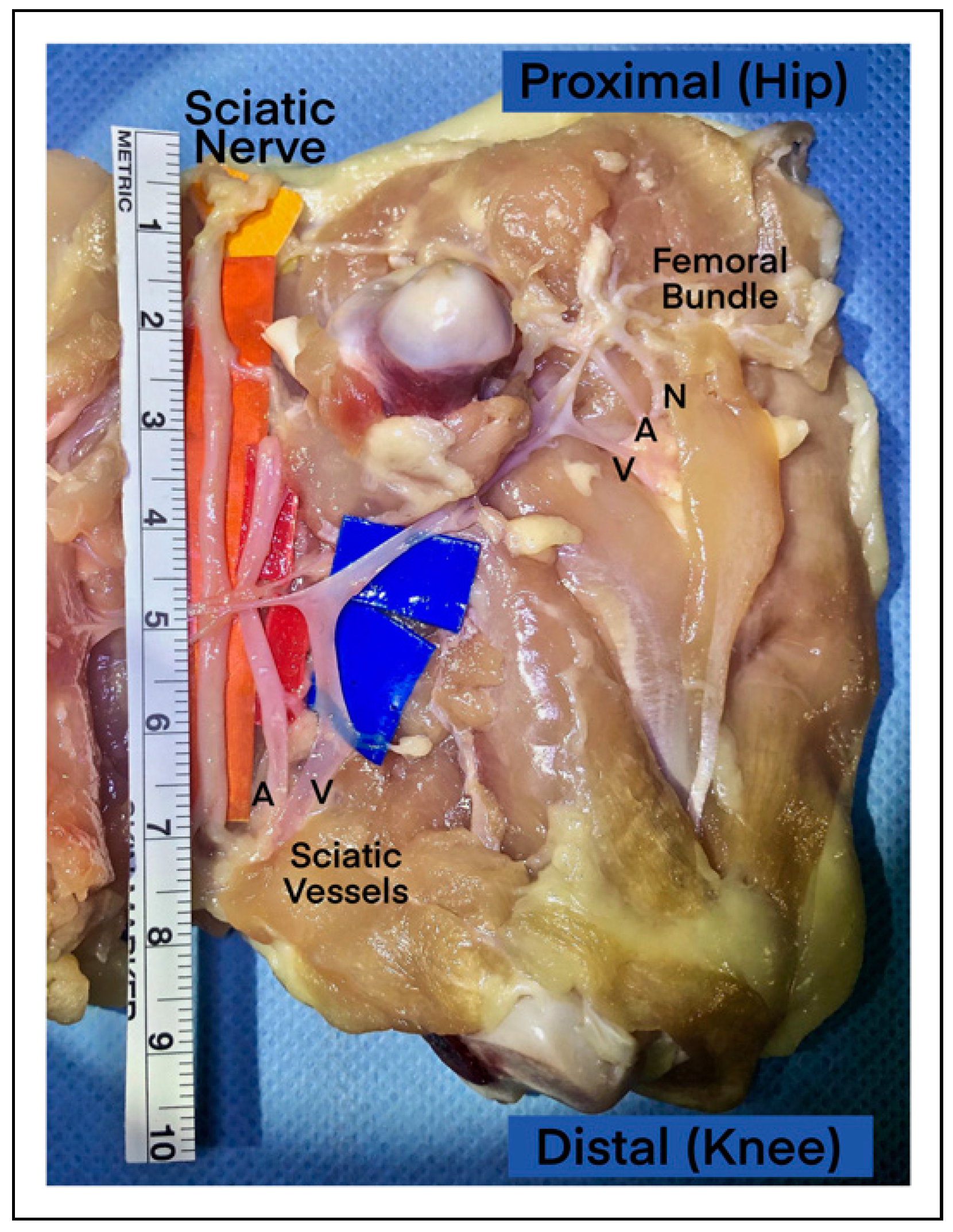
Figure 1. Postero-medial view of a dissected chicken thigh showing the sciatic nerve anatomy (A: Artery, V; Vein).
Cmtr 17 00043 g001
Figure 2. RIANOS model printed and mounted with a sciatic nerve at the right sagittal osteotomy.
Cmtr 17 00043 g002
Figure 3. RIANOS model mounted inside a glove box.
Cmtr 17 00043 g003
Figure 4. RIANOS model mounted inside a dental phantom.
Cmtr 17 00043 g004
Table 1. Likert Survey Assessing Face and Content Validity.
Strongly Strongly
Statement \ Answers (N = 12)DisagreeDisagreeNeutralAgreeAgree
Face Validity
1) Does the appearance and manipulation of the chicken sciatic nerve properly simulate the IAN?0 (0%)0 (0%)0 (0%)1 (8.3%)11 (91.7%)
2) Does the visual appearance of the mandible with its bilateral sagittal split osteotomies look realistic?0 (0%)0 (0%)0 (0%)4 (33.3%)8 (66.7%)
3) Does the box of gloves properly simulate the depthness and shape of the human mouth?0 (0%)1 (8.3%)2 (16.7%)5 (41.7%)4 (33.3%)
4) Does the face phantom properly simulate the depthness and shape of a human mouth?0 (0%)0 (0%)0 (0%)2 (16.7%)10 (83.3%)
5) Did you prefer practicing intraoral microsurgical techniques using loupes?0 (0%)0 (0%)4 (33.3%)4 (33.3%)4 (33.3%)
6) Did you prefer practicing intraoral microsurgical techniques using the optic microscope?0 (0%)2 (16.7%)0 (0%)3 (25%)7 (58.3%)
Content Validity
7) Does RIANOS achieve overall realism for practicing intraoral microsurgical techniques compared to training in a flat surface?0 (0%)0 (0%)0 (0%)2 (16.7%)10 (83.3%)
8) Is RIANOS a useful tool for residents to learn nerve repair of the IAN during orthognathic surgery?0 (0%)0 (0%)0 (0%)0 (0%)12 (100%)
9) Is RIANOS a useful tool for training surgeons with previous orthognathic surgery and nerve repair experience?0 (0%)0 (0%)0 (0%)1 (8.3%)11 (91.7%)
10) Would you consider implementing RIANOS as a training model for plastic surgery and/or maxillofacial surgery residents?0 (0%)0 (0%)0 (0%)0 (0%)12 (100%)