Next Article in Journal
A Review of 13,470 Head and Neck Injuries from Trampoline Jumping
Previous Article in Journal
Appropriate Use of Preoperative Imaging in Feminization Cranioplasty
 
 
Craniomaxillofacial Trauma & Reconstruction is published by MDPI from Volume 18 Issue 1 (2025). Previous articles were published by another publisher in Open Access under a CC-BY (or CC-BY-NC-ND) licence, and they are hosted by MDPI on mdpi.com as a courtesy and upon agreement with Sage.
Article

Trends in Maxillomandibular Fixation Technique at a Single Academic Institution

by
Heather Schopper
1,*,
Natalie A. Krane
2,
Kevin J. Sykes
1,
Katherine Yu
1,
J. David Kriet
1 and
Clinton D. Humphrey
1,*
1
Department of Otolaryngology, Head and Neck Surgery, University of Kansas Medical Center, 3901 Rainbow Blvd, Kansas City, KS 66160, USA
2
Department of Otolaryngology, Head and Neck Surgery, Oregon Health & Science University, Portland, OR, USA
*
Authors to whom correspondence should be addressed.
Craniomaxillofac. Trauma Reconstr. 2024, 17(2), 119-123; https://doi.org/10.1177/19433875231176339
Submission received: 1 November 2022 / Revised: 1 December 2022 / Accepted: 1 January 2023 / Published: 13 May 2023

Abstract

:
Abstract: Study Design: Retrospective chart review. Objective: Restoration of premorbid occlusion is a key goal in the treatment of mandibular fractures. Placement of the patient in maxillomandibular fixation (MMF) is performed during mandibular fracture repair to help establish occlusion. A number of techniques are available to achieve MMF. We sought to examine trends in MMF technique at our institution. Methods: A retrospective chart review was conducted to evaluate patients who underwent surgical treatment of mandibular fractures between January 1, 2011 and March 31, 2021. Data including fracture characteristics, mechanism of injury, patient demographics, complication rates, and MMF technique utilized were collected. Results: One hundred sixty-three patients underwent MMF (132 males). The most common etiology of fracture was assault (34%). There was an increasing preference for rapid MMF techniques over time, as opposed to standard Erich arch bars. No significant difference in obtaining adequate fracture reduction as determined by postoperative imaging or complications were noted between those who underwent MMF with newer rapid techniques vs traditional MMF techniques. Conclusions: Our institution has demonstrated changing trends in the technique utilized for establishing occlusion intraoperatively, more recently favoring rapid MMF techniques, with similar rates of complications and ability to adequately reduce fractures.

Introduction

The restoration of form and function is the fundamental goal when treating mandibular fractures. Maxillomandibular fixation has been used to re-establish premorbid occlusion for almost 100 years [1,2,3]. There are a wide variety of established methods for MMF, including Erich arch bars, Ivy loops, intermaxillary fixation (IMF) screws, and the newer hybrid systems. Each technique has its benefits and drawbacks, but with the same primary goals: to easily establish occlusion in a short amount of time with limited risk of stick injury to the surgeon and a low risk of postoperative complications. This study reviews trends in methods of establishing MMF at a single academic university hospital and level 1 trauma center over the past 10 years, with a focus on the increasing use of more contemporary devices such as intermaxillary fixation screws, hybrid arch bars, and wireless dental occlusion ties (Minne Ties®) for intraoperative stabilization of occlusion [4]. We began using the newest device, wireless dental occlusion ties (WDOT) at our institution in 2017. WDOT are a patented blunt tipped “zip tie”-type device that are used to secure intraoperative MMF in a similar manner to embrasure wires. Our objective was to compare rates of adequate postoperative reduction and complications between different devices used to establish MMF. We hypothesized that newer rapid techniques including WDOT demonstrate equivalent rates of adequate fracture reduction without an increase in postoperative complications.

Materials and Methods

This study was approved by the University of Kansas Medical Center (KUMC) Institutional Review Board. A list of patients who underwent surgical treatment of mandibular fractures at our institution over the last 10 years was collected using CPT codes 21450, 21451, 21452, 21453, 21454, 21461, 21462, 21465, and 21470. Patient electronic medical records were retrospectively reviewed and data were collected, including demographic and comorbidity data, mandibular fracture type/pattern, use of intraoperative MMF, use of postoperative MMF, ability to obtain appropriate reduction as determined by postoperative imaging, length of follow-up, and complications. Complications included infection, hematoma, nonunion/malunion, hardware failure, need for additional procedures, bony sequestra, wound dehiscence, and other.
Patients who did not undergo intraoperative MMF were excluded from analysis. The remaining patients were categorized by type of MMF technique used and subcategorized by type of mandibular fracture treated. Mean length of follow-up was calculated. Nominal variables were compared between the 2 groups using the χ2 test with a P value <.05 used for significance.

Results

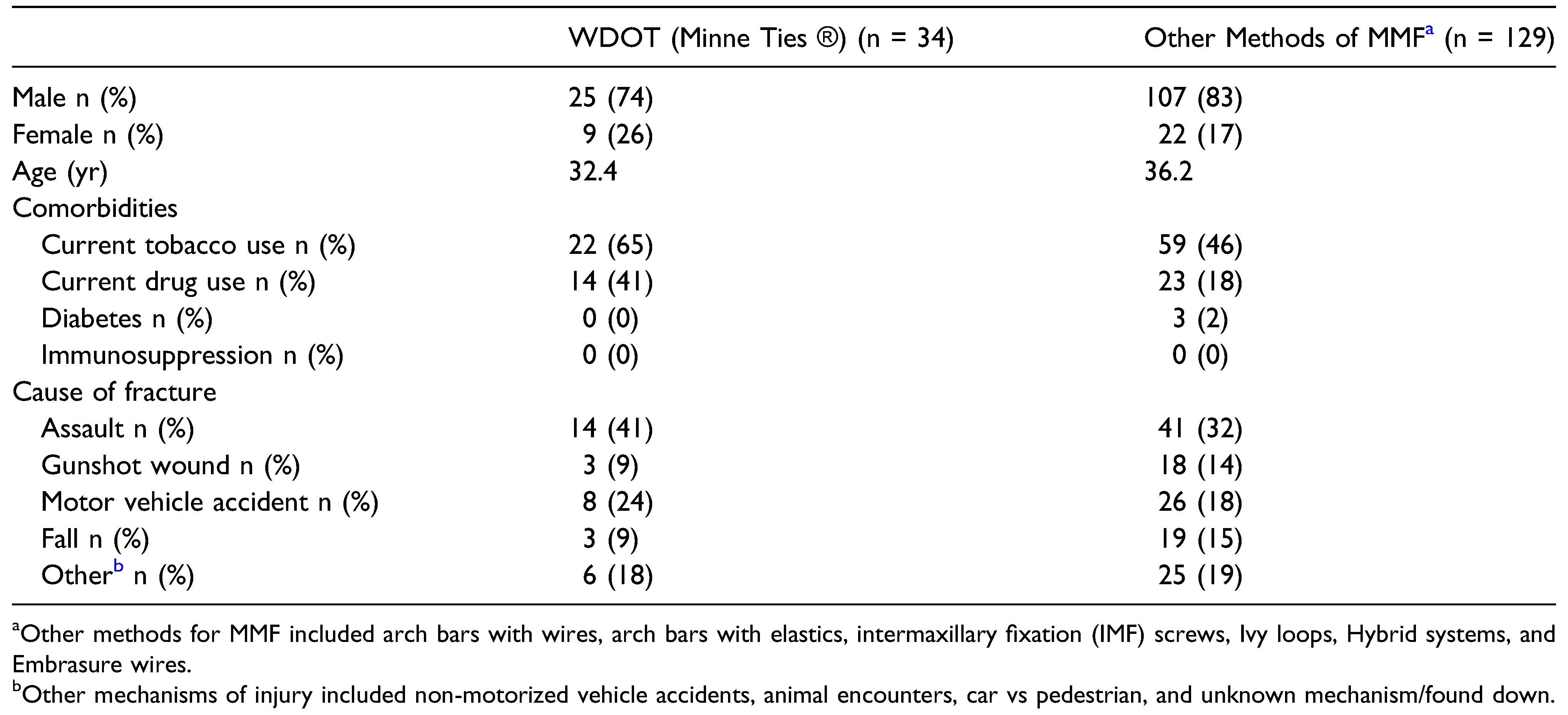
A total of 229 patients were treated surgically for mandibular fractures between January 1, 2011 and March 1, 2021. Twenty patients were treated with MMF alone and 66 were treated with open reduction internal fixation (ORIF) alone. Patients in which ORIF was utilized without MMF were excluded from subsequent analysis, resulting in 163 patients eligible for analysis. Eighty-nine were treated with intraoperative MMF, and 74 were left in MMF postoperatively. The mechanism of injury for all patients was traumatic, 25 of whom sustained complex fractures secondary to gunshot wounds. Demographic, comorbidity, and fracture etiology data can be found in Table 1.
The techniques used for MMF included WDOT (n = 34), arch bars with wires (n = 42), arch bars with elastics (n = 23), intermaxillary fixation (IMF) screws (n = 31), Ivy loops (n = 5), hybrid systems (n = 6), and embrasure wires (n = 34). Multiple types of MMF may have been used in a single patient. Use of specific methods of MMF by year is shown in Figure 1. WDOT use began in 2017 and followed an increasing rate of use from 14.3% of cases with intraoperative MMF in 2017 to 42.9% of cases in the first 3 months of 2021.
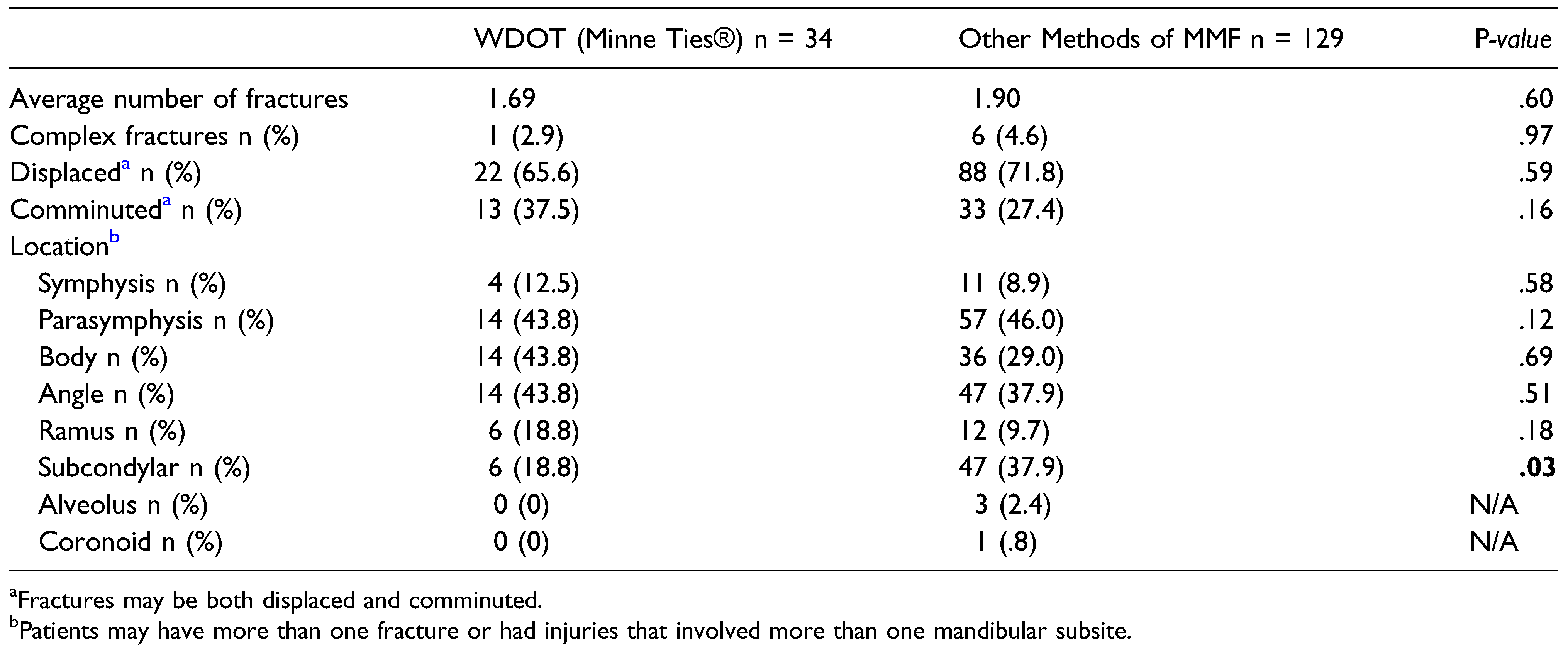
There was no significant difference in the number of fractures, rates of displacement or comminution, or location of fracture when comparing WDOT use to other methods to establish MMF (Table 2). The one exception to this was subcondylar fractures; WDOT were used significantly less frequently in these patients, likely because a common treatment for subcondylar fractures is postoperative MMF, for which WDOT are not currently indicated. Based on postoperative imaging, 92.6% (25/27) of patients in whom WDOT were used demonstrated adequate reduction, while 94.9% (131/138) who underwent other means of MMF demonstrated adequate reduction (P = .625). 2% (n = 1) and 4% (n = 6) of patients, respectively, were not expected to obtain reduction of their fractures based on the nature of the fracture (e.g., subcondylar fractures) and were therefore excluded from the calculations of reduction rates.
The average time to repair was 3.9 days (SD 5.2 days) and 5.4 days (SD 7.6 days) in the WDOT and non-WDOT groups, respectively. Overall, 75.6% (123/163) of patients presented for at least one postoperative visit (follow-up duration ranged from 7 days to 57.5 months [SD 10.2 months]).
The most common complication among the WDOT group was wound dehiscence (18.2%), while in the non-WDOT group it was the need for additional procedures (15.9%). There was no statistically significant difference in complication rates between WDOT and other forms of MMF (36.4% WDOT vs 37.8% in others, respectively; P = .900) (Table 3).

Discussion

Our review of MMF techniques at our institution over the last 10 years shows an increasing frequency of use of the more rapid forms of MMF, including novel devices, such as WDOT, with no significant difference in rates of establishing adequate reduction or complications. This is the first study to compare the use of WDOT to other forms of MMF, with few articles in the literature reporting the use of WDOT for this purpose [4,5]. When reviewing the current literature, Erich arch bars remain the standard of care for MMF in most patients with complex fractures, those requiring closed treatment of subcondylar fractures, and/or for stabilization of alveolar fractures where the arch bar acts as a splint. There is no conclusive objective evidence arch bars are superior to other forms of MMF, though they are generally the most familiar for many surgeons [6]. There are well described downsides to arch bars, including the time needed to apply them and the high risk of stick injuries with the use of wires [7,8,9,10]. Some of these concerns can be overcome with the use of newer systems, such as the hybrid arch bars, which are both faster to apply and have a lower risk of stick injury [8,11]. Similarly, other techniques, such as IMF screws and embrasure wires, require less time for application than arch bars and have lower risk of stick injury as fewer wires overall are used. They also tend to be less costly [10,12,13,14,15]. More costly techniques like hybrid systems may still offer overall cost benefit due to decreased OR time needed. Fewer points of fixation do lend the risk of inadequate strength to maintain occlusion, screw loosening, and the inability to use elastics to guide occlusion in the postoperative period [16]. One patient in our study did require application of arch bars after they fractured the wires of their IMF screws. Overall, our institution has been moving towards more rapid techniques for intraoperative MMF, such as embrasure wires, hybrid systems, and WDOT. When considering the benefits of these aforementioned methods of MMF, including less time for application and lower risk for sharps injury, WDOT offer a facile means to establish occlusion, while the blunt tip provides little risk for sharps injury of the surgeon and limited periodontal trauma for the patient. Similar to embrasure wires, often only 2 or 4 WDOT were needed, which made application and removal both simple and time efficient. WDOT is similar in nature to traditional embrasure wires allowing for a rapid transfer in skills. Although not addressed in this study, WDOT can also be utilized to establish intraoperative occlusion for midface fracture repair. In this study, we demonstrated that WDOT function well in both simple and complicated fractures in terms of adequate fracture reduction, as determined by postoperative imaging and complication rates were comparable to other forms of MMF.
Patients must be appropriately selected when considering the use of WDOT. Similar to embrasure wires, WDOT rely on sufficient native dentition with close contacts between teeth to create secure MMF. Patients without sufficient dentition pose a challenge and may benefit from techniques such as IMF screws or hybrid systems that are not secured directly to the teeth. The authors use WDOT only for intraoperative MMF, as WDOT do not provide the rigid splint functionality of an arch bar, may loosen with movement of the mandible, and cannot currently be used with elastics to guide occlusion. However, others have proposed using WDOT in some patients requiring prolonged MMF for closed treatment of mandible fractures [4].
Limitations of the study include its retrospective nature and inclusion of only a single institution’s data. Multiple surgeons were included with different personal preferences and levels of familiarity with the various MMF techniques, which could introduce bias in patient selection for each MMF technique. Additionally, patients lost to follow-up may result in underreporting of complications.
In summary, contemporary devices such as WDOT offer a rapid and simple means to establish MMF with similar rates of adequate fracture reduction and complications to that seen with other traditional forms of MMF. WDOT also carry a lower risk of stick injury than techniques requiring the use of wires. WDOT should be considered a viable option for intraoperative MMF in patients with good dentition to establish occlusion during mandible fracture repair.

Funding

The author(s) received no financial support for the research, authorship, and/or publication of this article.

Author’s Note

This study has been approved by the Institutional Review Board of The University of Kansas Medical Center.

Declaration of conflicting interests

The author(s) declared the following potential conflicts of interest with respect to the research, authorship, and/or publication of this article: C. Humphrey received a consulting fee from Invisian Medical LLC in 2021. All other authors deny any potential conflicts of interest.

References

  1. Capizzi, P.J.; Bite, U.; Arnold, P.G.; Woods, J.E. John B. Erich, D.D.S., M.D. Plast Reconstr Surg. 1997, 99, 1473–1475. [Google Scholar] [CrossRef]
  2. Convers, J.M. Robert H. Ivy, M.D., D.D.S. A tribute. Plast Reconstr Surg. 1973, 51, 411–412. [Google Scholar] [CrossRef]
  3. Ivy, R. Observations of fractures of the mandible. JAMA 1922, 79, 295. [Google Scholar] [CrossRef]
  4. Johnson, A.W. Dental occlusion ties: a rapid, safe, and non-invasive maxillo-mandibular fixation technology. Laryngoscope Investig Otolaryngol. 2017, 2, 178–183. [Google Scholar] [CrossRef] [PubMed]
  5. Rai, A.; Jain, A. A technique for intraoperative maxillomandibular fixation. Oral Maxillofac Surg. 2017, 21, 485–486. [Google Scholar] [CrossRef] [PubMed]
  6. Falci, S.G.; Douglas-de-Oliveira, D.W.; Stella, P.E.; Santos, C.R. Is the Erich arch bar the best intermaxillary fixation method in maxillofacial fractures? A systematic review. Med Oral Patol Oral Cir Bucal. 2015, 20, e494–e499. [Google Scholar] [CrossRef] [PubMed]
  7. Farber, S.J.; Snyder-Warwick, A.K.; Skolnick, G.B.; Woo, A.S.; Patel, K.B. Maxillomandibular fixation by plastic surgeons: cost analysis and utilization of resources. Ann Plast Surg. 2016, 77, 305–307. [Google Scholar] [CrossRef] [PubMed]
  8. Kendrick, D.E.; Park, C.M.; Fa, J.M.; Barber, J.S.; Indresano, A.T. Stryker SMARTLock hybrid maxillomandibular fixation system: clinical application, complications, and radiographic findings. Plast Reconstr Surg. 2016, 137, 142e–150e. [Google Scholar] [CrossRef] [PubMed]
  9. Rai, A.; Datarkar, A.; Borle, R.M. Are maxillomandibular fixation screws a better option than Erich arch bars in achieving maxillomandibular fixation? A randomized clinical study. J Oral Maxillofac Surg. 2011, 69, 3015–3018. [Google Scholar] [CrossRef] [PubMed]
  10. Roeder, R.A.; Guo, L.; Lim, A.A. Is the SMARTLock hybrid maxillomandibular fixation system comparable to intermaxillary fixation screws in closed reduction of condylar fractures? Ann Plast Surg. 2018, 81, S35–S38. [Google Scholar] [CrossRef] [PubMed]
  11. King, B.J.; Christense, B.J. Hybrid arch bars reduce placement time and glove perforations compared with erich arch bars during the application of intermaxillary fixation: a randomized controlled trial. J Oral Maxillofac Surg. 2019, 77, e1228. [Google Scholar] [CrossRef] [PubMed]
  12. Ali, A.S.; Graham, R.M. Perils of intermaxillary fixation screws. Br J Oral Maxillofac Surg. 2020, 58, 728–730. [Google Scholar] [CrossRef] [PubMed]
  13. Engelstad, M.E.; Kelly, P. Embrasure wires for intraoperative maxillomandibular fixation are rapid and effective. J Oral Maxillofac Surg. 2011, 69, 120–124. [Google Scholar] [CrossRef] [PubMed]
  14. Satpute, A.S.; Mohiuddin, S.A.; Doiphode, A.M.; Kulkarni, S.S.; Qureshi, A.A.; Jadhav, S.B. Comparison of Erich arch bar versus embrasure wires for intraoperative intermaxillary fixation in mandibular fractures. Oral Maxillofac Surg. 2018, 22, 419–428. [Google Scholar] [CrossRef] [PubMed]
  15. Tracy, K.; Gutta, R. Are embrasure wires better than arch bars for intermaxillary fixation? J Oral Maxillofac Surg. 2015, 73, 117–122. [Google Scholar] [CrossRef] [PubMed]
  16. Aslam-Pervez, N.; Caccamese, J.F., Jr.; Warburton, G. A randomized prospective comparison of maxillomandibular fixation (MMF) techniques: “SMARTLock” hybrid MMF versus MMF screws. Oral Surg Oral Med Oral Pathol Oral Radiol. 2020, 130, 640–644. [Google Scholar] [CrossRef]
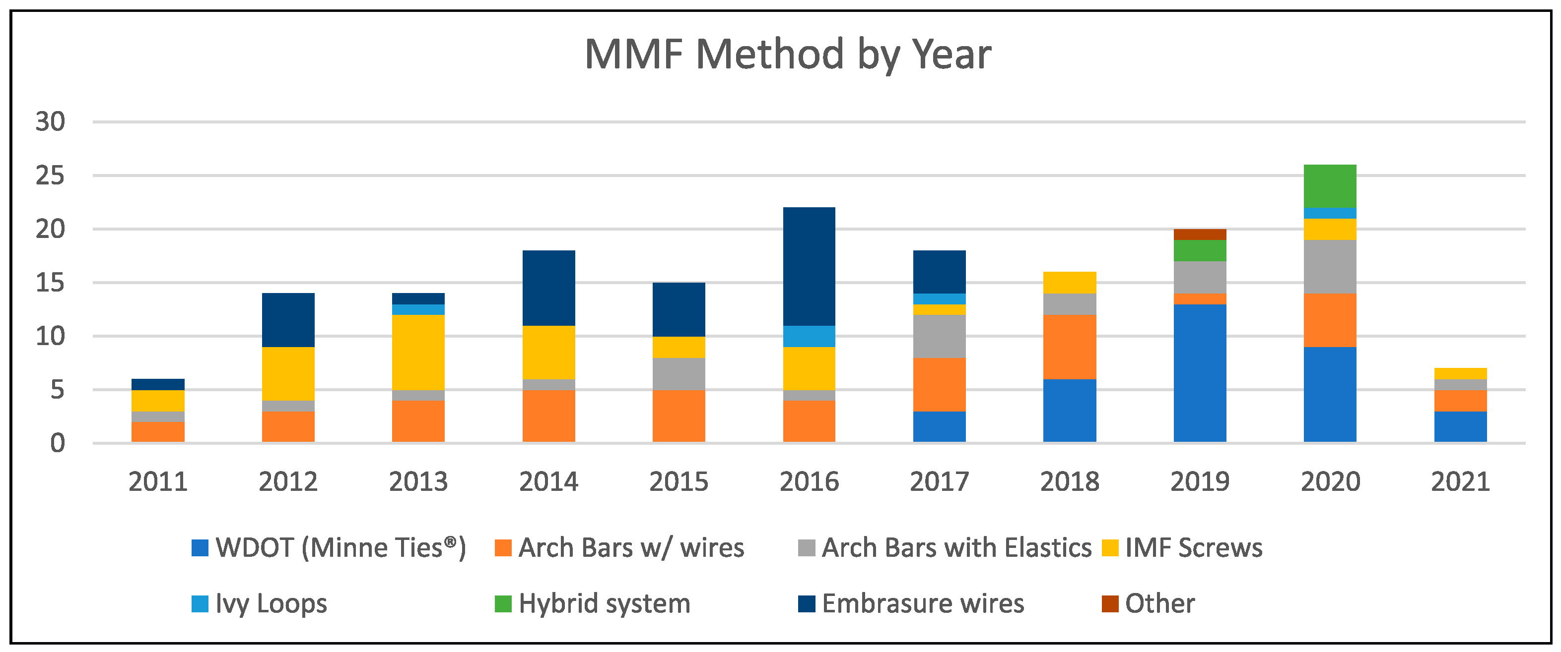
Figure 1. Intraoperative MMF method by year. 2021 is an incomplete year and includes cases through 3/31/2021.
Cmtr 17 00018 g001
Table 1. Demographic, comorbidity, and etiology of fracture data.
Cmtr 17 00018 i001
Table 2. Fracture patterns.
Cmtr 17 00018 i002
Table 3. Complication rates.
Cmtr 17 00018 i003