Next Article in Journal
Is Using the Harmonic Scalpel Better than Conventional Hemostasis in Neck Dissection? A Meta-Analysis
Previous Article in Journal
Sliding Spine Relocation Surgery with Anterior Septal Reconstruction
 
 
Craniomaxillofacial Trauma & Reconstruction is published by MDPI from Volume 18 Issue 1 (2025). Previous articles were published by another publisher in Open Access under a CC-BY (or CC-BY-NC-ND) licence, and they are hosted by MDPI on mdpi.com as a courtesy and upon agreement with Sage.
Review

In Vitro Enhanced Osteogenic Potential of Human Mesenchymal Stem Cells Seeded in a Poly (Lactic-co-Glycolic) Acid Scaffold: A Systematic Review

by
Karla C. Maita
1,
Francisco R. Avila
1,
Ricardo A. Torres-Guzman
1,
Rachel Sarabia-Estrada
2,
Abba C. Zubair
3,
Alfredo Quinones-Hinojosa
2 and
Antonio J. Forte
2,*
1
Division of Plastic Surgery, Mayo Clinic, 4500 San Pablo Rd, Jacksonville, FL 32224, USA
2
Department of Neurosurgery, Mayo Clinic, Jacksonville, FL, USA
3
Department of Laboratory Medicine and Pathology, Mayo Clinic, Jacksonville, FL, USA
*
Author to whom correspondence should be addressed.
Craniomaxillofac. Trauma Reconstr. 2024, 17(1), 61-73; https://doi.org/10.1177/19433875231157454
Submission received: 1 November 2022 / Revised: 1 December 2022 / Accepted: 1 January 2023 / Published: 13 February 2023

Abstract

:
Study Design: Human bone marrow stem cells (hBMSCs) and human adipose-derived stem cells (hADSCs) have demonstrated the capability to regenerate bone once they have differentiated into osteoblasts. Objective: This systematic review aimed to evaluate the in vitro osteogenic differentiation potential of these cells when seeded in a poly (lactic-co-glycolic) acid (PLGA) scaffold. Methods: A literature search of 4 databases following Preferred Reporting Items for Systematic Reviews and Meta-Analyses (PRISMA) guidelines was conducted in January 2021 for studies evaluating the osteogenic differentiation potential of hBMSCs and hADSCs seeded in a PLGA scaffold. Only in vitro models were included. Studies in languages other than English were excluded. Results: A total of 257 studies were identified after the removal of duplicates. Seven articles fulfilled our inclusion and exclusion criteria. Four of these reviews used hADSCs and three used hBMSCs in the scaffold. Upregulation in osteogenic gene expression was seen in all the cells seeded in a 3-dimensional scaffold compared with 2-dimensional films. High angiogenic gene expression was found in hADSCs. Addition of inorganic material to the scaffold material affected cell performance. Conclusions: Viability, proliferation, and differentiation of cells strongly depend on the environment where they grow. There are several factors that can enhance the differentiation capacity of stem cells. A PLGA scaffold proved to be a biocompatible material capable of boosting the osteogenic differentiation potential and mineralization capacity in hBMSCs and hADSCs.

Introduction

Bone tissue engineering (BTE) has become a promising strategy for achieving bone regeneration to treat skeletal defects caused by trauma, congenital malformations, bone tumor resections, and infections, among other pathologies [1]. An estimated 2 million bone graft procedures were done in the United States in a 15-year period [2]. Bone grafts are recognized as the gold standard for bone repair [3], even though there is a high incidence of associated complications. Limited availability in the donor site, bone reabsorption, remodeling issues, and harvest site morbidity, such as pain, bleeding, and infections [4], are some of the complications that can increase hospital stays and costs related to this procedure. Looking to balance patient outcomes and costs, BTE is emerging as a great option to address the several hindrances of autologous bone grafts [5]. Human mesenchymal stem cells (hMSCs), biomaterial scaffolds, and growth factors are the principal components of BTE. Combining these 3 main components provides an ideal environment for new bone formation before implantation in the patient (Figure 1) [6]. Scaffolds must have a 3-dimensional (3D) morphology similar to human bone [7], and they must also be biocompatible, biodegradable, and have osteoconductive properties to guide cells in the formation of new bone tissue [8]. Poly (lactic-co-glycolic acid) (PLGA), a synthetic polymer with all these characteristics, is a common organic component used in BTE [9].
Bone marrow, adipose-derived tissue, and periosteum-derived tissue are principal sources for harvesting adult hMSCs [1]. Bone marrow, the first source discovered, is used most often to obtain human bone marrow stem cells (hBMSCs) because it has been well characterized and widely studied [10]. Still, some limitations have been described, such as low stem cell yield, long in vitro expansion, and complications related to the harvesting procedure (e.g., pain, bleeding, infections) [6]. On the other hand, human adipose-derived stem cells (hADSCs) are gaining interest as a good source of hMSCs since they have demonstrated high yields from lipoaspirates, multilineage potential, faster cell proliferation, and easier harvesting procedures [11,12].
We hypothesize that hADSCs seeded in a PLGA scaffold can achieve a successful in vitro osteogenic differentiation potential, as shown by hBMSCs related to bone regeneration. We aim to review the available literature on in vitro osteogenic differentiation potential in these 2 cell groups seeded in a PLGA scaffold.

Methods

Study Selection

This systematic review included articles evaluating the in vitro osteogenic differentiation potential for bone regeneration of hBMSCs and hADSCs seeded in a PLGA scaffold. We followed Preferred Reporting Items for Systematic Reviews and Meta-Analyses (PRISMA) guidelines for article identification and final selection. To be included, articles had to be written in English. In addition, studies were included if hBMSCs and hADSCs were used and seeded in a PLGA scaffold or if they objectively measured in vitro osteogenic differentiation or bone mineralization capacity of the cells using techniques such as real-time polymerase chain reaction, alkaline phosphatase (ALP) activity, histologic evaluation, or immunohistochemical analysis, among others. No specific publication status was considered. Studies were excluded if they used only in vivo models; cells other than hMSCs; hMSCs from an unknown source; or if they seeded in a type of material other than a PLGA scaffold.

Data Source and Search Strategy

This systematic review was conducted on January 2021, by querying the electronic databases MEDLINE, Embase, CINAHL, and Web of Science. A search strategy was generated using the following Medical Subject Headings terms: “adipose tissue derived mesenchymal stem cells” OR “mesenchymal stem cells, adipose-derived” OR “mesenchymal stem cells, adipose derived” OR “adipose tissue-derived mesenchymal stromal cells” OR “adipose tissue derived mesenchymal stromal cells” OR “adipose-derived mesenchymal stromal cells” OR “adipose derived mesenchymal stromal cells” OR “adipose-derived mesenchymal stem cell” OR “derived mesenchymal stem cell” AND “stem cell, mesenchymal” OR “stem cells, mesenchymal” OR “mesenchymal stem cell” OR “mesenchymal stem cells” OR “bone marrow stromal stem cells” OR “bone marrow mesenchymal stem cells” OR “bone marrow mesenchymal stem cell” OR “bone marrow stromal cell” OR “bone marrow stromal cells, multipotent” OR “multipotent bone marrow stromal cell” OR “multipotent bone marrow stromal cells” AND “bone mineralization” OR “mineralization, bone” OR “bone regeneration” OR “regeneration, bone” AND “PLGA scaffold.”

Data Collection Process
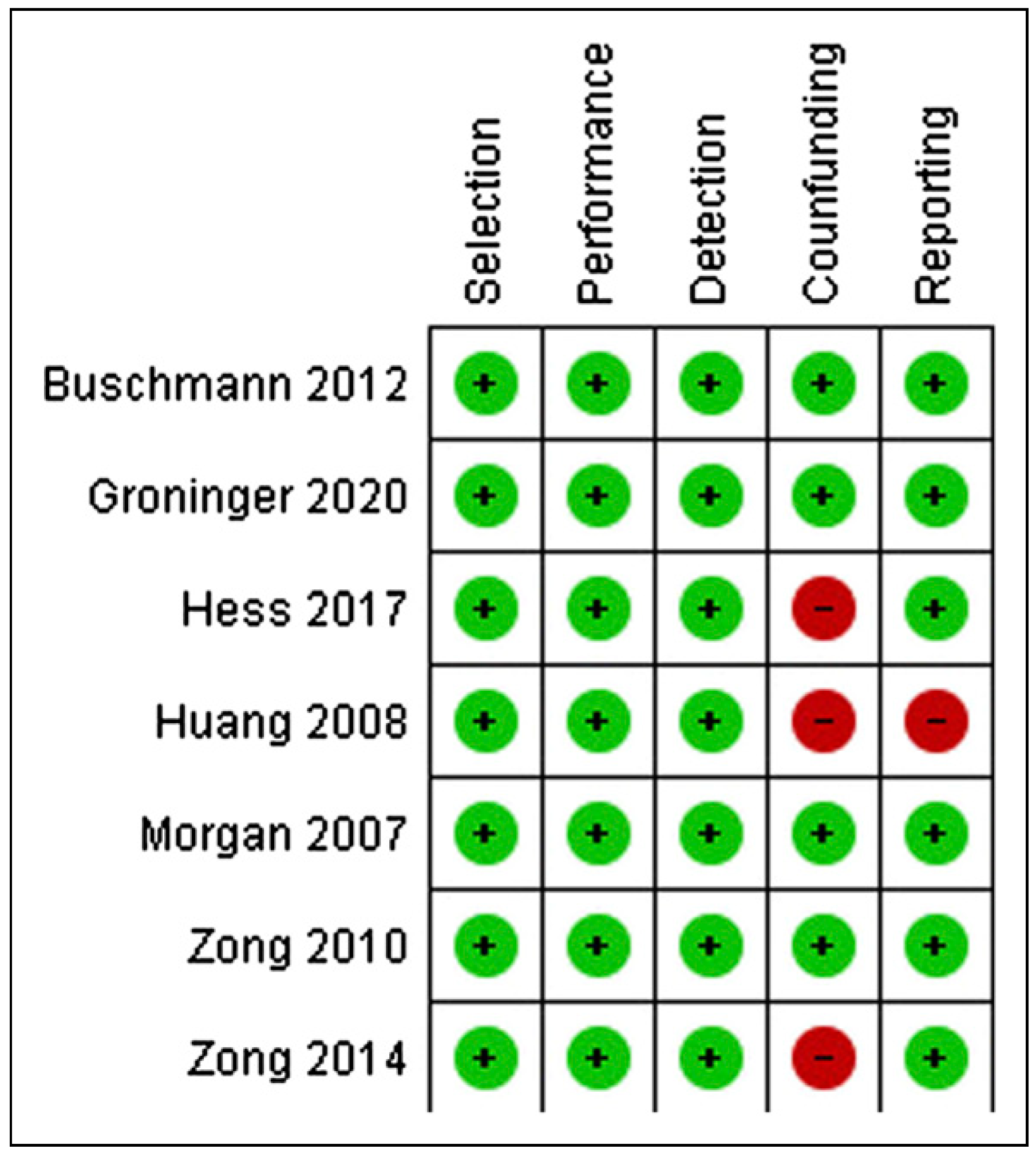
The first author undertook a broad literature search and filtered studies based on titles and abstracts following the inclusion and exclusion criteria described above. Then the full text of the selected studies was screened for the final selection. The second author conducted an independent search. Following an exhaustive discussion about the discrepancies, the authors attained a consensus.

Risk of Bias

The Risk Of Bias In Non-Randomized Studies—of Interventions (ROBINS-I) tool (Cochrane Library) was used to assess the risk of bias [13]. A bias summary and graph were created using RevMan 5.3 (Cochrane Library). Descriptions of individualized bias and cross-sectional studies bias are shown in Figure 2 and Figure 3, respectively. Thus, considering the authors’ criteria, three studies demonstrated factors that could generate bias, mainly associated with confounder variables and reporting outcomes. Hence, studies adding osteoconductive material as hydroxyapatite microparticles (Zong et al. [14]) could overestimate their results related to new bone volume due to the similarity displayed between these materials and the host’s bone in the microcomputed tomography. Additionally, seeding cells on different surfaces (Hess et al. [15] and Huang et al. [16]) might bias the experiment results since cell-to-cell interaction, morphology, and extracellular matrix are not similar. Finally, the risk of reporting bias is high in case of assumption of results with no direct evidence. For example, Huang et al. [16] reported a decrease in gene expression associated with dexamethasone doses added to osteogenic differentiation media, which was not proven directly with a control group.

Results

Study Selection and Characteristics

Based on the 257 articles identified, 7 articles fulfilled the inclusion criteria. The study selection process is outlined in Figure 4. The most relevant outcomes of the 7 studies are described below and are summarized in Table 1.
Four articles used hADSCs harvested by lipoaspiration and processed the tissue following the methods described by Zuk et al. [21] Of these, 3 studies used cells taken from women aged 29 [15,18] and 45 years old [17]. hBMSCs were used in 3 studies. One study obtained the cells from hematologically normal patients undergoing routine hip replacement surgery [19]. One study obtained them from healthy donors aged 24 to 28 years old [14]. Lastly, 1 of the studies obtained the cells from healthy donors of unspecified ages [20]. The processing of the cells was similar in all the studies. This process is characterized by keeping the cells on Minimal Essential Medium—alpha modification until they reach approximately 80% of confluence, which in some of the studies was achieved after 3, 5, or 7 days.

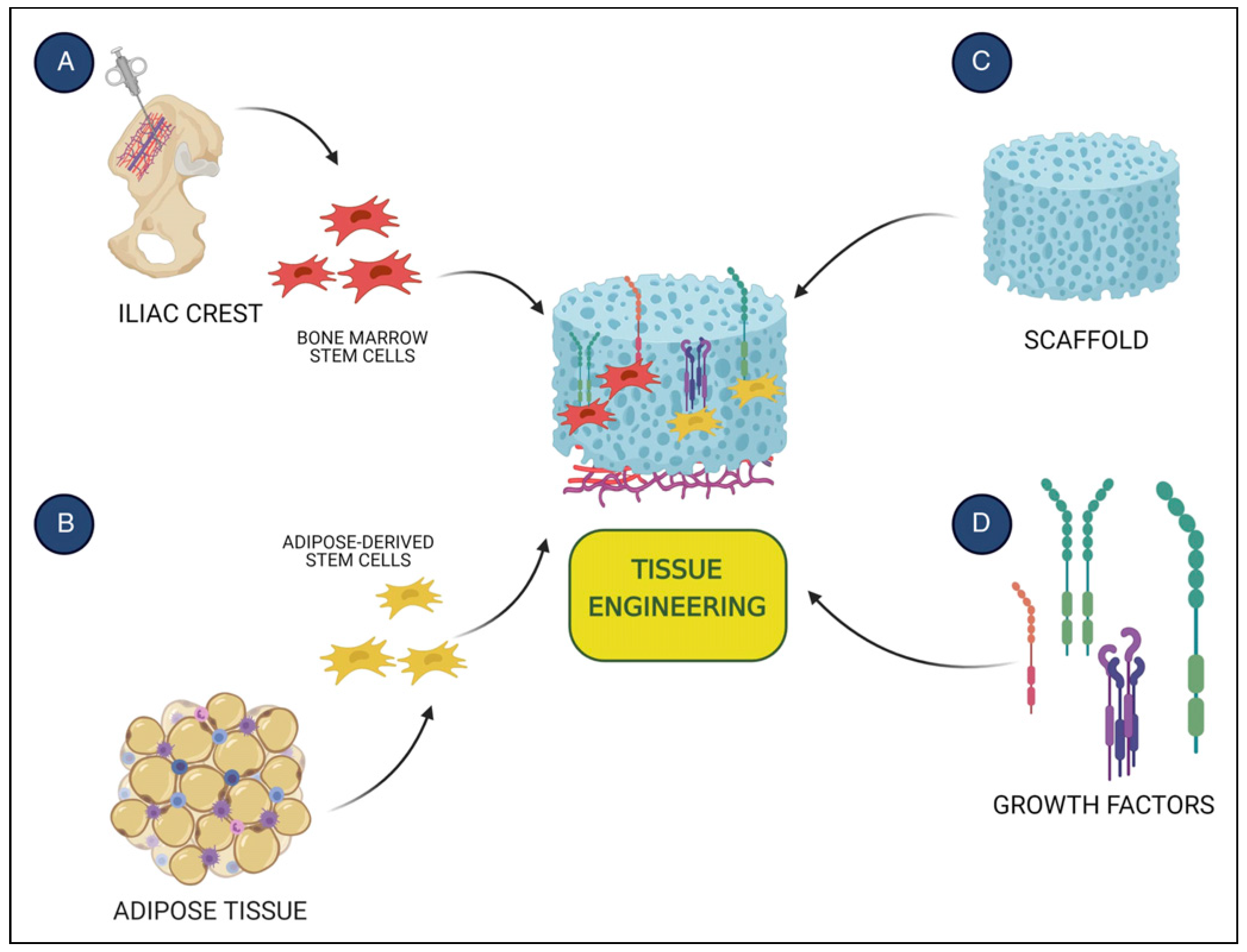
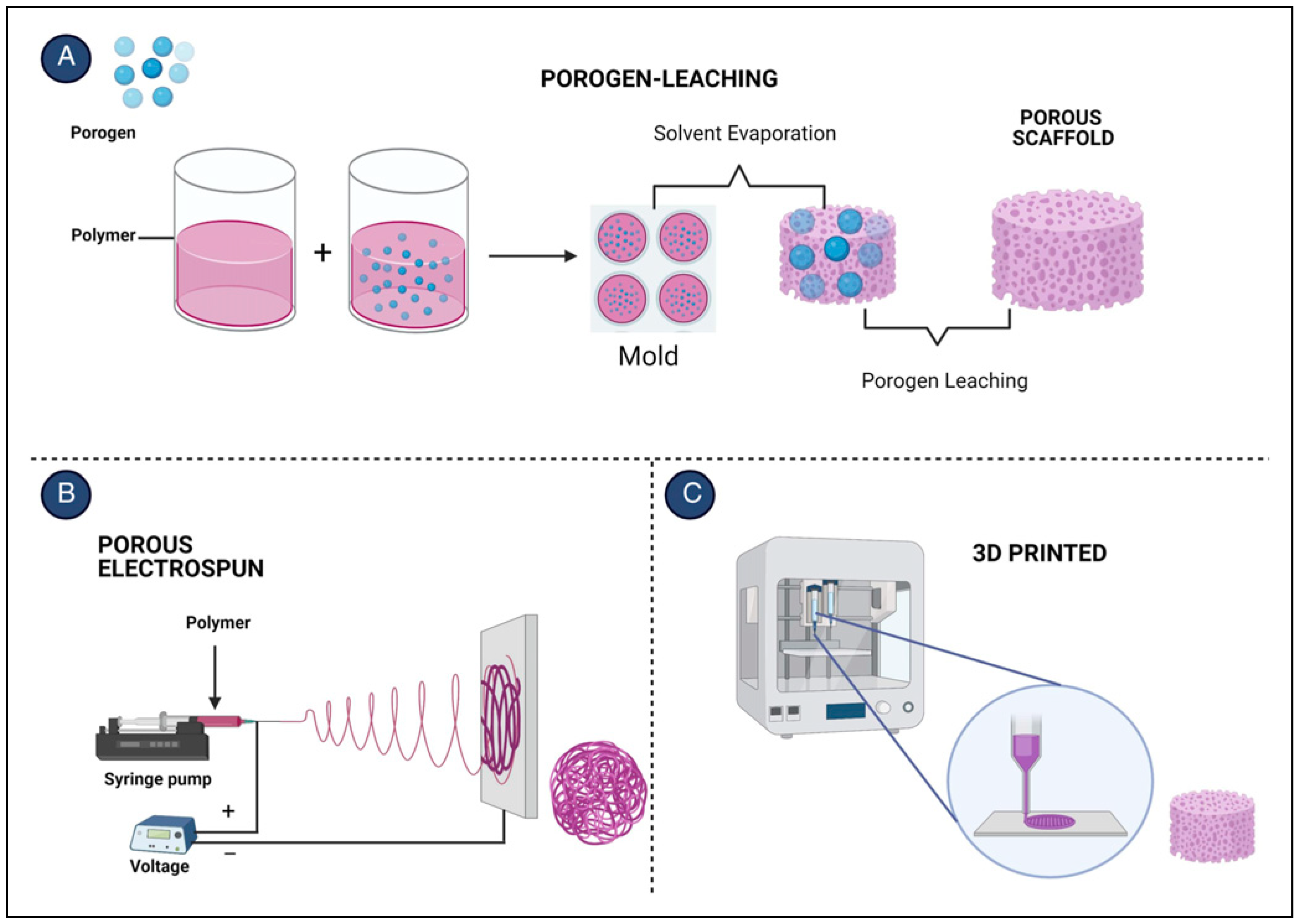
Poly (Lactic-co-Glycolic) Acid Scaffold Features

Clinically approved PLGA with a lactic:glycolic ratio of 85: 15 [15,16,17,18] was used in more than half of the studies, followed by 75:25 [19]. The scaffold’s pore size was well characterized by Zong et al. [14,20], with an average of 280–450 µm determined by scanning electron microscopy (SEM) images. The authors also measured the porosity of the scaffold by ethanol inhalation, reaching 85% porosity. Similar characteristics were described by Huang et al. [16].
Regarding the degradation rate of the material, Morgan et al. [19] found microcavitations on the inner surface as well as higher roughness of the scaffolds using SEM images after 6 weeks of incubation. A modest increase in percentage of mass loss over time showed an increased degradation rate at 26 days, without impact on cell biocompatibility. Zong et al. [14] also calculated the percentage of mass loss of the scaffold. After 8 weeks of incubation, almost 50% of the weight was lost. However, when hydroxyapatite nanoparticles (nHAP) were added to the scaffold, nearly 90% of initial mass was retained. Other relevant scaffold features are summarized in Table 2.

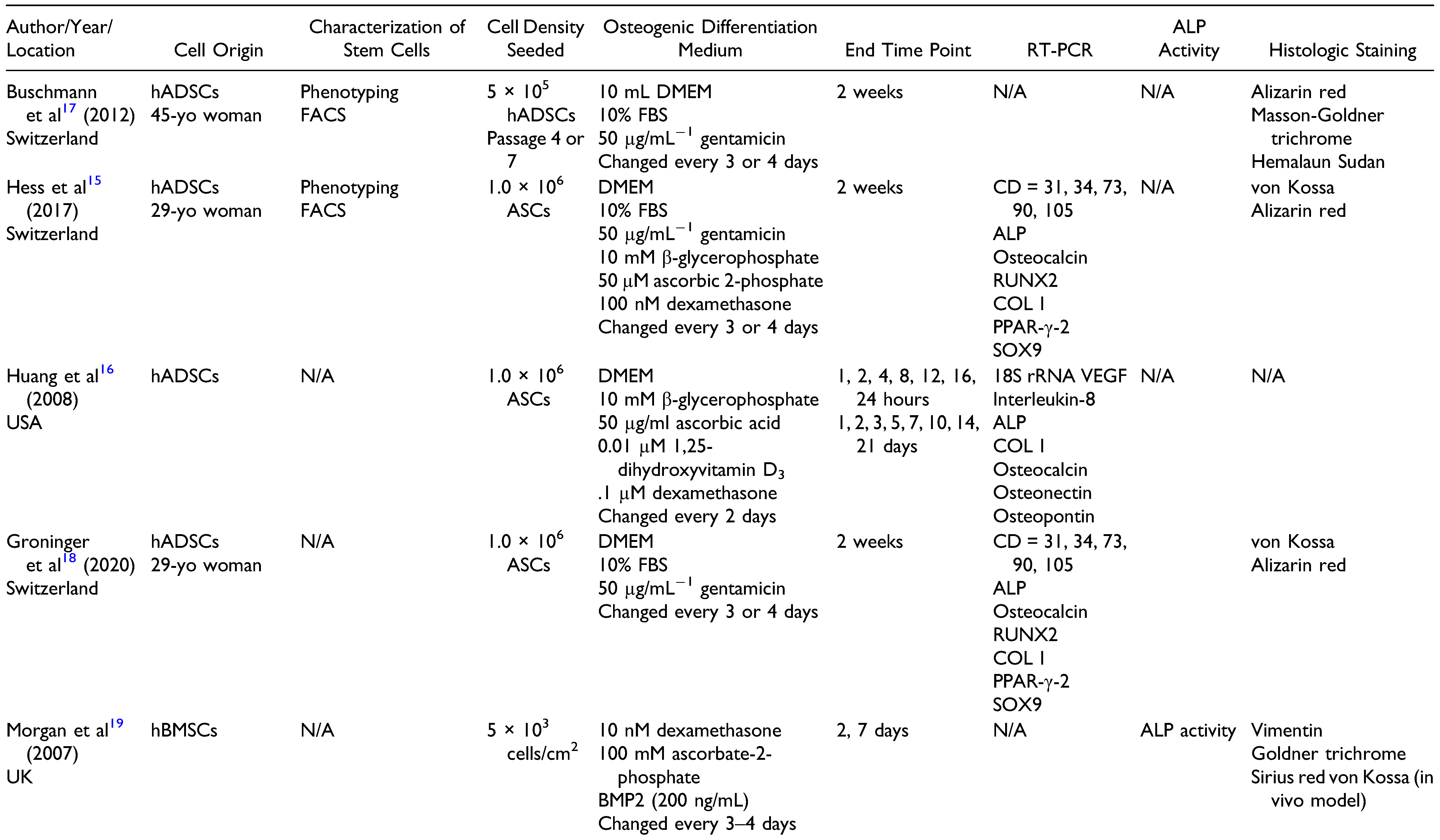
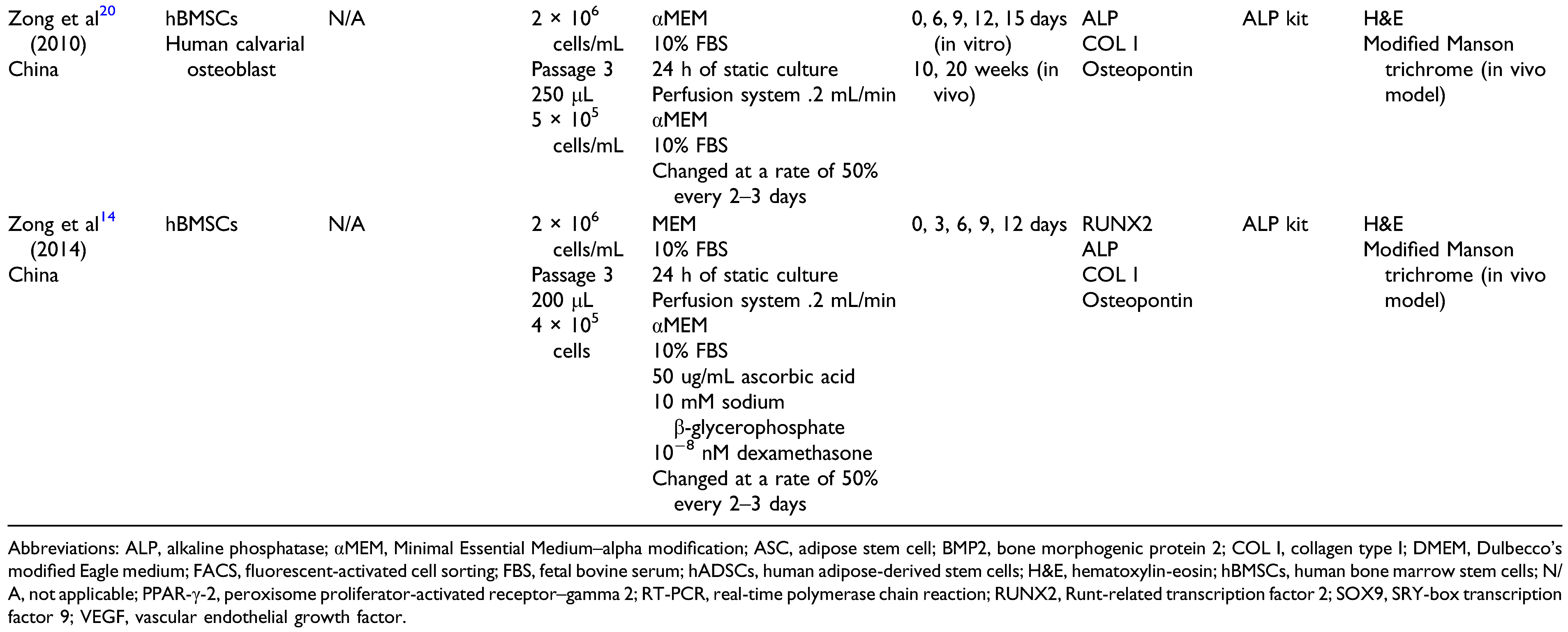
Characterization of hBMSCs in a PLGA Scaffold

Seeded density, viability, adhesion, and cell proliferation. After seeding hBMSCs in PLGA scaffold and plastic (control group) at a concentration of 5 × 10³ cells/cm2 in osteogenic conditions, Morgan et al. [19] demonstrated cell viability by assessing the DNA content of propidium iodine in hBMSCs by flow cytometry analysis. Sub-G1 DNA content identified apoptotic cells. After 6 days in the culture medium, decreased levels of cell death were found in both populations. Additional evidence of cell viability was obtained through the measurement of CellTracker Green and ethidium homodimer for viable and necrotic cells, respectively, with absence of substantial cell death.
Zong et al. [14] added 200 µL of hBMSC suspension to the scaffold, allowing cells to attach for 2 hours. Afterward, the scaffold was put in an osteogenic medium, using a perfusion system. Two scaffold types were studied, PLGA alone and nHAP/PLGA. Cell viability was determined by staining with fluorescein diacetate after culture at 0, 6, and 12 days. Cells in the scaffold were visualized with confocal laser scanning microscopy. Distribution of cells in both scaffolds was similar. On day 0, most of the cells were distributed to the upper surface. However, after being cultured in the perfusion system for 12 days, the distribution in both models became more uniform in the entire scaffold.
After having assessed biocompatibility, Morgan et al. [19] evaluated cell proliferation rate by quantifying DNA levels and detecting carboxy fluorescein succinimidyl ester–labeled cells by flow cytometry. At 120 hours, 25-fold and 26-fold increases in DNA levels were identified on PLGA and plastic, respectively.
In 2010, Zong et al. [20] used a continuous perfusion system as a culture medium. After 24 hours in a static culture, cell density was .42 ± .08 × 106 hBMSCs/scaffold. During perfusion culture, cell density increased gradually until it reached 2.27 ± .27 × 106 hBMSCs/scaffold after 12 days, 5 times the initial number (250 µL = 5 × 10⁵ cell/mL).
In 2014, the same authors compared cell density in a PLGA scaffold to a PLGA/nHAP scaffold seeded in a perfusion system [14]. They found no significant difference in cell density between days 9 and 12 (P > .05) in both scaffolds. Morphology of hBMSCs in the scaffold was evaluated through SEM images, without significant differences between either scaffolds (P> .05). After day 12, cells overlapped, and some extracellular matrix components were observed in both scaffolds.
Osteogenic differentiation assay. Zong et al. [14] performed RT-PCR to analyze osteogenic genes expressed by hBMSCs seeded in a PLGA scaffold after 14 days in an osteogenic medium. Osteogenic genes included were Runt-related transcription factor 2 (RUNX2), also referred to as core-binding factor α-1, collagen type I (COL I), ALP, and osteopontin. All of them were expressed, with higher genic expression when hydroxyapatite was added to the scaffold material (P < .05). Alkaline phosphatase activity was also performed on days 7 and 14, increasing during osteogenic induction. In contrast, genic levels of cells cultured in a growth medium remained low. Significantly higher levels of ALP activity were observed in the PLGA/n-HAP scaffolds (P < .05).
In 2010, the same authors compared the osteogenic activity of undifferentiated hBMSCs (hMSC construct) and osteoblast-like cells derived from human bone marrow (osteoblast construct), seeding the same cell density in a PLGA scaffold and measuring ALP activity [20]. The results showed higher ALP levels in the osteoblast construct scaffold than in the hMSC construct scaffold (P < .01). Likewise, the osteogenic expression of ALP, COL I, and osteopontin was highest in the osteoblast construct, concluding that undifferentiated hBMSCs could be induced into osteoblast-like cells after 14 days in a perfusion culture with an osteogenic medium.

Characterization of hADSCs in a PLGA Scaffold

Seeded density, adhesion, and cell proliferation. The majority of the authors selected the cell density of 1 × 10⁶ hADSCs [15,16,18]. Before being seeded in a PLGA scaffold, Buschmann et al. [17] cultured hADSCs in an osteogenic medium and performed cell phenotyping by fluorescent-activated cell sorting, which demonstrated that typical stem cell markers were expressed with high mean fluorescence intensity. Afterward, 2 PLGA scaffolds, 1 with only PLGA and the other with PLGA plus amorphous calcium phosphate (aCaP), were cultured with 5 × 10⁵ hADSCs. The authors were able to demonstrate uniform 3D ingrowth in both materials. Histologic evaluation showed that hADSCs seeded in PLGA/aCaP retained their morphology. Cell width was significantly different after 1 week in the culture (P < .01).
In 2020, Hess et al. [15] cultured hADSCs in PLGA alone and PLGA/aCaP disk scaffolds (3D) with a cell density of 1.0 × 10⁶ cells during 2 weeks in an osteogenic medium. The same quantity was seeded in flat films (2D) to compare the behavior of hADSCs on different structures. After 2 weeks, cell growth capacity was assessed by hematoxylin-eosin staining and showed that cells seeded in 3D scaffolds migrated and distributed equitably through the fibers. In contrast, cells seeded in 2D films were principally on the surface of the films.
Osteogenic differentiation assay. Groninger et al. [18] compared the genic expression of hADSCs seeded in PLGA alone and PLGA/aCaP scaffolds after 2 weeks, finding downregulation of osteogenic markers in the PLGA/aCap scaffold. Statistically significant upregulation of ALP was found in 3D PLGA scaffold seeded in Dulbecco’s modified Eagle medium (DMEM) (P < .01).
Conversely, Hess et al. [15] seeded hADSCs in 2D films or 3D scaffolds. Human adipose-derived stem cells cultured in 3D scaffolds demonstrated lower ALP levels compared with 2D films, regardless of the medium. Interestingly, if the 3D scaffold was cocultured with 2D films, the gene expression increased. Only RUNX2 was downregulated in the coculture medium. The opposite was seen with osteocalcin, which was upregulated under all conditions. Also, upregulation of CD31 and CD34 in the coculture system was observed.
Huang et al. [16] compared hADSC performance on 2D films to 3D scaffolds and found relatively constant low ALP activity levels over the first 24 hours. Afterward, the expression increased substantially in the 3D scaffold cultures, reaching levels approximately 3.5 times greater at day 14. Also, COL I and osteonectin levels followed similar patterns in both cultures, characterized by low levels of gene expression during the first 14 days with a slight increase after that. There was a nearly 3.5-times greater expression of COL I and osteonectin in day 21 in the 3D scaffold. High expression of angiogenic markers such as vascular endothelial growth factor (VEGF) and interleukin-8 was also reported.
Only Groninger et al. [18] used microcomputed tomography to scan bone tissue formation based on bone volume fraction (bone volume/total volume). After 14 days cultured in vitro in 2 different mediums, DMEM and osteogenic medium, they found an increased bone volume fraction in osteogenic medium compared to DMEM.

Discussion

The ideal components of engineered bone tissue used for bone regeneration are yet to be identified [22]. What has been proven so far is that tissue must have osteogenic (hMSCs), osteoinductive (growth factors), and osteoconductive (scaffold) properties to be able to imitate the complex structure of human bone [23].
The Mesenchymal Stromal Cell Committee of the International Society for Cellular Therapy established several criteria to define hMSCs. One of these criteria is to have the biological capacity to differentiate to osteoblasts, adipocytes, and chondroblasts cultured in vitro. The osteogenic differentiation capacity of hMSCs can be demonstrated by the presence of specific stains, such as alizarin red or von Kossa, in the newly formed bone [24].
Bone marrow is recognized as the central location of hMSCs. However, many other sources that contain these prodigious cells have been identified, such as adipose tissue [25]. Researchers in bone regeneration have demonstrated that hMSCs obtained from these 2 sources have, both in vivo and in vitro, a high capability to be used as the primary source to regenerate bone tissue, with the vast majority favoring hBMSCs [26,27,28,29,30].
Although the superior performance of bone marrow has been more than demonstrated, some advantages of adipose tissue have been identified, redirecting the focus of some researchers towards this resource. In 2006, Kern et al. [31] found that hADSCs showed a high proliferative capacity, the longest in vitro culture period, and were not affected by donor age compared with hBMSCs. Similar results were found by Chen et al., [32] who concluded that patient age and multiple cell passages have less impact on cell proliferation and osteogenic differentiation of hADSCs compared with hBMSCs.
In 2016, El-Badawy et al. [33], based on the emergent use of adipose tissue as a stem cell source due to its abundance and easy harvesting procedure, evaluated the regenerative potency of hADSCs compared with hBMSCs in an animal model, using a novel dielectrophoresis microfluid platform based on printed circuit board technology. They showed that hADSCs were more effective at promoting neovascularization and exhibited greater resistance to senescence related to oxidative stress and apoptosis induced by hypoxia, with stronger angiogenic activity characterized by higher gene expression of the angiogenic markers Oct-3/4 and VEGF (P < .01). Similar results were found by Huang et al. [16].
Accordingly, to potentiate the capacity of hMSCs to regenerate bone, they must be seeded in a similar bone structure. A 3D scaffold offers a structure similar to bone to home the cells (Figure 5). A wide range of materials has been studied, including natural (e.g., collagen, gelatin, hyaluronic acid) or synthetic ceramics (eg hydroxyapatite, β-tricalcium phosphate), and polymers (poly (lactic), poly (glycolic acid), poly (ε caprolactone), poly (propylene fumarate)), each with distinctly unique features in terms of porosity, pore size [34], and surface, which play fundamental roles in the regeneration process [5].
Poly (lactic-co-glycolic) acid is a copolymer approved by the US Food and Drug Administration for clinical use due to its biodegradable, biocompatible, and nontoxic features [35]. Primarily, the 3D PLGA scaffold has a high capability of allowing cell adhesion, proliferation, and ingrowth, as shown by all studies evaluated in this systematic review.
To contribute to this matter, Huang et al. [16] discovered an increase in osteogenic and angiogenic marker expression on hADSCs when they were seeded in a 3D PLGA scaffold. High levels of VEGF and interleukin-8 were evident 1 hour after cell seeding. Vascular endothelial growth factor has shown an essential role in the migration and proliferation of endothelial cells, thus contributing to the angiogenesis process [36].
Similarly, Kim et al. [37] compared the angiogenic potential of hADSCs and hBMSCs in an animal model with a hindlimb vascular injury. Reverse transcriptase PCR and real-time PCR showed higher expression of angiogenic-related genes such as metalloproteinases 3 or 9 in the hADSC-treated groups, with significantly increased blood flow in the ischemic leg 14 days after injury/cell injection compared with the control group.
Another finding that favors the angiogenic effect of hADSCs was found by Hess et al. [15] when high levels of CD34 were detected in cells seeded in a 3D scaffold in osteogenic medium, indicative of the endothelial cell differentiation capacity of hADSCs. This property help to ensure the neovascularization of the scaffold in BTE, that is required to guarantee the presence of oxygen, nutrients, and clearance of waste, all of these essential to cell viability and bone development [38].
Related to osteogenic potential, Hess et al. [15] reported comparable results with Huang et al. [16], showing high expression of osteocalcin and RUNX2 in cells seeded in a 3D scaffold compared with 2D films. Additionally, they cultured 2D films with a 3D scaffold (coculture system) on standard (DMEM) and osteogenic medium. Cells in the coculture system demonstrated higher expression of ALP, COL I, RUNX2, and osteocalcin, and the paracrine effect was clear, where not only cell performance was affected by the material in which they were seeded but also by cellular crosstalk via cytokines and growth factors, called the leveling effect.
There is a tendency to combine different materials to find the ideal scaffold that perfectly mimics natural bone. Different combinations of biopolymers and bioceramics are commonly used, [39,40,41,42] even though they are difficult mechanisms to process [43].
In 2020, Groninger et al. [18] demonstrated that changing the scaffold material surface or the culture medium affected in vitro osteogenic gene expression. Significant downregulation on all osteogenic genes was found when aCaP nanoparticles were added to the scaffold. The cell-material interaction and the material-medium interaction are fundamental factors that should be evaluated in the tissue bioengineering field.
On the other hand, addition of aCaP nanoparticles increased the roughness of the scaffold surface, [44] as shown by Buschmann et al. [19] They compared hADSCs seeded in PLGA to PLGA/aCap showing well cell adhesion and fast, and uniform ingrowth of the cells in both materials. Although, cell morphology modified in a PLGA scaffold became wider and roundish.
Zong et al. [14] demonstrated that the addition of nHAP to the scaffold surface increased osteoconductive property and cell adhesion. It might be attributable to an enhanced pore wall roughness. Also, the increase in osteogenic markers and extracellular matrix speaks of better osteoinductive properties, which were noticed only in the presence of the osteogenic medium.
Biodegradability of the material also should be considered. Biodegradability is the gradual disappearance of the scaffold by the body’s cells over time, producing easy to excrete and nontoxic products. The degradation rate often depends on enzyme activity, especially in natural source materials, making it difficult to predict. The rate of degradation is different for polymers, where the process relies on hydrolysis. By adjusting the composition of the polymer, the hydrophilic part allows control of the degradation rate of the material [45]. Morgan et al., [20] using PLGA with a ratio of 75:25, showed evidence of degradation after 6 weeks of incubation, with no adverse effect on cell performance. A degradation rate increase after 8 weeks was found by Zong et al. [14] They used a PLGA ratio of 85:15 combined with hydroxyapatite, observing a weight loss rate lower in PLGA combined with hydroxyapatite than PLGA alone.
All this confirms the complexity involved in BTE. Many factors must be considered when deciding which components will be part of new bone. Efforts must be oriented towards the balance between biocompatibility, osteoinductivity, osteoconductivity, and biodegradation properties. We found that the PLGA scaffold has demonstrated to enhance the osteogenic and osteoconductive properties of both hBMSCs and hADSCs.

Future Clinical Implications

Bone defects represent one of the most feared complications of any surgeon due to the long time to heal that these entail and the high degree of morbidities associated with the gold standard treatment option as the autologous bone graft. Since PLGA is a biomaterial already FDA-approved, it facilitates the translational ability of clinical practice. Furthermore, the regenerative capacity shown by the hMSC on several experimental in vivo animal models potentiates their use as a therapeutic strategy in humans. The preclinical studies included in this systematic review support the implementation of tissue engineering for future clinical trials to find an excellent bone substitute as an option in place of the autologous bone graft. Thus, the fabrication of a new bone in the bench will contribute to finding an alternative to decrease patient morbidities related to autologous bone grafting procedures and using allografts.
Several limitations are present in this study. Since only studies published in English were included, studies in other languages were not analyzed. Also, there was a scarcity of studies comparing human stem cells performance from different origin and seeded in a specific PLGA scaffold material, as well as the diversity of protocols used. Additionally, there was potential bias in the selection process, and in misinterpreting data and results, all of these being a potential source of bias common to systematic reviews.

Conclusions

This systematic review showed that the osteogenic potential of hBMSCs and hADSCs is enhanced when they are seeded in a PLGA scaffold. Several in vitro studies have established the biocompatibility of cells with this material, increasing their osteogenic and even angiogenic potential when a 3D structure is used as a niche. Different strategies, such as scaffold features, the addition of some inorganic materials, coculture systems, dynamic cultures, or perfusion culture systems during the cellular expansion before in vivo transplantation, have been shown to enhance cellular performance of bone regeneration. Despite not finding a study comparing cells under precisely the same conditions, most of the protocols evaluated in this systematic review showed similarities in cell processing, seeded cell density, scaffold characteristics, and measurement techniques. Even though upregulation of osteogenic gene expression was observed in both cellular linages, the superiority of one vs the other could not be established due to limited data and studies heterogenicity. Future studies that compare the performance of cells under strictly the same conditions considering number of cells seeded, scaffold characteristics and culture features must be carried out.

Funding

The author(s) disclosed receipt of the following financial support for the research, authorship, and/or publication of this article: This study was supported in part by the Mayo Clinic Clinical Research Operations Group and Mayo Clinic Center for Regenerative Medicine. These entities had no role in the study design, in the collection, analysis and interpretation of data; in the writing of the manuscript, or the decision to submit the manuscript for publication.

Declaration of Conflicting Interests

The author(s) declared no potential conflicts of interest with respect to the research, authorship, and/or publication of this article.

References

  1. Ho-Shui-Ling, A.; Bolander, J.; Rustom, L.E.; Johnson, A.W.; Luyten, F.P.; Picart, C. Bone regeneration strategies: Engineered scaffolds, bioactive molecules and stem cells current stage and future perspectives. Biomaterials. 2018, 180, 143–162. [Google Scholar]
  2. Kinaci, A.; Neuhaus, V.; Ring, D.C. Trends in bone graft use in the United States. Orthopedics. 2014, 37, e783–e788. [Google Scholar]
  3. Crist, T.E.; Mathew, P.J.; Plotsker, E.L.; Sevilla, A.C.; Thaller, S.R. biomaterials in craniomaxillofacial reconstruction: Past, present, and future. J Craniofac Surg. 2020, 32, 535–540. [Google Scholar]
  4. Ebraheim, N.A.; Elgafy, H.; Xu, R. Bone-graft harvesting from iliac and fibular donor sites: Techniques and complications. J Am Acad Orthop Surg. 2001, 9, 210–218. [Google Scholar] [PubMed]
  5. Amini, A.R.; Laurencin, C.T.; Nukavarapu, S.P. Bone tissue engineering: Recent advances and challenges. Crit Rev Biomed Eng. 2012, 40, 363–408. [Google Scholar]
  6. Liao, H.T.; Chen, C.T. Osteogenic potential: Comparison between bone marrow and adipose-derived mesenchymal stem cells. World J Stem Cells. 2014, 6, 288–295. [Google Scholar] [PubMed]
  7. Chen, G.; Kawazoe, N. Porous scaffolds for regeneration of cartilage, bone and osteochondral tissue. Adv Exp Med Biol. 2018, 1058, 171–191. [Google Scholar]
  8. Casagrande, S.; Tiribuzi, R.; Cassetti, E.; et al. Biodegradable composite porous poly(dl-lactide-co-glycolide) scaffold supports mesenchymal stem cell differentiation and calcium phosphate deposition. Artif Cells Nanomed Biotechnol. 2018, 46 (Suppl. 1), 219–229. [Google Scholar]
  9. Bharadwaz, A.; Jayasuriya, A.C. Recent trends in the application of widely used natural and synthetic polymer nanocomposites in bone tissue regeneration. Mater Sci Eng C Mater Biol Appl. 2020, 110, 110698. [Google Scholar]
  10. Friedenstein, A.J.; Petrakova, K.V.; Kurolesova, A.I.; Frolova, G.P. Heterotopic of bone marrow. Analysis of precursor cells for osteogenic and hematopoietic tissues. Transplantation. 1968, 6, 230–247. [Google Scholar]
  11. Mizuno, H.; Tobita, M.; Uysal, A.C. Concise review: Adipose-derived stem cells as a novel tool for future regenerative medicine. Stem Cells. 2012, 30, 804–810. [Google Scholar] [CrossRef] [PubMed]
  12. Pendleton, C.; Li, Q.; Chesler, D.A.; Yuan, K.; Guerrero-Cazares, H.; Quinones-Hinojosa, A. Mesenchymal stem cells derived from adipose tissue vs bone marrow: In vitro comparison of their tropism towards gliomas. PLoS One. 2013, 8, e58198. [Google Scholar] [CrossRef]
  13. Sterne, J.A.; Hernán, M.A.; Reeves, B.C.; et al. ROBINS-I: A tool for assessing risk of bias in non-randomised studies of interventions. BMJ. 2016, 355, i4919. [Google Scholar] [CrossRef]
  14. Zong, C.; Qian, X.; Tang, Z.; et al. Biocompatibility and bone-repairing effects: Comparison between porous poly-lactic-co-glycolic acid and nano-hydroxyapatite/poly(lactic acid) scaffolds. J Biomed Nanotechnol. 2014, 10, 1091–1104. [Google Scholar] [PubMed]
  15. Hess, S.C.; Stark, W.J.; Mohn, D.; et al. Gene expression in human adipose-derived stem cells: Comparison of 2D films, 3D electrospun meshes or co-cultured scaffolds with two-way paracrine effects. Eur Cell Mater. 2017, 34, 232–248. [Google Scholar] [CrossRef] [PubMed]
  16. Huang, C.K.; Huang, W.; Zuk, P.; et al. Genetic markers of osteogenesis and angiogenesis are altered in processed lipoaspirate cells when cultured on three-dimensional scaffolds. Plast Reconstr Surg. 2008, 121, 411–423. [Google Scholar] [CrossRef]
  17. Buschmann, J.; Harter, L.; Gao, S.; et al. Tissue engineered bone grafts based on biomimetic nanocomposite PLGA/amorphous calcium phosphate scaffold and human adipose-derived stem cells. Injury. 2012, 43, 1689–1697. [Google Scholar]
  18. Groninger, O.; Hess, S.; Mohn, D.; et al. Directing stem cell commitment by amorphous calcium phosphate nanoparticles incorporated in PLGA: Relevance of the free calcium ion concentration. Int J Mol Sci. 2020, 21, 2627. [Google Scholar]
  19. Morgan, S.M.; Tilley, S.; Perera, S.; et al. Expansion of human bone marrow stromal cells on poly-(DL-lactide-coglycolide) (PDL LGA) hollow fibres designed for use in skeletal tissue engineering. Biomaterials. 2007, 28, 5332–5343. [Google Scholar] [CrossRef]
  20. Zong, C.; Xue, D.; Yuan, W.; et al. Reconstruction of rat calvarial defects with human mesenchymal stem cells and osteoblast-like cells in poly-lactic-co-glycolic acid scaffolds. Eur Cell Mater. 2010, 20, 109–120. [Google Scholar]
  21. Zuk, P.A.; Zhu, M.; Mizuno, H.; et al. Multilineage cells from human adipose tissue: Implications for cell-based therapies. Tissue Eng. 2001, 7, 211–228. [Google Scholar] [CrossRef]
  22. Oliveira, É.R.; Nie, L.; Podstawczyk, D.; et al. Advances in growth factor delivery for bone tissue engineering. Int J Mol Sci. 2021, 22, 903. [Google Scholar] [CrossRef]
  23. Pereira, H.F.; Cengiz, I.F.; Silva, F.S.; Reis, R.L.; Oliveira, J.M. Scaffolds and coatings for bone regeneration. J Mater Sci Mater Med. 2020, 31, 27. [Google Scholar]
  24. Dominici, M.; Le Blanc, K.; Mueller, I.; et al. Minimal criteria for defining multipotent mesenchymal stromal cells. The International society for cellular therapy position statement. Cytotherapy. 2006, 8, 315–317. [Google Scholar] [CrossRef] [PubMed]
  25. Satija, N.K.; Singh, V.K.; Verma, Y.K.; et al. Mesenchymal stem cell-based therapy: A new paradigm in regenerative medicine. J Cell Mol Med. 2009, 13, 4385–4402. [Google Scholar] [PubMed]
  26. Im, G.I.; Shin, Y.W.; Lee, K.B. Do adipose tissue-derived mesenchymal stem cells have the same osteogenic and chondrogenic potential as bone marrow-derived cells? Osteoarthritis Cartilage. 2005, 13, 845–853. [Google Scholar]
  27. Liu, T.M.; Martina, M.; Hutmacher, D.W.; Hui, J.H.P.; Lee, E.H.; Lim, B. Identification of common pathways mediating differentiation of bone marrow- and adipose tissue-derived human mesenchymal stem cells into three mesenchymal lineages. Stem Cells. 2007, 25, 750–760. [Google Scholar]
  28. Vishnubalaji, R.; Al-Nbaheen, M.; Kadalmani, B.; Aldahmash, A.; Ramesh, T. Comparative investigation of the differentiation capability of bone-marrow- and adipose-derived mesenchymal stem cells by qualitative and quantitative analysis. Cell Tissue Res. 2012, 347, 419–427. [Google Scholar] [CrossRef]
  29. Park, S.H.; Sim, W.Y.; Min, B.H.; Yang, S.S.; Khademhosseini, A.; Kaplan, D.L. Chip-based comparison of the osteogenesis of human bone marrow- and adipose tissue-derived mesenchymal stem cells under mechanical stimulation. PLoS One. 2012, 7, e46689. [Google Scholar] [CrossRef]
  30. Shafiee, A.; Seyedjafari, E.; Soleimani, M.; Ahmadbeigi, N.; Dinarvand, P.; Ghaemi, N. A comparison between osteogenic differentiation of human unrestricted somatic stem cells and mesenchymal stem cells from bone marrow and adipose tissue. Biotechnol Lett. 2011, 33, 1257–1264. [Google Scholar]
  31. Kern, S.; Eichler, H.; Stoeve, J.; Klüter, H.; Bieback, K. Comparative analysis of mesenchymal stem cells from bone marrow, umbilical cord blood, or adipose tissue. Stem Cells. 2006, 24, 1294–1301. [Google Scholar] [CrossRef] [PubMed]
  32. Chen, H.T.; Lee, M.J.; Chen, C.H.; et al. Proliferation and differentiation potential of human adipose-derived mesenchymal stem cells isolated from elderly patients with osteoporotic fractures. J Cell Mol Med. 2012, 16, 582–593. [Google Scholar] [CrossRef]
  33. El-Badawy, A.; Amer, M.; Abdelbaset, R.; et al. Adipose stem cells display higher regenerative capacities and more adaptable electro-kinetic properties compared to bone marrow-derived mesenchymal stromal cells. Sci Rep. 2016, 6, 37801. [Google Scholar]
  34. Gosain, A.K.; Riordan, P.A.; Song, L.; et al. A 1-year study of osteoinduction in hydroxyapatite-derived biomaterials in an adult sheep model: Part, I.I. Bioengineering implants to optimize bone replacement in reconstruction of cranial defects. Plast Reconstr Surg. 2004, 114, 1155–1163; discussion 1164–1155. [Google Scholar]
  35. Danhier, F.; Ansorena, E.; Silva, J.M.; Coco, R.; Le Breton, A.; Préat, V. PLGA-based nanoparticles: An overview of biomedical applications. J Control Release. 2012, 161, 505–522. [Google Scholar] [PubMed]
  36. Ferrara, N.; Bunting, S. Vascular endothelial growth factor, a specific regulator of angiogenesis. Curr Opin Nephrol Hypertens. 1996, 5, 35–44. [Google Scholar] [PubMed]
  37. Kim, Y.; Kim, H.; Cho, H.; Bae, Y.; Suh, K.; Jung, J. Direct comparison of human mesenchymal stem cells derived from adipose tissues and bone marrow in mediating neovascularization in response to vascular ischemia. Cell Physiol Biochem. 2007, 20, 867–876. [Google Scholar]
  38. Mokhtari-Jafari, F.; Amoabediny, G.; Dehghan, M.M. Role of biomechanics in vascularization of tissue-engineered bones. J Biomech. 2020, 110, 109920. [Google Scholar]
  39. Bhuiyan, D.B.; Middleton, J.C.; Tannenbaum, R.; Wick, T.M. Mechanical properties and osteogenic potential of hydroxyapatite-PLGA-collagen biomaterial for bone regeneration. J Biomater Sci Polym Ed. 2016, 27, 1139–1154. [Google Scholar]
  40. Bhuiyan, D.B.; Middleton, J.C.; Tannenbaum, R.; Wick, T.M. Bone regeneration from human mesenchymal stem cells on porous hydroxyapatite-PLGA-collagen bioactive polymer scaffolds. Biomed Mater Eng. 2017, 28, 671–685. [Google Scholar]
  41. Qi, H.; Ye, Z.; Ren, H.; et al. Bioactivity assessment of PLLA/PCL/HAP electrospun nanofibrous scaffolds for bone tissue engineering. Life Sci. 2016, 148, 139–144. [Google Scholar] [CrossRef] [PubMed]
  42. Zhao, B.; Xu, H.; Gao, Y.; et al. Promoting osteoblast proliferation on polymer bone substitutes with bone-like structure by combining hydroxyapatite and bioactive glass. Mater Sci Eng C Mater Biol Appl. 2019, 96, 1–9. [Google Scholar] [CrossRef] [PubMed]
  43. Shuai, C.; Yu, L.; Feng, P.; Gao, C.; Peng, S. Interfacial reinforcement in bioceramic/biopolymer composite bone scaffold: The role of coupling agent. Colloids Surf B Biointerfaces. 2020, 193, 111083. [Google Scholar] [CrossRef] [PubMed]
  44. Boyan, B.D.; Hummert, T.W.; Dean, D.D.; Schwartz, Z. Role of material surfaces in regulating bone and cartilage cell response. Biomaterials. 1996, 17, 137–146. [Google Scholar] [CrossRef]
  45. Du, B.; Yin, H.; Chen, Y.; et al. A waterborne polyurethane 3D scaffold containing PLGAwith a controllable degradation rate and an anti-inflammatory effect for potential applications in neural tissue repair. J Mater Chem B. 2020, 8, 4434–4446. [Google Scholar] [CrossRef]
Figure 1. The 3 main components of the tissue engineering include: stem cells from different sources such as (A) bone marrow or (B) adipose tissue, (C) the structure that serves as support for these cells and (D) biological molecules that enhance cell’s performance. Created with BioRender.
Cmtr 17 00012 g001
Figure 2. Individualized bias. Green color represents low risk and red color represents high risk of bias.
Cmtr 17 00012 g002
Figure 3. Cross-sectional studies bias. (+) indicates absence, and (—) indicates presence of bias.
Cmtr 17 00012 g003
Figure 4. Preferred reporting items for systematic reviews and meta-analyses (PRISMA) flow diagram. Included and excluded studies. hMSC indicates human mesenchymal stem cell; PLGA, poly (lactic-co-glycolic) acid.
Cmtr 17 00012 g004
Figure 5. Fabrication techniques applied in the scaffold building (A) porogen leaching (B) electrospinning (C) 3-Dimensional bio-printed. Created with BioRender.
Cmtr 17 00012 g005
Table 1. Summary of Characteristics of Included Articles.
Cmtr 17 00012 i001Cmtr 17 00012 i002
Table 2. Poly (Lactic-co-Glycolic Acid) Scaffold Features.
Cmtr 17 00012 i003

