Next Article in Journal
Systematic Mapping Review of Orthognathic Surgery (Protocol)
Previous Article in Journal
Extracorporealization of the Mandibular Condyle: Effects on Viability and Function
 
 
Craniomaxillofacial Trauma & Reconstruction is published by MDPI from Volume 18 Issue 1 (2025). Previous articles were published by another publisher in Open Access under a CC-BY (or CC-BY-NC-ND) licence, and they are hosted by MDPI on mdpi.com as a courtesy and upon agreement with Sage.
Article

Nine Years of Retrospective Study of Mandibular Fractures in Semi-urban Teaching Hospital, Shimla, Himachal Pradesh, India

by
Narotam K. Ghezta
*,
Yogesh Bhardwaj
,
Rangila Ram
,
Monika Parmar
,
Rowena N. Basi
and
Pooja Thakur
Department of Oral and Maxillofacial Surgery, Himachal Pradesh Government Dental College and Hospital, Room no 506, Shimla 171001, India
*
Author to whom correspondence should be addressed.
Craniomaxillofac. Trauma Reconstr. 2023, 16(2), 138-146; https://doi.org/10.1177/19433875221095984
Submission received: 1 November 2021 / Revised: 1 December 2021 / Accepted: 1 January 2022 / Published: 3 May 2022

Abstract

:
Study Design: Retrospective study. Objective: To evaluate current trends in maxillofacial trauma, a retrospective study of mandibular fractures at Government Dental College and Hospital Shimla H.P was carried out. Methods: In this retrospective study, records of 910 patients with mandibular fractures were reviewed between 2007 and 2015 in the Department of Oral and Maxillofacial Surgery out of total 1656 facial fractures. These mandibular fractures were assessed according to age, sex, aetiology in addition to monthly and yearly distribution. Post-operative complications such as malocclusion, neurosensory disturbances and infection were recorded. Results: It was observed that mandibular fractures were most frequent in males (67.5%) and in the age group of 21–30 years; accidental fall was the most common etiological factor (43.8%) in the present study and in stark difference to already published reports. The most common fracture site was that of condylar region 239 (26.2%). Open reduction and internal fixation (ORIF) were done in 67.3% cases whereas 32.6% were managed by maxillomandibular fixation and circummandibular wiring. Miniplate osteosynthesis was the most favoured technique. The complication with ORIF was 16%. Conclusions: To treat mandibular fractures, currently there are many techniques. However, in minimizing complications and in achieving satisfactory functional and aesthetic results experienced surgical team plays an important role.

Introduction

The face is one of the most commonly injured areas of the facial skeleton that both aesthetically and functionally can have long term consequence [1,2]. Around the globe various studies on the incidence of maxillofacial trauma have been previously reported, and the main cause of facial trauma worldwide are assaults and road traffic accidents, but can vary from one nation to another [3,4]. Mandible is a U-shaped bone with thick buccal and lingual cortices and thin medullary cavity. Despite the fact that mandible is one of the largest and strongest bone of the facial region, it is one of the most commonly fractured maxillofacial bone due to its prominent position on the face [1,5]. Mandible is the only mobile bone of an otherwise fixed facial skeleton, due to which a fractured part cannot be left unnoticed because it is very painful and worsens with mastication and phonation movements [6]. These injuries may lead to severe consequences if not identified or treated inappropriately mainly in the form of facial asymmetry, speech impairment, airway reduction, difficulty in swallowing, deranged occlusion or temporo-mandibular joint disorder [7]. Moreover better understanding of the demographic pattern regarding mandibular fracture can help health care provider to plan the management of injuries of maxillo-facial region and such demographic information can be used for funding of public health programs and prevention of such injuries in the future [8]. Previously, numerous studies on population groups from every continent had been conducted by independent investigators, with a common goal to know the nature of mandibular injuries. This retrospective study was done in an effort to evaluate incidence, aetiology, anatomical distribution and trends in the management of maxillofacial trauma from 2007 to 2015 at GDC Shimla. The Government Dental College (GDC) and Hospital, Shimla is the only teaching institute and a tertiary care center for the state of Himachal Pradesh, rendering the treatment to a hilly and remote area and receives patients from the entire state.

Materials and Methods

In the nine years of this retrospective study, the records, radiographs and computed tomograms (CT. Scan) of 910 patients presenting with mandibular fractures were reviewed from the year 2007 to 2015 in the Department of Oral and Maxillofacial Surgery GDC, Shimla, Himachal Pradesh, India. Data regarding age, gender, cause of injury, location of the fracture, type of treatment and post-operative complications were recorded. Treatment of the fractured bone was done by open reduction and internal fixation by an intra-oral or extra-oral approach and osteosynthesis done with the 2.0 titanium mini-plate systems and lag screws in 613 (67.3%) patients, whereas in 297 (32.6%) patients the mandibular fracture was treated by closed reduction and maxillo-mandibular fixation (MMF). The location of the fracture was evaluated on conventional radiographs, computed tomograms and by surgical procedures. All patients treated for mandibular fractures whether admitted to hospital or seen on an out-door basis were included in this study. This retrospective study was exempted from Institutional Review Board approval and was conducted in accordance with the Declaration of Helsinki.

Results

Mandibular Fracture Prevalence

During the nine years study, 910 patients were found to have at least one mandibular fracture as part of their injury, with a total of 1656 maxillo-facial fractures. Thus, of the entire sample of maxillo-facial injuries, 54.9% of patients had mandibular fractures. The cause of injury was classified as accidental fall, road traffic accident (RTA), interpersonal violence, animal assault, pathological, sports-related injuries and others. Anatomically, mandibular fractures were classified as canine, body, angle, condyle, coronoid process, supra-angular and alveolar process.

Age and Gender Distribution of Mandibular Fracture Patients

Patient with mandibular fracture ranged from 36-month-old boy involved in a fall while playing to a 72-year-old female who sustained mandibular fractures in road traffic accident (RTA). There were 67.5% (n = 615) males of all mandibular fractures; whereas female accounted for 32.4% (n = 295). The majority of the mandibular fracture patients were male in the age group of 21–40 years, with the peak incidence occurring at the age group of 21 to 30 years (19.5%). However, the peak incidence for women occurred in the age group of 31–40 years (6.3%) (Table 1, Figure 1).

Mandibular Fracture Distribution According to Year and Month

The total number of mandibular fractures per year was constant except in the year 2012. Approximately 90–115 patients were seen each year except in 2012 when only 73 patients were seen (Table 1, Figure 1). Incidence was highest in the month of April (12.9%) and lowest in the month of February and December (5.3%) (Table 2, Figure 2).

Mandibular Fracture Aetiology

The major aetiology of mandibular fractures was fall in our study comprising of 399 cases (43.8%) of the entire sample (910 patients). Road traffic accident (RTA) was the second major cause (32.5%, 296 patients), followed by interpersonal violence (10%, 91 patients), sports (2.4%, 22 patients), animal assault (3.9%, 36 patients), pathological fractures (4.5%, 41 patients) and others (2.7%, 25 patients Table 3, Figure 3). Fractures from fall were more common in males (63.4%, 253 patients) than females (36.5%, 146 patients), and the peak incidence of fall in both male and female was in the age group of 21–30 years. 63.4% of the male and 36.5% of the female were involved in road traffic accidents. The majority of mandibular fractures occurred in twenty-one to thirty years of age group in both genders. Alleged Inter personnel violence accounted for only 10% (91 patients) in which 6.9%% of males (63 patients) and 3% of the females (28 patients) were involved. Of the patients whose injuries were related to sports activity (2.4%, 22 patients), 1.7% of the male and .7% of the female sustained fractured mandible. Patients sustaining fractures related to animal assault (3.9% 36 patients) occurred predominately in male (2.9%) than .9% in female patients. Other factors related to mandible fractures include iatrogenic (4 patients), epilepsy (11 patients) and gunshot injury (10 patients) that comprised of 2.7% of the total mandibular fractures. Gunshot injury-related mandibular fractures occurred only in males in the age group of 31–40 years. Most of the patients with cysts and tumors presented with pathological fracture of mandible and occurred predominantly in male patients (29,70.7%) in the age group of 51–60 years, and less commonly in females (12, 29.2%) with peak incidence occurring in 41–70 years of age.

Location of Mandibular Fractures

There were total of 910 mandibular fractures in the 1656 facial fractures averaging 1.8 mandibular fractures of total facial fractures. The most common fractures in this study were that of condyle, followed by the canine region of mandible. Angle fractures of the mandible were third in prevalence. Seven hundred eleven patients had a single mandibular fracture, whereas one hundred ninety-nine patients had two fractures. Among the 910 patients who sustained one fracture only, 186 (20.4%) were in the canine region followed by 169 (18.5%) in the angle region and 156 (17.1%) in the condylar region. Among bilateral fractures, 83 (9.1) were in the condylar region, 36 (3.9%) in the angle region and 31 (3.4%) in the canine region (Table 4, Figure 4).

Other Fractures Associated with Mandibular Fractures

Mandibular fractures occurred in 54.9% (910) of the total 1656 patients in this study. Other fractures of the facial region that occurred in association with mandibular fractures were 746 (45%) of the total sample. The most common facial fractures associated with fractured mandible were lefort fractures followed by fractures of zygomatic complex and naso-orbito-ethmoid region (Table 5, Figure 5).

Treatment of Mandibular Fractures

Six hundred and thirteen cases (67.3%) were treated with internal fixation technique whereas two hundred ninety-seven (32.6%) cases were treated by maxillo-mandibular fixation (MMF) and circummandibular wiring out of total 910 patients (Table 6, Figure 6). The treatment modality chosen in 67.3% of the 910 cases operated with internal fixation was mini plate osteosynthesis and cortical screw by intra-oral or extra-oral method. Majority of the patients were operated in the first week of admission or the same day except medically compromised patients until anesthetic clearance. Post-operatively, 67% of the patients did not require maxillo-mandibular fixation. Guiding elastics were used in 21% of cases for an average of 7–10 days and heavy elastic traction or wire maxilla-mandibular fixation was used in 12% of cases for a period of 2–4 weeks particularly in bilateral condylar fractures where only one side of the condyle was operated. The conservatively managed cases were treated using wire or elastic MMF for a period of 1–4 weeks followed by active jaw exercises and physiotherapy. In case of pediatric patients open reduction and internal fixation was avoided unless anatomical reduction was required which could not be achieved by using conservative treatment modalities. In long term follow-up, post-operative infection was the most common complication recorded in this series; however, these patients responded and resolved to appropriate antimicrobial therapies after culture and sensitivity test and removal of the plates. Plate removal was performed under local anesthesia in most of the infected cases. In the postoperative period, nerve-related problems were found in 4.3% (40 cases) which later resolved over time in 3 weeks to 3 months except in three cases where permanent neuro-sensory deficit occurred mainly due to overenthusiastic and inexperienced surgeons. The occlusal discrepancy occurred in 43 cases (4.7%) which got corrected by a minor occlusal adjustment in the majority of cases except in eleven cases (1.2%) where there was non-union of the bony fragments. The non-union occurred either because of infection or plate fracture. Sialocele occurred in seventeen (1.8%) post-surgical cases in the condylar region which got resolved after conservative management such as pressure bandage within a time period of 7–10 days. Complication of mandibular fractures is shown in Table 7, Figure 7.

Discussion

World Health Organization (WHO) statistics indicate that 1 million people die, and between 15 and 20 million are injured annually in road traffic accidents [9]. The cause of the injury is influenced by geographic and socioeconomic conditions, culture, religion and era which varies from nation to nation [8,10,11]. The results of our investigations particularly with regard to age and gender are by enlarge in agreement with the previous reports [12]. Males were injured commonly between the age group of 21–30 years, with a permanence of male subjects reported widely mainly due to involvement in dangerous sport activities and reckless motor vehicle driving [8]. Although in regards to the fracture of the facial skeleton, various causative factors have been mentioned. Some studies have reported motor vehicle related accidents as the major etiology of facial fractures,[8,11,13,14,15]. whereas others show that assault as the most frequent cause [12,16,17,
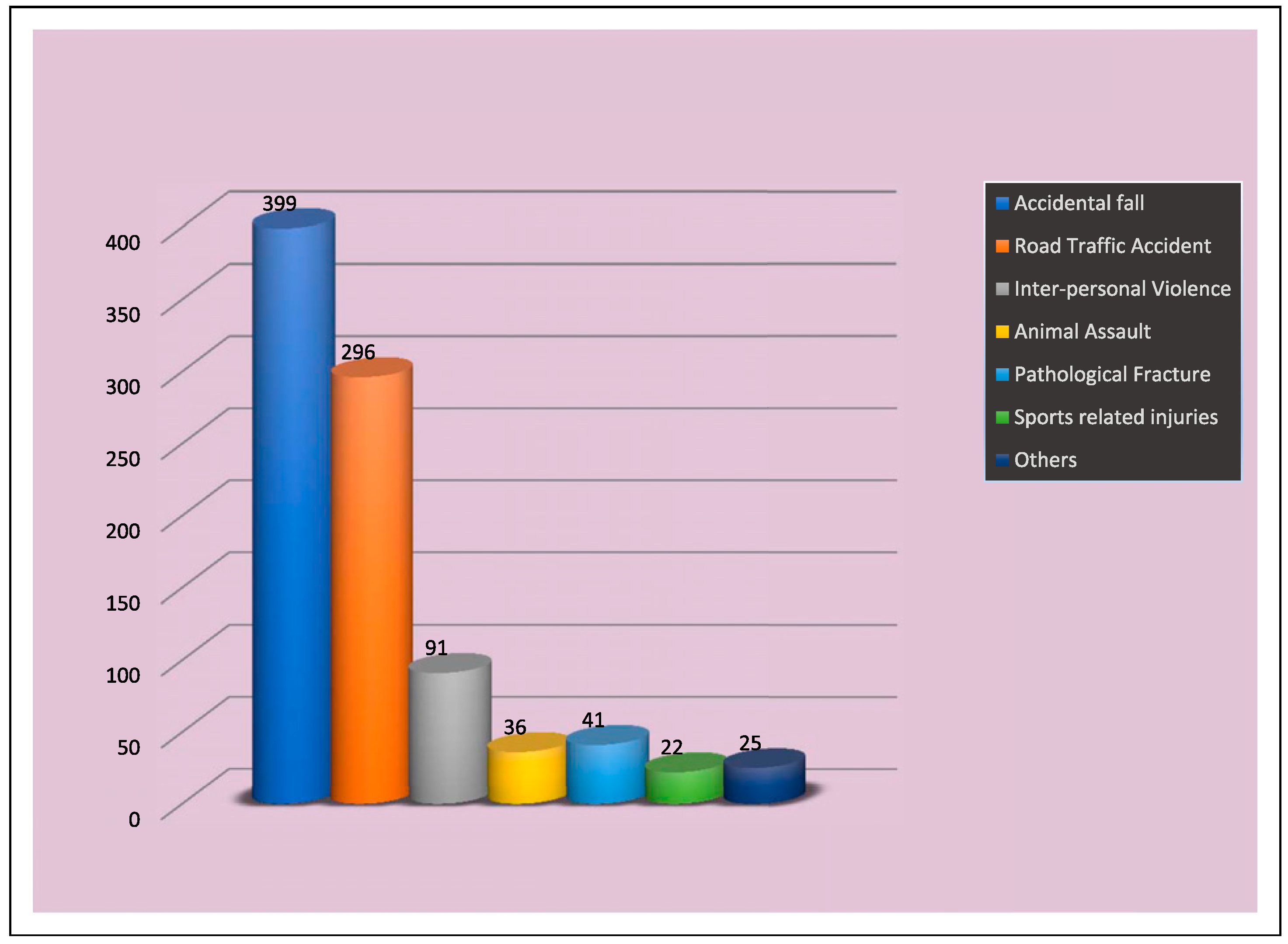
18]. The result of the present retrospective study was in stark difference to already published reports because, in our series 399 (43.8%) patients sustained mandibular fractures mainly due to fall (Table 3, Figure 3). This may be explained by the fact that the area of the current study is the Himalayan hilly terrain. The increased propensity to fall may be explained by the fact that motorable roads in the hilly region are scanty, especially in the rural areas, and people use temporary walking pathways to go from one place to another. These narrow non-mettle pathways on steep hills are responsible for the increased susceptibility of fall by slipping. With Himachal Pradesh being one of the most northern parts of the country with long winters, fall on the snow may explain the situation in this part of the country. The reported incidence of mandibular fractures in the literature ranges from 3 to 20% whereas already explained in our study, the incidence of mandibular fractures due to fall as high as 43.8%. Another significant etiological factor was road traffic accident consisting of 296 (32.5%) of the total mandibular fractures. In Himachal Pradesh, the factors involved are bad conditions of road, less effective law enforcement (especially for over speeding and drunk driving), increase in traffic, less use of helmets and seat belts, deep curved roads and less tolerance among youngsters, which is confirmed by the pre-eminent cause of their mandibular fractures. In the literature, fight or interpersonal violence (IPV) are reported being the most common causes of mandibular fractures in rural and farming population and in various ethnic groups [17,
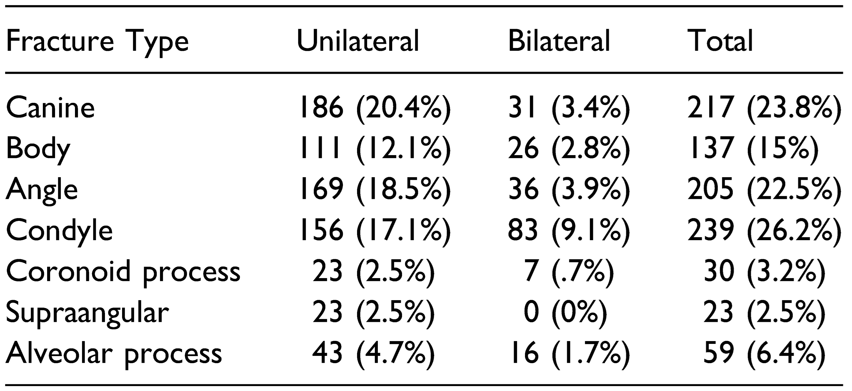
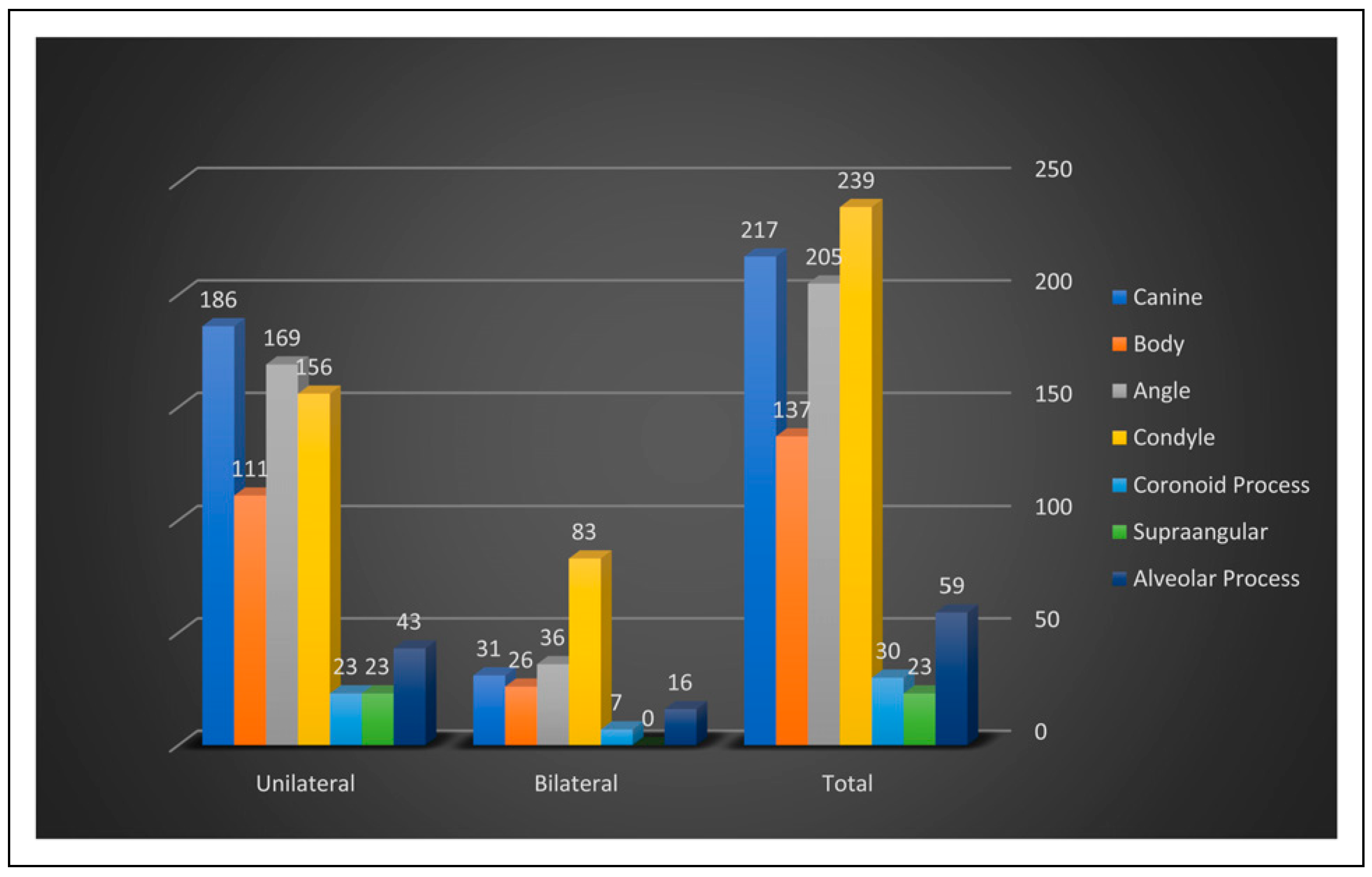
18]. Contrary to this, it is interesting that only a small percentage of the mandibular fractures were caused by I.P.V 91 (10%) in our study. The reason may be that Himachal is a small and peaceful state with low population density compared to other parts of the world. Although the fractures of the mandible have been studied vastly, studies depicting the relation between fracture site and cause are rare [17,19]. The location of the fracture seems to bank on the cause. The major portion of the fall-related fractures of the mandible in our study involved the condyle and canine, which are largely in agreement with the previous reports [4,10,16], particularly that a fall on the chin results in a high incidence of such fractures [20]. On the other hand, the fractures sustained in road traffic accidents (RTA) generally, were fractures of the condylar region and angle of the mandible and also include few fractures through the canine and body region which is in accordance with the finding of previous published work [17]. In assault, there was a predominance of angle and body fracture in the present study and the least to be affected was coronoid which was consistent with the earlier reports [
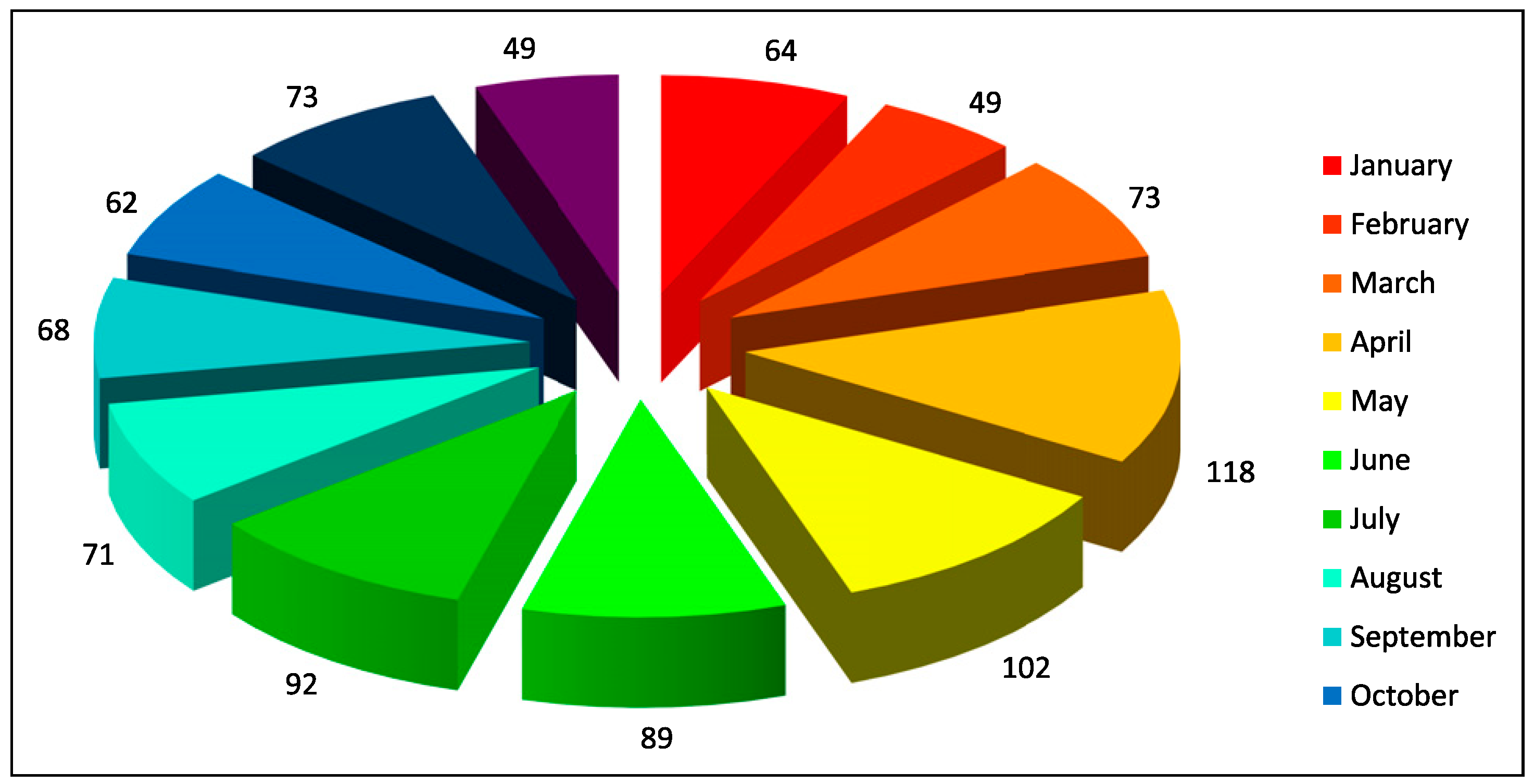
21]. Sports-related mandibular fractures were common in many countries, but they were few in our series. The monthly incidence of fractures of mandible was fairly constant with seasonal variations as reported in earlier studies [
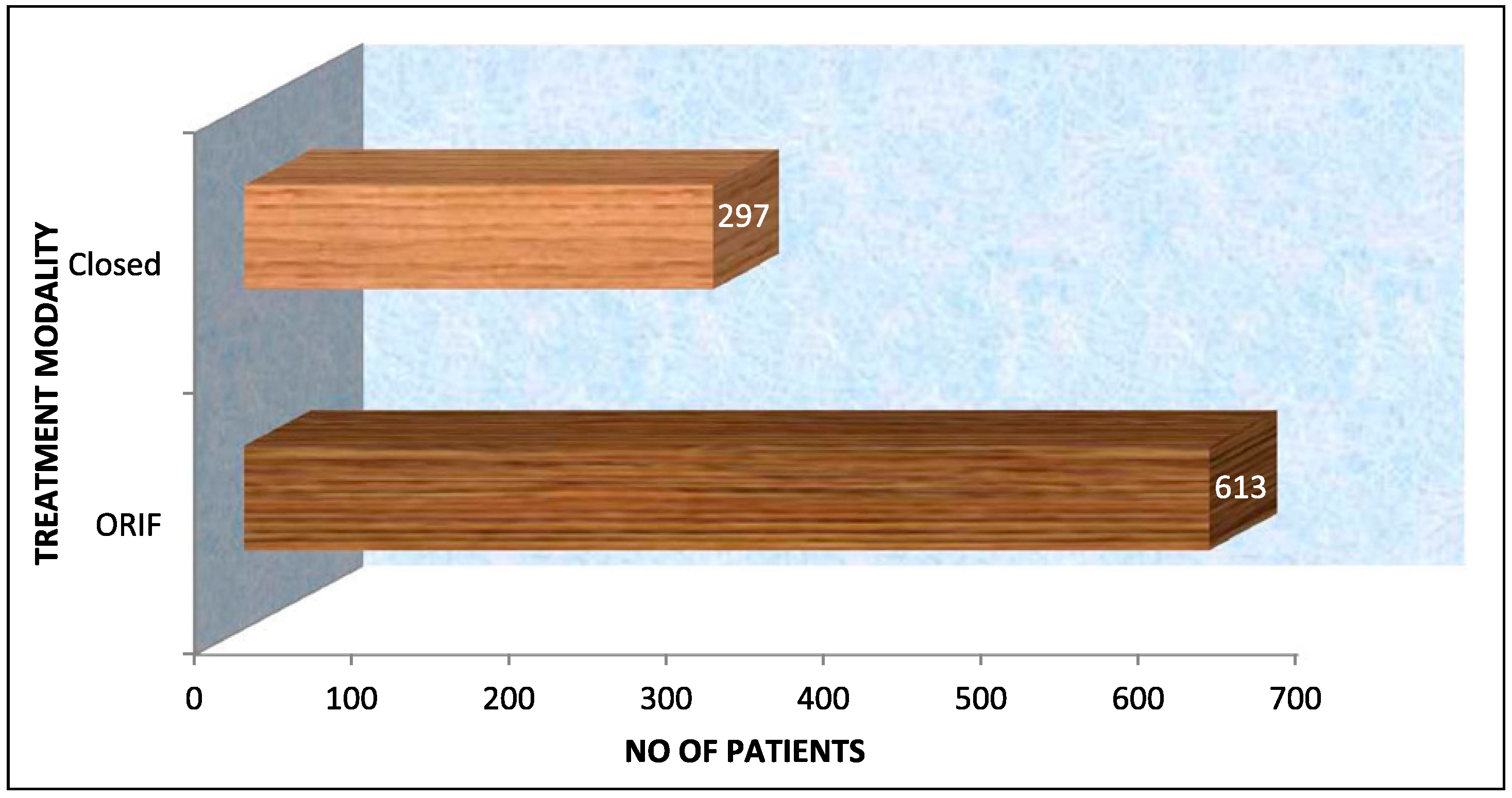
8]. The month from April to July, respectively, was the busiest in our study as in these month people from all over the country visit Himachal Pradesh because of good weather, and vacations provide an opportunity for travel and outdoor activities whilst also increasing the incidence of automobile crashes and interpersonal activities due to alcohol misuse. In recent years, there has been an inclination towards the open reduction and internal fixation (ORIF) as the choice of treatment of mandibular fractures after the advent of miniplate osteosynthesis. Our choices were no different, with ORIF being the treatment of choice, in this study. The main advantage of these mini-plates is reliable, the convenience of application, rapid recovery of normal jaw function and maintenance of normal body weight [22].

Conclusion

All around the globe, even though data on mandibular fractures may show trends, it is observable that there are enough variations to make direct comparisons difficult. Socioeconomic, environmental and cultural factors all contribute to playing a part in determining the type of patients involved, site of trauma and cause of injury. It is clear that there may also be differences within a nation as well as between nations, reflecting the influence of local factors. Despite such limitations of epidemiologic studies, it is still important to monitor these trends overtime in an attempt to detect an area of concern, as well as determine the effectiveness of the method used in accident prevention.

Funding

The author(s) received no financial support for the research, authorship, and/or publication of this article.

Acknowledgments

We would like to acknowledge the help rendered by the faculty, staff and residents of the Department of Oral and Maxillofacial Surgery, Government Dental College and Hospital, Shimla in the treatment and care of the patients involved in this series and preparation of this manuscript.

Conflicts of Interest

The author(s) declared no potential conflicts of interest with respect to the research, authorship, and/or publication of this article.

References

  1. Dongas, P.; Hall, G. Mandibular fracture patterns in Tasmania, Australia. Aust. Dent. J. 2002, 47, 131–137. [Google Scholar] [CrossRef]
  2. Lida, S.; Kongo, M.; et al. Retrospective analysis of 1502 patients with facial fractures. Int. J. Oral Maxillofac. Surg. 2001, 30, 286–290. [Google Scholar]
  3. Simsek, S.; Simsek, B.; Abubaker, A.O.; Laskin, D.M. A comparative study of mandibular fractures in the United States and Turkey. Int. J. Oral Maxillofac. Surg. 2007, 36, 395–397. [Google Scholar] [CrossRef]
  4. Bormann, K.H.; Wild, S.; Gellrich, N.C.; et al. Five-year retrospective study of mandibular fractures in Freiburg, Germany: Incidence, etiology, treatment, and complications. J. Oral Maxillofac. Surg. 2009, 67, 1251–1255. [Google Scholar] [CrossRef]
  5. Subhashraj, K.; Ramkumar, S.; Ravindran, C. Pattern of mandibular fractures in Chennai, India. Br. J. Oral Maxillofac. Surg. 2008, 46, 126–127. [Google Scholar] [CrossRef] [PubMed]
  6. Patrocinio, L.G.; Patrocinio, J.A.; Borba, B.H.; et al. Mandibular fracture: Analysis of 293 patients treated in the Hospital of Clinics, Federal University of Uberlandia. Rev. Bras. Otorrinolaringol. 2005, 71, 560–565. [Google Scholar] [CrossRef]
  7. Knoll, W.-D.; Gaida, A.; Maurer, P. Analysis of mechanical stress in reconstruction plates for bridging mandibular angle defects. J. Cranio-Maxillofacial Surg. 2006, 34, 201–209. [Google Scholar] [CrossRef]
  8. Sakr, K.; Farag, I.A.; Zeitoun, I.M. Review of 509 mandibular fractures treated at the University Hospital, Alexandria, Egypt. Br. J. Oral Maxillofac. Surg. 2006, 44, 107–111. [Google Scholar] [CrossRef]
  9. Ahmed, H.E.A.; Jaber, M.A.; Abu Fanas, S.H.; Karas, M. The pattern of maxillofacial fractures in Sharjah, United Arab Emirates: A review of 230 cases. Oral Surg. Oral Med. Oral Pathol. Oral Radiol. Endod. 2004, 98, 166–170. [Google Scholar] [CrossRef]
  10. Iida, S.; Hassfeld, S.; Reuther, T.; et al. Maxillofacial fractures resulting from falls. J. Cranio-Maxillofac. Surg. 2003, 31, 278–283. [Google Scholar] [CrossRef]
  11. Tanaka, N.; Tomitsuka, K.; Shionoya, K.; et al. Aetiology of maxillofacial fracture. Br. J. Oral Maxillofac. Surg. 1994, 32, 19–23. [Google Scholar]
  12. Fridrich, K.L.; Pena-Velasco, G.; Olson, R.A.J.; Olson, J. Changing trends with mandibular fractures: A review of 1,067 cases. J. Oral Maxillofac. Surg. 1992, 50, 586–589. [Google Scholar]
  13. Erol, B.; Tanrikulu, R.; Görgün, B. Maxillofacial Fractures. Analysis of demographic distribution and treatment in 2901patients (25-year experience). J Cranio-Maxillofac. Surg. 2004, 32, 308–313. [Google Scholar]
  14. Haug, R.H.; Foss, J. Maxillofacial injuries in the pediatric patient. Oral Surg. Oral Med. Oral Pathol. Oral Radiol. Endod. 2000, 90, 126–134. [Google Scholar] [PubMed]
  15. Ugboko, V.I.; Odusanya, S.A.; Fagade, O.O. Maxillofacial fractures in a semi-urban Nigerian teaching hospital. Int. J. Oral Maxillofac. Surg. 1998, 27, 286–289. [Google Scholar] [PubMed]
  16. Maladiere, E.; Bado, F.; Meningaud, J.-P.; Guilbert, F.; Bertrand, J.-C. Aetiology and incidence of facial fractures sustained during sports: A prospective study of 140 patients. Int. J. Oral Maxillofac. Surg. 2001, 30, 291–295. [Google Scholar] [CrossRef]
  17. Ellis, E.; Moos, K.F.; El-Attar, A. Ten years of mandibular fractures: An analysis of 2,137 cases. Oral Surg. Oral Med. Oral Pathol. 1985, 59, 120–129. [Google Scholar]
  18. Schön, R.; Rowda, S.I.L.; Carter, B. Mandibular fractures in Townsville, Australia: Incidence, aetiology andtreatment using the 2.0 AO/ASIF miniplate system. Br. J. Oral Maxillofac. Surg. 2001, 39, 145–148. [Google Scholar] [CrossRef]
  19. Olson, R.A.; Fonseca, R.J.; Zeitler, D.L.; Oshon, D.B. Fractures of the mandible: A review of 580 cases. J. Oral Maxillofac. Surg. 1982, 40, 23–28. [Google Scholar]
  20. Lindqvist, C.; Sorsa, S.; Hyrkäs, T.; Santavirta, S. Maxillofacial fractures sustained in bicycle accidents. Int. J. Oral. Maxillofac. Surg. 1986, 15, 12–18. [Google Scholar] [PubMed]
  21. Vetter, J.D.; Topazian, R.G.; Goldberg, M.H.; Smith, D.G. Facial fractures occurring in a medium-sized metropolitan area: Recent trends. Int. J. Oral Maxillofac. Surg. 1991, 20, 214–216. [Google Scholar] [CrossRef] [PubMed]
  22. Cawood JL Small plate osteosynthesis of mandibular fractures. Br. J. Oral Maxillofac. Surg. 1985, 23, 77–91. [CrossRef] [PubMed]
Figure 1. Bar diagram showing distribution of mandibular fractures by year and sex.
Cmtr 16 00019 g001
Figure 2. Pie diagram showing monthly distribution of mandibular Fractures.
Cmtr 16 00019 g002
Figure 3. Bar diagram showing etiological factors.
Cmtr 16 00019 g003
Figure 4. Bar diagram showing fracture distribution among various anatomic site.
Cmtr 16 00019 g004
Figure 5. Pie diagram showing maxillofacial fractures associated with mandibular fractures.
Cmtr 16 00019 g005
Figure 6. Bar diagram showing treatment modalities for mandibular fractures.
Cmtr 16 00019 g006
Figure 7. Patient with complications of mandibular fractures.
Cmtr 16 00019 g007
Table 1. Distribution of mandibular fractures by year and sex.
Cmtr 16 00019 i001
Table 2. Monthly distribution of mandibular fractures.
Cmtr 16 00019 i002
Table 3. Aetiological factors.
Cmtr 16 00019 i003
Table 4. Fracture distribution among various anatomic sites.
Cmtr 16 00019 i004
Table 5. Maxillofacial fractures associated with mandibular fractures.
Cmtr 16 00019 i005
Table 6. Treatment modalities mandibular fractures.
Cmtr 16 00019 i006
Table 7. Patient with complications of mandibular fractures.
Cmtr 16 00019 i007

