Next Article in Journal
New Beginnings
Previous Article in Journal
Recent Advances in Nasoalveolar Molding Therapy Using 3D Technology
 
 
Craniomaxillofacial Trauma & Reconstruction is published by MDPI from Volume 18 Issue 1 (2025). Previous articles were published by another publisher in Open Access under a CC-BY (or CC-BY-NC-ND) licence, and they are hosted by MDPI on mdpi.com as a courtesy and upon agreement with Sage.
Review

Titanium Mesh Exposure After Bone Grafting: Treatment Approaches—A Systematic Review

by
Giovanni Cunha
1,2,*,
Pedro Henrique de Azambuja Carvalho
1,
Lílian Caldas Quirino
1,
Luiz Henrique Soares Torres
1,
Valfrido Antônio Pereira Filho
1,
Mario Francisco Real Gabrielli
1 and
Marisa Aparecida Cabrini Gabrielli
1
1
Department of Diagnosis and Surgery, Division of Oral and Maxillofacial Surgery, School of Dentistry, São Paulo State University (Unesp), Araraquara, Brazil
2
Department of Oral and Cranio-Maxillofacial Surgery, University Hospital Basel, Switzerland
*
Author to whom correspondence should be addressed.
Craniomaxillofac. Trauma Reconstr. 2022, 15(4), 397-405; https://doi.org/10.1177/19433875211046114
Submission received: 1 November 2020 / Revised: 1 December 2020 / Accepted: 1 January 2021 / Published: 14 September 2021

Abstract

:
Study Design: A systematic review according to PRISMA statement has been designed to answer the preliminary question: “In titanium mesh exposures, is there a treatment alternative which leads to an increased graft maintenance?” and fill the PICO assessment out. Objective: To review studies published in the past 20 years (1999-2019) in which mesh exposure has occurred, detecting the suitable approaches to treat exposure allowing the graft maintenance. Methods: Initial search on PUBMED, SCOPUS, and COCHRANE databases resulted in 777 articles, and hand-searching identified 6 articles. After removing duplicates and unrelated articles, eligibility criteria were applied, and 31 studies were selected (randomized clinical trials, retrospective/prospective clinical trials, and case series). Results: A total of 677 surgical sites and 225 cases of mesh exposure were identified. Eleven treatments have been identified. Chlorhexidine was the primary approach in 46% of cases, followed by oral hygiene instructions and follow-up with 22.5% of occurrences. In 21% of clinical situations, titanium mesh removal was the treatment of choice, associated with other measures (i.e., antibiotic prescription). There seems to be a consensus in cases of infection. When this complication was associated with tissue dehiscence, the primary treatment was mesh removal. The same does not occur when the site needs to be cleaned for long-term periods. Conclusion: In 2 decades of use of titanium meshes, the available treatments do not seem to have evolved, and there is not enough data to establish a guideline.

Introduction

The long-standing edentulism until oral rehabilitation may render the atrophy of the alveolar bone ridge, making it insufficient for implant placement. Thus, professionals must use bone grafting procedures to restore the required volume, and, in many cases, the intraoral donor sites are not enough.
In this background, the availability and association of different biomaterials may allow the functional rehabilitation of large areas. Autogenous, xenogeneic, and alloplastic grafts could be combined since they are considered safe and present satisfactory clinical results.[1,2]
In cases of severe resorption, especially when the shape of the ridge is lost, with compromised arch height and width, reconstruction becomes challenging. A framework will then be necessary to hold grafting materials, and reestablish the alveolar ridge’s contour. In such situations, the stabilization of block grafts with screws is usually not possible. Among the frameworks described in the literature for alveolar reconstructions, titanium meshes stand out since the late 1990s.[3,4]
Those meshes allow the desired 3-dimensional modeling of the ridge by manual approach or by CAD-CAM technology.[5,6,7] They can be used with various biomaterials and techniques available,[6,8] being employed with human recombinant morphogenetic protein-2 (RHBMP-2),[9] platelet aggregates,[10] and collagen membranes allow blood to flow through the periosteum and endosteum to the adjacent graft.[8]
Although the titanium meshes have several applications and advantages, a recurring inconvenience has been reported when used for guided bone regeneration (GBR), which is the exposure through the soft tissue during the healing period.[11,12,13] A Systematic Review published in 2014 showed exposure as the most frequent complication when titanium mesh was used for GBR; of the 246 surgical sites analyzed, there was exposure in 51 of them (21%).[14] Corroborating, Trento et al 2018 found as the significant complication the mesh exposure.[9] In 2019, another systematic review described the exposure as the most undesired presentation in the healing period, with 81 dehiscences described.[15] This is undoubtedly an essential factor for graft vitality and alveolar ridge reconstruction, which may become vulnerable to the oral flora, increasing the risk of infection and consequent revascularization failure. Also, it may lead to the invasion of epithelial cells into the graft through the pores of the mesh.[5,16]
Unfortunately, the current literature focuses on the possible measures to avoid mesh exposure, and less is know about how to treat it. Many authors have suggested different approaches to decrease the incidence of mesh exposure, such as avoiding the use of dental prostheses in the early postoperative period,[5] applying medications to the flap sutures,[17,18] preoperative soft tissue expansion to increasing the amount of mucosa available,[19,20] variations in the incision[21] and use of platelet aggregates (PRF or PRP).[10] It is also available to studies on how to handle exposure and keep the site clean[5,7,22,23] instead of treatment to enhance the soft tissue phenotype, improving the promote better nutrition to the graft.
However, after the occurrence of the exposure, the treatment can be challenging. The literature lacks resolutive techniques that present efficacy and predictability, being restricted to palliative care and follow-up. Approaches such as the debridement of the exposed tissue,[24] removal of the entire mesh or just a part of it,[24] chlorhexidine gel, and mouthwash use,[25,26] have been suggested for treatment after exposure and their effectiveness remains uncertain. In recent literature searches, studies reviewing this topic were scarce.[27] Even systematic reviews and clinical trials are limited to reporting the complication rates after titanium mesh placement rather than treatment options.[9,14,28] Recently randomized clinical trials[28] and systematic reviews[14,15] have reported similar management as used at the end of the 1990s: Clorexidine gel and mouthwash, oral hygiene instructions, and titanium mesh removal. Suggesting that’s this topic possibly has not evolved over the years.
Unlike the prevention of exposure, the approaches available for treatment after exposure are limited. It is unclear whether the techniques described in recent decades are effective. This study aims to systematically investigate articles in which titanium meshes were used for GBR and shows exposure through the soft tissue to identify the methods of treatment employed and their effectiveness.

Materials and Methods

Protocol e Registration

This study considered the preliminary question: This initial question allowed to development of the PICO assessment according to PRISMA statement,[29] as follows: “In titanium mesh exposures, is there a treatment alternative which leads to an increased graft maintenance?
Population: Patients with titanium mesh exposure after alveolar bone augmentation. Intervention: Treatment of Mesh exposure. Comparison: Different options of treatment. Outcome: the success of mesh exposure treatment is assessed by bone graft maintenance and soft and hard tissue viability up to implant placement. This means that no specific analysis was carried out on the graft, as this study focus on the treatment options.
This systematic review is registered at PROSPERO under number CRD42020177792.

Study Selection

Selected modalities of study for inclusion in this systematic review: Randomized clinical trial (RCT), retrospective and prospective clinical trial (CT), Transversal (T) and Cohort (CO), and Case series (CS) that included at least 7 reports in each series.

Eligibility Criteria

-
Studies which was described:
-
alveolar bone reconstruction/augmentation and subsequent exposure of the titanium mesh.
-
Treatment approach and the treatment outcome as well (i.e., if it was possible to keep up the graft up to the implant phase, if the surgical site gets infected if the approach needed to be replaced).
-
Human studies.
-
Published in the English language from 1999 to 2019.
-
Reporting at least 6 months of follow-up from titanium mesh placement.
-
Informed consent document signed by the patients and registered at respective ethics committee (when applicable).
The inclusion criteria described above were searched in the title and abstract of the articles.

Exclusion Criteria

-
Absence of titanium mesh exposure.
-
Presence of infection at the time of bone grafting.
-
Less than 5 years after cancer treatment by head and neck irradiation and surgical resection.
-
Immediate bone reconstruction with titanium mesh in patients undergoing removal of malignant lesions of the jaws.
-
Patients with full or partial maxillectomy.
-
Patients with uncontrolled diabetes and/or other metabolic diseases.
-
Use of drugs that affect the bone quality (e.g., bisphosphonates or chemotherapy).
-
Pregnancy at the time of surgery or postoperative healing period.
The exclusion criteria described above were searched in the full reading of the articles.

Search Strategy

The review was conducted with bibliographic search in the PUBMED MEDLINE, SCOPUS, and COCHRANE databases using the following descriptors: “Titanium mesh/cover/shell,” “alveolar ridge augmentation/grafting/graft,” “bone augmentation/graft/reconstruction/particulate,” “xenograft” “autogenous graft,” “exposure,” “dehiscence,” “treatment,” “approach,” “resolution,” “outcome.” Thus, it was possible to build the search strategies below.
  • ((((((Titanium cover) OR Titanium Shell) OR Titanium MEsh)) AND (((Graft Failure) OR dehiscence) OR Mesh exposure))) AND ((((treatment) OR approach) OR resolution) OR outcome)
  • (Mesh OR titanium shell OR titanium mesh) AND (exposure OR dehiscence) AND (alveolar ridge augmentation OR alveolar ridge grafting OR alveolar ridge graft OR alveolar bone graft OR xenograft OR autogenous graft)
  • “titanium” AND “mesh” AND “exposure” AND “bone” AND “augmentation.”
  • “titanium” AND “mesh” AND “exposure” AND “bone” “reconstruction.”
  • “titanium” AND “mesh” AND “exposure” AND “bone” AND “particulate.”
Most importantly, the first search string was automatically generated by the PUBMED MEDLINE search engine. The others were manually constructed to find the most significant number of studies matching the selection criteria.
For the selection, the title and abstract of the articles were analyzed, delimiting the following criteria:

Data Extraction

The titles and abstracts of the articles identified were assessed by 2 independent researchers (LCQ and LHST) and the other 2 to solve disagreements (GC and PHAC). Three senior surgeons supported the research group in this process (VAPF, MFRG, and MACG). The following data were extracted after a complete reading of the articles: year of study, author, type of study, amount of graft, type of graft (block, particulate, block-particulate—without distinction among autogenous, xenogeneic, or allograft), graft region, number of patients, the onset of exposure (how many days after surgery), method of treatment, complications associated with the approach employed (infection, graft loss, tissue necrosis), and success rate (graft vitality).

Risk of Bias Assessment

Supplemental Table 1 shows the methodological quality assessment using the PRISMA statement[29] criteria to verify the strength of scientific evidence available in the current literature for use in clinical decision making. The classification of the potential risk of bias for each study was assessed considering the following criteria used in previous reviews[9,30,31,32]: Randomized sample selection, the definition of inclusion/exclusion criteria, follow-up report, validated measurements, and statistical analysis. Presence of all the criteria above: low risk of bias; 1 missing criterion: moderate risk of bias; 2 or more missing criteria: high risk of bias.

Results

According to Figure 1, the initial search with the 5 descriptors inserted in the 3 databases resulted in 777 articles. Six additional studies were found by hand-search in the reference list of the selected articles because those papers had titles and abstracts potentially in agreement with the inclusion requirements. After removing duplicates, the number was updated to 440 studies. By applying the inclusion criteria, 39 studies were selected for a full reading. Among them, 8 studies were removed according to the exclusion criteria. One was excluded since it concerned reconstruction after partial maxillectomy, which is beyond the scope of this review; 1 presented immediate alveolar reconstruction using titanium mesh after resection of benign tumors and squamous cell carcinoma; in 1 titanium mesh was not used in the reconstruction; 2 were excluded due to the short period elapsed between cancer treatment by surgical resection/radiation therapy and bone augmentation; 2 did not mention the treatment used for the mesh exposure; 1 study did not involve mesh exposure. Two studies[33,34] were included because they used non-resorbable membranes reinforced by titanium and also presented exposure and treatment employed. Thus, 31 studies were selected for full reading and data extraction. Among those, there were 5 RCT, 13 CT, 9 T, 1 CO, and 3 CS.
In the quality evaluation, 4 articles showed low risk, 8 showed moderate risk, and 19 showed a high risk of bias (Supplemental Table 1). The collected data and results included in this review are shown in Supplemental Table 2.
In this review, a total of 643 patients with 677 surgical sites and 225 titanium mesh exposures (33.23%) were identified, which represents an arithmetic average of 33.23% of exposures. According to the articles selected, the cases of mesh exposure presented an arithmetic average of 92.5% of success rate. The appointments reported after the GBR procedure occurred for at least 6 months. The maximum follow-up informed was 13 months[35] (last return).
Eleven different clinical approaches have been identified and the data can be consulted in Supplemental Table 3. Chlorhexidine was the main treatment, representing 46% of the cases, it was prescribed in mouthwash solution,[13,18,25,36,37,38] topic application of gel in different concentrations (0,12%,[23] 0,2%[5,22,33] and 1%[19,25,26,39] or not report,[40,41]) spray[42] and for mesh brushing[7,38] in order to avoid infection. The second most often intervention was oral hygiene instructions and follow up (OHF)[2,12,16,33,35,36,42,43,44] with 22.5% of occurrences. In 21% of clinical situations, titanium mesh removal was the treatment of choice,[2,4,11,12,22,23,26,34,37,39,41,45,46,47,48] associated with other measures (i.e. antibiotic prescription (ATB),[2,34,47] collagen membrane[39] or resuture[39,45]). Other approaches were described in lesser extent: Mesh ajustment,[16] curretage[5,22] and prosthesis removal.[49]
Regarding the onset of the mesh exposure, the articles were classified as early exposure when it occurred up to 21 days postoperatively. Exposure was considered late when occurring after more than 21 days, the expected period for surgical site epithelialization. Three studies[19,34,40] described early exposure. Eighteen[4,5,7,11,12,13,18,22,23,25,26,36,39,42,43,46,48,49] found late exposure. Eight articles described episodes of dehiscence in both periods.[2,16,33,38,41,44,45,47] In 2 studies, there was no description of the exposure onset period.[35,37]
Complications after treatment of mesh exposures are described. Some authors referred that surgical sites with increased exposure[26] or episodes of infection during the CHX use[37] or after resuturing,[45] resulting in the need to remove the mesh. Another study showed a 50% loss of graft volume after removal of the mesh, collagen membrane, resuturing, and use of chlorhexidine gel.[39]
Due to the heterogeneity of the data collected among the selected articles, a meta-analysis cannot be performed. Therefore, the data were compiled and presented descriptively.

Discussion

The incidence of exposure of titanium meshes used in alveolar grafting procedures may differ according to the study selected; some articles have reported a high exposure rate showing up to 70 to 80% of the sample with soft tissue dehiscence.[5,48] Rasia dal Polo et al[14] in 2014 have found an incidence of 51 cases of dehiscences in 246 treated patients (21%). In 2019, another systematic review[15] described 81 cases in 175 GBR sites, representing an exposure rate of 46.5%. In this review, after analyzing articles from 1999 to 2019 in 3 high-quality databases (PUBMED MEDLINE, SCOPUS, and COCHRANE), the results show an average of 33.23% during the last 2 decades.
The exposure may cause damage to the underlying graft and the covering and supporting tissues. A lower volume of newly formed bone in mesh exposure areas is also described.[16,35] Data show a positive correlation between the exposure area and the reduction in the planned bone volume. Approximately 16% of bone loss occurs for every 2 cm[2] of exposed mesh.[5] In addition, the success rate for grafts with early mesh exposure and subsequent local infection may be as low as 30%.[46] The success rate reported was higher where the mesh exposure did not lead to infection. However, the number of patients with exposure and with no infectious signs is low. In addition, there was a higher prevalence of infection in free extremities and alveolar ridges with severe atrophy, possibly because of the smaller amount of soft tissue and the need for a large volume of biomaterial.[48]
In contrast, several studies displayed high bone graft success rates. Even with expressive numbers of mesh exposure in their samples, some authors even reported 100% success and no need for a secondary procedure. One hypothesis that would explain this result is that many of the studies reported a tissue layer called pseudo periosteum or dense connective tissue without clinical signs of inflammation,[12,13,22,23,26,35,45] which is formed below the mesh and typically occupies the area after partial graft resorption. In 2019, Cucchi et al[50] investigated the histological features of the pseudo-periosteum. The authors have found 3 types according to the thickness layer. When titanium mesh was used for GBR, the sample shows a thicker layer. Although its clinical importance is unclear, it could protect the remaining graft until it is incorporated.[45]
Some studies described the onset of exposure as early or late.[7,22,23] However, due to the variability of the term “early” and “late,” this review considered 21 days necessary for soft tissue epithelialization to classify the onset of the exposure. Those occurring up to 3 weeks postoperatively were considered as early exposures. Some articles described the “postoperative period without complications,” even if there was titanium mesh exposure. This undoubtedly refers to the final success of the graft.
The mean follow-up period for the grafts was 6 to 8 months, with reports of up to 13 months.[35] However, when the implant installation and prosthesis longevity are considered, more extended periods can be found, which are not within the scope of this study. When part of the sample received titanium meshes compared to a control group or to patients who received titanium-reinforced barriers,[33] only the group that received the mesh/reinforced membrane was considered for analysis and verification of success rate.
Eleven treatment approaches were described. The TiMesh repair refers to cutting or polishing the edges of the mesh to improve soft tissue healing. The use of 0.9% saline solution as a cleaning fluid has been reported in many studies, associated or not with chlorhexidine. Chlorhexidine was adopted as a treatment option in almost all studies, either at 0.12% and 0.2% (mouthwash) or 1% (gel) concentration. In some cases, the use of brushes soaked in chlorhexidine was adopted.[7,37] The period of chlorhexidine use also varied. Some authors prescribed its use for fixed periods,[13] while others maintained the application until soft tissue closure or moment of implant placement. Besides the use as a gel or mouthwash, chlorhexidine has also been used as a brushing cream for the exposed mesh region.[7,37] However, the utilization of this pharmacological compound is not intended to allow soft tissue repair with epithelial proliferation or angiogenesis. Chlorhexidine reduces bacterial contamination of the surgical site and consequently attenuates the chances of local infection. Thus, it may increase success rates.
However, as for the surrounding soft tissue and the graft itself, chlorhexidine could act negatively. It has already been associated with cellular cytotoxicity, causing necrosis and apoptosis events in endothelial cells, fibroblasts, and alveolar osteoblasts, making its safety and efficacy for preventing infection in tissue exposures controversial. It is important to note that this in vitro investigation used doses of 0.12% or lower.[51] There is a possibility that the chlorhexidine cytotoxic potential may cause increased exposure of the grafted area during the postoperative period.
The treatments used according to different exposure periods do not seem to have considerably changed. Miyamoto et al[45] and Pieri et al[39] used resuturing when exposures appeared within 7 days and 60 days. The first did not report the chlorhexidine use and described 4 sites with infection and required for TiMesh removal. The latter associated the collagen membrane and CHX after TiMesh removal. Several listed authors have used a similar combination: TiMesh Removal, ATB, CHX, and OHF. Marx et al[52] reported the onset of exposure in 7 days. Except for the resuturing, the authors have used a similar approach as described previously by Miyamoto et al.[45] TiMesh removal and antibiotic therapy a Proussaefs and Lozada[44] and Abrahamsson et al[19] presented a sample with exposure up to 14 days. While the first give OHF to keep the site clean, and the second administered CHX 1%. Merli et al,[34] Zita Gomes et al,[38] and Khanna et al[40] have described the dehiscence within 21 days with removal associated an ATB and CHX were the treatments used. In long periods, the same appears to occur. Poli et al[13] (120 days), Maiorana et al[36] (150 days), and Uehara et al[48] (180 days) have reported the CHX use, OHF, and Ti Mesh removal.
The frequency of appointments after the exposure could be an essential factor to establish a treatment plan and avoid the site infection. Unfortunately, according to the data available was not possible to quantify the number of appointments.
The comparison of the term “success rates” obtained for each article needs to be done carefully because this term might have different meanings among authors. In some studies, individuals in the same sample were treated differently, considering the onset of mesh exposure or the presence of infection. However, the clinical success rate was determined in general. These studies did not correlate each treatment used and the individual success rate.
It is also important to point out that the complications and approaches were considered as reported in the articles. However, in clinical practice, some treatments are inseparable from others. They follow surgical principles, such as curettage of infected tissue or removal of the mesh with the association of saline solution for irrigation and mechanical removal of microorganisms. Thus, it is necessary to be cautious when analyzing separately the treatment options reported in the literature (Supplemental Table 3).
The exposure associated with early use of prosthesis over the grafted region has also been described.[49] The pressure exerted on the mucosa underlying the prosthesis could lead to the decreased blood supply and tissue necrosis. The literature recommends not to use removable dentures over the surgical site for up to 30 days.[22,23] The type of incision can also be considered a factor for early exposure of titanium meshes. Most studies have horizontal and vertical incisions, which present considerable postoperative exposure rates, possibly due to lower blood supply during the healing period.[21] Similarly, Miyamoto et al[45] found higher exposure incidence in more complex bone defects, such as those in which the bone volume to be restored is higher. Pellegrino et al[23] described the largest exposure area in late postoperative periods in their sample.
However, this study aimed to investigate how many treatment options are available and the possibility of graft maintenance, meaning the possibility of dental implant placement on grafted areas, it means that no evaluation was carried out on the grafts once this study just focused only on the approaches used to handle with the exposure. The potential causes for TiMesh exposure, such as anatomical location and bone thickness/height of the alveolar ridge, the extension of the grafted area, type of bone defect, features of the biomaterial used, number of implants installed, mesh fixation, incision, and suture technique, pre and postoperative medication and combination of mesh with PRF or collagen membranes were not considered on this analysis. However, some data are presented to suggest further studies. Likewise, the shape and geometry of the meshes were not considered as analysis variables. Mesh configuration may be a major factor in graft exposure. Studies were demonstrating early tissue dehiscence when meshes without pores were used[53] because the presence of pores could allow adequate blood perfusion, which means graft nutrition and immunologic system cells. In addition, the type of biomaterial used in the graft was not the focus of this investigation.
The assessment and approaches to performing gingival biotype/phenotype changes before essentialusing titanium mesh could be an important factor in avoiding exposure because of thicker gingiva providing reliable periimplantar mucosa.[54] New studies need to access and correlate soft tissue augmentation and mesh exposure.
The level of scientific evidence also varied, with predominance for non-randomized clinical trials and retrospective studies in which there was no control group. The studies included did not present homogeneity in the methodologies and treatments. The data variability will allow a second study to correlate the possible etiologies for the TiMesh exposure. New reviews with RCT design are necessary to clarify this issue.

Conclusions

There seems to be a consensus in the literature on cases of infection. When this complication was associated with tissue dehiscence, the treatment of choice was removing the titanium mesh and exhaustive irrigation. The same is not true when it is necessary to keep the grafted area clean for long periods, although many studies have used chlorhexidine. In 2 decades of titanium meshes, the available treatments do not seem to have evolved, and there is not enough data to establish a guideline.

Supplementary Materials

Supplemental material for this article is available online.

Funding

The author(s) disclosed receipt of the following financial support for the research, authorship, and/or publication of this article: This study was financed by the Coordenação de Aperfeiçoamento de Pessoal de Nível Superior–Brasil (CAPES)—Finance Code 001.

Acknowledgments

The Division of Oral and Maxillofacial Surgery, Araraquara School of Dentistry, São Paulo State University (UNESP), Brazil.

Conflicts of Interest

The author(s) declared no potential conflicts of interest with respect to the research, authorship, and/or publication of this article.

References

  1. Brkovic, B.M.B.; Prasad, H.S.; Rohrer, M.D.; et al. Beta-tricalcium phosphate/type I collagen cones with or without a barrier membrane in human extraction socket healing: clinical, histologic, histomorphometric, and immunohistochemical evaluation. Clin Oral Investig. 2012, 16, 581–590. [Google Scholar] [PubMed]
  2. Marx, R.E.; Armentano, L.; Olavarria, A.; Samaniego, J. rhBMP2/ACS grafts versus autogenous cancellous marrow grafts in large vertical defects of the maxilla: an unsponsored randomized open-label clinical trial. Int J Oral Maxillofac Implants. 2013, 28, e243–e251. [Google Scholar] [PubMed]
  3. von Arx, T.; Kurt, B. Implant placement and simultaneous periimplant bone grafting using a micro titanium mesh for graft stabilization. Int J Periodontics Restorative Dent. 1998, 18, 117–127. [Google Scholar] [PubMed]
  4. von Arx, T.; Kurt, B. Implant placement and simultaneous ridge augmentation using autogenous bone and a micro titanium mesh: a prospective clinical study with 20 implants. Clin Oral Implants Res. 1999, 10, 24–33. [Google Scholar]
  5. Lizio, G.; Corinaldesi, G.; Marchetti, C. Alveolar ridge reconstruction with titanium mesh: a three-dimensional evaluation of factors affecting bone augmentation. Int J Oral Maxillofac Implants. 2014, 29, 1354–1363. [Google Scholar]
  6. Yamada, H.; Nakaoka, K.; Horiuchi, T.; et al. Mandibular reconstruction using custom-made titanium mesh tray and particulate cancellous bone and marrow harvested from bilateral posterior ilia. J Plast Surg Hand Surg. 2014, 48, 183–190. [Google Scholar]
  7. Ciocca, L.; Lizio, G.; Baldissara, P.; Sambuco, A.; Scotti, R.; Corinaldesi, G. Prosthetically CAD-CAM-guided bone augmentation of atrophic jaws using customized titanium mesh: preliminary results of an open prospective study. J Oral Implantol. 2018, 44, 131–137. [Google Scholar]
  8. Malchiodi, L.; Scarano, A.; Quaranta, M.; Piattelli, A. Rigid fixation by means of titanium mesh in edentulous ridge expansion for horizontal ridge augmentation in the maxilla. Int J Oral Maxillofac Implants. 1998, 13, 701–705. [Google Scholar]
  9. Trento, G.S.; Carvalho, P.H.A.; Macedo, D.V.; Gabrielli, M.A.C.; Monnazzi, M.S.; Pereira-Filho, V.A. Titanium mesh associated with rhBMP-2 in alveolar ridge reconstruction. Int J Oral Maxillofac Surg. 2018, 48, 546–553. [Google Scholar]
  10. Torres, J.; Tamimi, F.; Alkhraisat, M.H.; et al. Platelet-rich plasma may prevent titanium-mesh exposure in alveolar ridge augmentation with anorganic bovine bone. J Clin Periodontol. 2010, 37, 943–951. [Google Scholar]
  11. Assenza, B.; Piattelli, M.; Scarano, A.; Iezzi, G.; Petrone, G.; Piattelli, A. Localized ridge augmentation using titanium micromesh. J Oral Implantol. 2001, 27, 287–292. [Google Scholar] [CrossRef]
  12. Khamees, J.; Darwiche, M.A.; Kochaji, N. Alveolar ridge augmentation using chin bone graft, bovine bone mineral, and titanium mesh: clinical, histological, and histomorphomtric study. J Indian Soc Periodontol. 2012, 16, 235–240. [Google Scholar] [CrossRef]
  13. Poli, P.P.; Beretta, M.; Cicciù, M.; Maiorana, C. Alveolar ridge augmentation with titanium mesh. A retrospective clinical study. Open Dent J. 2014, 8, 148–158. [Google Scholar] [CrossRef]
  14. Rasia dal Polo, M.; Poli, P.P.; Rancitelli, D.; Beretta, M.; Maiorana, C. Alveolar ridge reconstruction with titanium meshes: a systematic review of the literature. Med Oral Patol Oral Cir Bucal. 2014, 19, e639–e646. [Google Scholar] [CrossRef]
  15. Briguglio, F.; Falcomatà, D.; Marconcini, S.; Fiorillo, L.; Briguglio, R.; Farronato, D. The use of titanium mesh in guided bone regeneration: a systematic review. Int J Dent. 2019, 2019, 906542. [Google Scholar] [CrossRef]
  16. Her, S.; Kang, T.; Fien, M.J. Titanium mesh as an alternative to a membrane for ridge augmentation. J Oral Maxillofac Surg. 2012, 70, 803–810. [Google Scholar] [CrossRef]
  17. De Stavola, L.; Tunkel, J. The role played by a suspended external-internal suture in reducing marginal flap tension after bone reconstruction: a clinical prospective cohort study in the maxilla. Int J Oral Maxillofac Implants. 2014, 29, 921–926. [Google Scholar] [CrossRef]
  18. Sagheb, K.; Schiegnitz, E.; Moergel, M.; Walter, C.; Al-Nawas, B.; Wagner, W. Clinical outcome of alveolar ridge augmentation with individualized CAD-CAM-produced titanium mesh. Int J Implant Dent. 2017, 3, 36. [Google Scholar] [CrossRef]
  19. Abrahamsson, P.; Wälivaara, D.Å.; Isaksson, S.; Andersson, G. Periosteal expansion before local bone reconstruction using a new technique for measuring soft tissue profile stability: a clinical study. J Oral Maxillofac Surg. 2012, 70, e521–e530. [Google Scholar] [CrossRef]
  20. Mertens, C.; Thiele, O.; Engel, M.; Seeberger, R.; Hoffmann, J.; Freier, K. The use of self-inflating soft tissue expanders prior to bone augmentation of atrophied alveolar ridges. Clin Implant Dent Relat Res. 2015, 17, 44–51. [Google Scholar] [CrossRef]
  21. Altiparmak, N.; Uckan, S.; Bayram, B.; Soydan, S. Comparison of tunnel and crestal incision techniques in reconstruction of localized alveolar defects. Int J Oral Maxillofac Implants. 2017, 32, 1103–1110. [Google Scholar] [PubMed]
  22. Lizio, G.; Mazzone, N.; Corinaldesi, G.; Marchetti, C. Reconstruction of extended and morphologically varied alveolar ridge defects with the titanium mesh technique: clinical and dental implants outcomes. Int J Periodontics Restorative Dent. 2016, 36, 689–697. [Google Scholar]
  23. Pellegrino, G.; Lizio, G.; Corinaldesi, G.; Marchetti, C. Titanium mesh technique in rehabilitation of totally edentulous atrophic maxillae: a retrospective case series. J Periodontol. 2016, 87, 519–528. [Google Scholar] [PubMed]
  24. Al-Ardah, A.J.; AlHelal, A.; Proussaefs, P.; AlBader, B.; Al Humaidan, A.A.; Lozada, J. Managing titanium mesh exposure with partial removal of the exposed site: a case series study. J Oral Implantol. 2017, 43, 482–490. [Google Scholar] [PubMed]
  25. Roccuzzo, M.; Ramieri, G.; Spada, M.C.; Bianchi, S.D.; Berrone, S. Vertical alveolar ridge augmentation by means of a titanium mesh and autogenous bone grafts. Clin Oral Implants Res. 2004, 15, 73–81. [Google Scholar]
  26. Roccuzzo, M.; Ramieri, G.; Bunino, M.; Berrone, S. Autogenous bone graft alone or associated with titanium mesh for vertical alveolar ridge augmentation: a controlled clinical trial. Clin Oral Implants Res. 2007, 18, 286–294. [Google Scholar] [PubMed]
  27. Cucchi, A.; Vignudelli, E.; Napolitano, A.; Marchetti, C.; Corinaldesi, G. Evaluation of complication rates and vertical bone gain after guided bone regeneration with non-resorbable membranes versus titanium meshes and resorbable membranes. A randomized clinical trial. Clin Implant Dent Relat Res. 2017, 19, 821–832. [Google Scholar] [CrossRef]
  28. Atef, M.; Tarek, A.; Shaheen, M.; Alarawi, R.M.; Askar, N. Horizontal ridge augmentation using native collagen membrane vs. titanium mesh in atrophic maxillary ridges: randomized clinical trial. Clin Implant Dent Relat Res. 2020, 22, 156–166. [Google Scholar] [CrossRef]
  29. Moher, D.; Liberati, A.; Tetzlaff, J.; Altman, D.G.; PRISMA Group. Preferred reporting items for systematic reviews and meta-analyses: the PRISMA statement. PLoS Med. 2009, 6, e1000097. [Google Scholar]
  30. Moura, L.B.; Carvalho, P.H.A.; Xavier, C.B.; et al. Autogenous non-vascularized bone graft in segmental mandibular reconstruction: a systematic review. Int J Oral Maxillofac Surg. 2016, 45, 1388–1394. [Google Scholar]
  31. La Torre, G.; Chiaradia, G.; Gianfagna, F.; De Laurentis, A.; Boccia, S.; Ricciardi, W. Quality assessment in meta-analysis. Ital J Public Heal. 2006, 3, 44–50. [Google Scholar] [CrossRef] [PubMed]
  32. Clementini, M.; Morlupi, A.; Canullo, L.; Agrestini, C.; Barlattani, A. Success rate of dental implants inserted in horizontal and vertical guided bone regenerated areas: a systematic review. Int J Oral Maxillofac Surg. 2012, 41, 847–852. [Google Scholar]
  33. Naenni, N.; Schneider, D.; Jung, R.E.; Hüsler, J.; Hämmerle, C.H.F.; Thoma, D.S. Randomized clinical study assessing two membranes for guided bone regeneration of peri-implant bone defects: clinical and histological outcomes at 6 months. Clin Oral Implants Res. 2017, 28, 1309–1317. [Google Scholar]
  34. Merli, M.; Migani, M.; Esposito, M. Vertical ridge augmentation with autogenous bone grafts: resorbable barriers supported by ostheosynthesis plates versus titanium-reinforced barriers. A preliminary report of a blinded, randomized controlled clinical trial. Int J Oral Maxillofac Implants. 2007, 22, 373–382. [Google Scholar] [PubMed]
  35. Proussaefs, P.; Lozada, J.; Kleinman, A.; Rohrer, M.D.; McMillan, P.J. The use of titanium mesh in conjunction with autogenous bone graft and inorganic bovine bone mineral (bio-oss) for localized alveolar ridge augmentation: a human study. Int J Periodontics Restorative Dent. 2003, 23, 185–195. [Google Scholar]
  36. Maiorana, C.; Santoro, F.; Rabagliati, M.; Salina, S. Evaluation of the use of iliac cancellous bone and anorganic bovine bone in the reconstruction of the atrophic maxilla with titanium mesh: a clinical and histologic investigation. Int J Oral Maxillofac Implants. 2001, 16, 427–432. [Google Scholar] [PubMed]
  37. Louis, P.J.; Gutta, R.; Said-Al-Naief, N.; Bartolucci, A.A. Reconstruction of the maxilla and mandible with particulate bone graft and titanium mesh for implant placement. J Oral Maxillofac Surg. 2008, 66, 235–245. [Google Scholar]
  38. Zita Gomes, R.; Paraud Freixas, A.; Han, C.H.; Bechara, S.; Tawil, I. Alveolar ridge reconstruction with titanium meshes and simultaneous implant placement: a retrospective, multicenter clinical study. Biomed Res Int. 2016, 2016, 1–12. [Google Scholar]
  39. Pieri, F.; Corinaldesi, G.; Fini, M.; Aldini, N.N.; Giardino, R.; Marchetti, C. Alveolar ridge augmentation with titanium mesh and a combination of autogenous bone and anorganic bovine bone: a 2-year prospective study. J Periodontol. 2008, 79, 2093–2103. [Google Scholar]
  40. Khanna, R.; Khanna, R.; Pardhe, N.D.; Srivastava, N.; Bajpai, M.; Gupta, S. Pure titanium membrane (ultra-Ti®) in the treatment of periodontal osseous defects: a split-mouth comparative study. J Clin Diagn Res. 2016, 10, ZC47–ZC51. [Google Scholar]
  41. Hartmann, A.; Hildebrandt, H.; Schmohl, J.U.; Kämmerer, P.W. Evaluation of risk parameters in bone regeneration using a customized titanium mesh: results of a clinical study. Implant Dent. 2019, 28, 543–550. [Google Scholar] [PubMed]
  42. Tallarico, M.; Ceruso, F.M.; Muzzi, L.; et al. Effect of simultaneous immediate implant placement and guided bone reconstruction with ultra-fine titanium mesh membranes on radiographic and clinical parameters after 18 months of loading. Materials (Basel). 2019, 12, 1710. [Google Scholar] [CrossRef] [PubMed]
  43. Artzi, Z.; Dayan, D.; Alpern, Y.; Nemcovsky, C.E. Vertical ridge augmentation using xenogenic material supported by a configured titanium mesh: clinicohistopathologic and histochemical study. Int J Oral Maxillofac Implants. 2003, 18, 440–446. [Google Scholar]
  44. Proussaefs, P.; Lozada, J. Use of titanium mesh for staged localized alveolar ridge augmentation: clinical and histologic-histomorphometric evaluation. J Oral Implantol. 2006, 32, 237–247. [Google Scholar]
  45. Miyamoto, I.; Funaki, K.; Yamauchi, K.; Kodama, T.; Takahashi, T. Alveolar ridge reconstruction with titanium mesh and autogenous particulate bone graft: computed tomography-based evaluations of augmented bone quality and quantity. Clin Implant Dent Relat Res. 2012, 14, 304–311. [Google Scholar]
  46. Strietzel, F.P.; Reichart, P.A.; Graf, H.L. Lateral alveolar ridge augmentation using a synthetic nano-crystalline hydroxyapatite bone substitution material (Ostim): preliminary clinical and histological results. Clin Oral Implants Res. 2007, 18, 743–751. [Google Scholar]
  47. Funato, A.; Ishikawa, T.; Kitajima, H.; Yamada, M.; Moroi, H. A novel combined surgical approach to vertical alveolar ridge augmentation with titanium mesh, resorbable membrane, and rhPDGF-BB: a retrospective consecutive case series. Int J Periodontics Restorative Dent. 2013, 33, 437–445. [Google Scholar] [PubMed]
  48. Uehara, S.; Kurita, H.; Shimane, T.; et al. Predictability of staged localized alveolar ridge augmentation using a micro titanium mesh. Oral Maxillofac Surg. 2015, 19, 411–416. [Google Scholar]
  49. Kim, Y.; Kim, T.K.; Leem, D.H. Clinical study of a flap advancement technique without vertical incision for guided bone regeneration. Int J Oral Maxillofac Implants. 2015, 30, 1113–1118. [Google Scholar] [CrossRef]
  50. Cucchi, A.; Sartori, M.; Parrilli, A.; Aldini, N.N.; Vignudelli, E.; Corinaldesi, G. Histological and histomorphometric analysis of bone tissue after guided bone regeneration with nonresorbable membranes vs. resorbable membranes and titanium mesh. Clin Implant Dent Relat Res. 2019, 21, 693–701. [Google Scholar] [CrossRef]
  51. Giannelli, M.; Chellini, F.; Margheri, M.; Tonelli, P.; Tani, A. Effect of chlorhexidine digluconate on different cell types: a molecular and ultrastructural investigation. Toxicol Vitr. 2008, 22, 308–317. [Google Scholar]
  52. Marx, R.E.; Armentano, L.; Olavarria, A.; Samaniego, J. rhBMP-2/ACS grafts versus autogenous cancellous marrow grafts in large vertical defects of the maxilla: an unsponsored randomized open-label clinical trial. Int J Oral Maxillofac Implants. 2013, 28, e243–e251. [Google Scholar] [CrossRef] [PubMed]
  53. Celletti, R.; Davarpanah, M.; Etienne, D.; et al. Guided tissue regeneration around dental implants in immediate extraction sockets: comparison of e-PTFE and a new titanium membrane. Int J Periodontics Restorative Dent. 1994, 14, 242–253. [Google Scholar] [PubMed]
  54. Kan, J.Y.K.; Rungcharassaeng, K.; Lozada, J.L.; Zimmerman, G. Facial gingival tissue stability following immediate placement and provisionalization of maxillary anterior single implants: a 2- to 8-year follow-up. Int J Oral Maxillofac Implants. 2011, 26, 179–187. [Google Scholar]
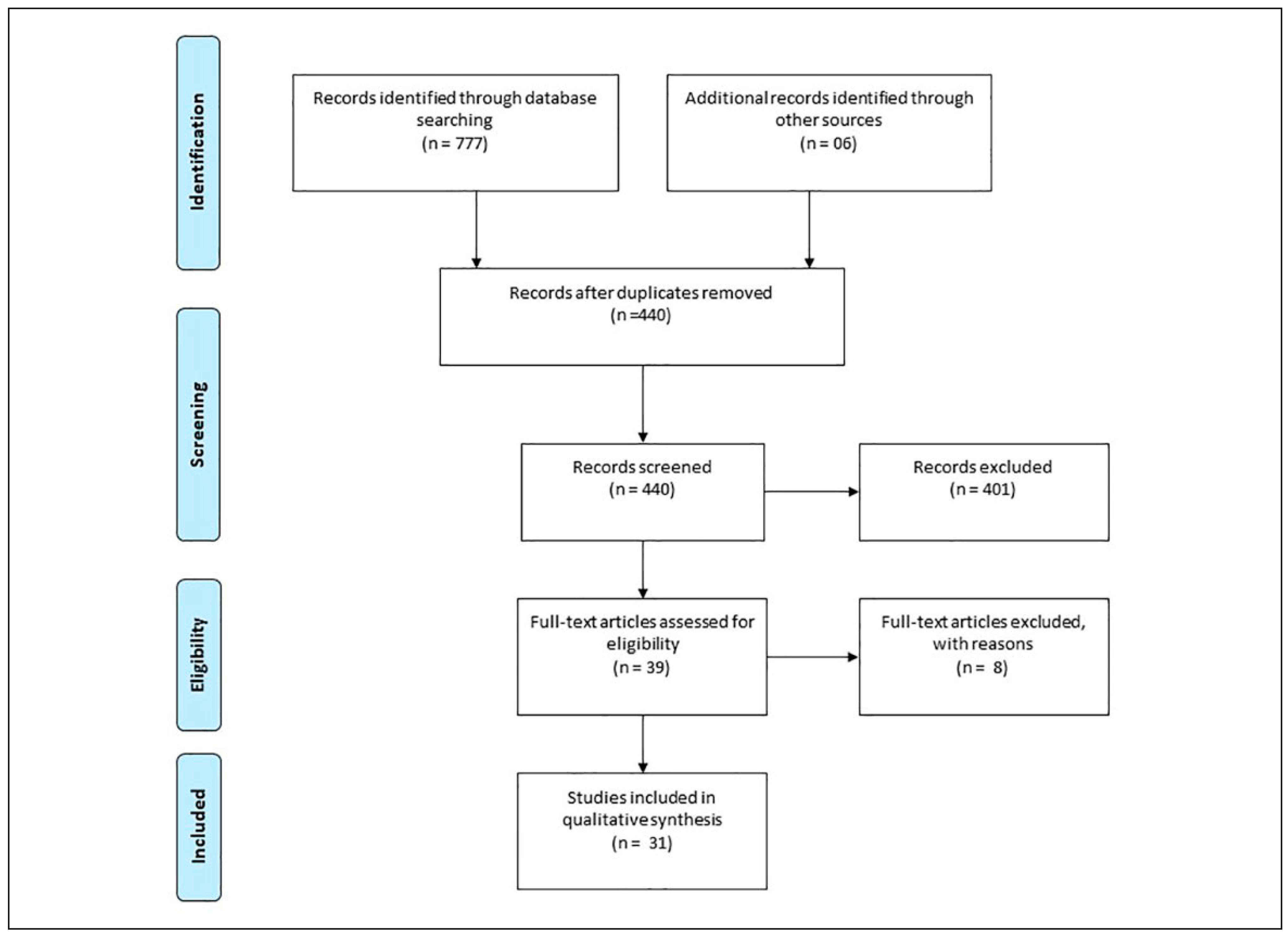
Figure 1. Data acquisition and selection in the PubMed, Scopus, and Cochrane. Source: According to Moher et al.[29].
Cmtr 15 00052 g001