Next Article in Journal
Should Degree of Third Molar Eruption Influence Operative Management of Mandibular Angle Fractures? A Systematic Review
Previous Article in Journal
Quantifying Aerosol Generation in Maxillofacial Trauma Repair Techniques
 
 
Craniomaxillofacial Trauma & Reconstruction is published by MDPI from Volume 18 Issue 1 (2025). Previous articles were published by another publisher in Open Access under a CC-BY (or CC-BY-NC-ND) licence, and they are hosted by MDPI on mdpi.com as a courtesy and upon agreement with Sage.
Case Report

Introduction of 3D Printing in a German Municipal Hospital—Practice Guide for CMF Surgery

by
H Gleissner
1,2,*,
G Castrillon-Oberndorfer
1,3 and
St Gehrlich
1
1
Klinik für Mund-, Kiefer- und plastische Gesichtschirurgie, Universitätsklinik der Paracelsus Medizinischen Privatuniversität Nürnberg, Bavaria, Germany
2
MKG Praxis Regensburg, Bahnhofstraße 13, 93047 Regensburg, Bavaria, Germany
3
ALB Fils Kliniken, Klinik für Mund-, Kiefer- und Gesichtschirurgie, Baden-Wuerttemberg, Germany
*
Author to whom correspondence should be addressed.
Craniomaxillofac. Trauma Reconstr. 2022, 15(4), 369-378; https://doi.org/10.1177/19433875211050721
Submission received: 1 November 2020 / Revised: 1 December 2020 / Accepted: 1 January 2021 / Published: 30 September 2021

Abstract

:
Study Design: This study aimed to introduce 3D printing in a municipal hospital to improve the treatment of craniomaxillofacial patients and optimize costs and operating time. Thus we describe the implementation of low-cost in-house 3D printing to facilitate orbitaland mandible reconstruction in CMF surgery. Moreover, we address legal requirements, safety at work, fireand data protection. Finally, we want to share our experiences using 3D printing and point out its advantages in providing better patient care. Methods: We outline the setup of in-house 3D printing and focus on obeying German health care regulations. We based our approach on a fused deposition modeling 3D printer and free software. As proof of concept, we treated 4 cases of severe orbital trauma and 1 case of mandibular reconstruction. We printed a 3D patient-specific model for each case and adapted a titanium mesh implant, respectively, a titanium reconstruction plate before performing the surgery. Results: Our approach reduced costs, duration of anesthesia, operating time, recovery time, and postoperative swelling and increased the revenue. Functional outcome in orbital reconstruction like eye movement and double vision, was improved compared to the conventional technique. No severe complications like loss-of-vision or surgical revision occurred. Likewise, mandibular reconstruction showed no plate loosening or plate fracture. Conclusion: The implementation of cost-efficient 3D printing resulted in successful patient treatment with excellent outcomes. Our practice guide offers a 3D printing workflow and could be adapted to fit the needs of other specialties like neurosurgery, orthopedic surgery as well.

1. Introduction

3D printing and 3D segmentation of bony structures of the skull play an increasingly important role in craniomaxillofacial-surgery as the anatomy of the craniomaxillofacial skeleton is complex. [1,2,3,4] Anatomically correct reduction of fractures or the restitution of accurate anatomy of the mandible or other bony defects is a challenging task [5,6] As the orbit is a complex three-dimensional structure with fragile bones, this is especially true. 3D segmentation and patient-specific implants, help to facilitate orbital repair. [7,8,9,10] It is also beneficial in head and neck surgery or severe trauma cases that involve other parts of the skull. [2,5,6,11,12,13,14,15,16,17,18,19,20,21] Unfortunately, 3D printing often requires highly specialized and costly hardware and ongoing software license fees. Outsourcing to medical service providers is quite common but expensive, and it takes time until the finished product arrives. Still, the classic freehand approach is frequently used but lacks accuracy and is prone to complications. We aim to present a complete in-house workflow for patient-specific 3D model creation, postprocessing, e.g., pre-bending of plates and orbital meshes, sterilization, and application in the operating theater, without any dependencies from medical service providers or else. The current healthcare system favors cost-efficient therapies, especially in global economic crises due to the COVID-19 pandemic. Therefore, we evaluate the feasibility of introducing in-house 3D printing with low-budget hardware and free software. Our study sets forth the legal requirements, like safety at work, fire protection, and data privacy in Germany. According to our knowledge, this is the first publication related to Germany that merges the clinical and legal aspects to successfully implement 3D printing in the existing infrastructure of a municipal hospital.

2. Requirements and Features

In order to implement 3D-printing, several legal requirements had to be considered, which are described in the following.
First, questions arise from the point of safety at work (ArbSchG), on how to implement a 3D Printing device in a workplace safely. The area of application for a 3D printer falls under the Product Safety Act (ProdSG) and Machinery Directive 2006/42/EG. This rather strict regime ensures that devices are allowed to enter the European, in particular, the German market only if they meet the requirements. So 3D printers purchased within Germany or the European Union should have already fulfilled this. In addition to formal demands, the material requirements must also be met, such as CE-Certificate and Declaration of Conformity.
In order to operate a 3D printer at work, the Industrial Safety Regulation (BetrsichV) and the Ordinance on Hazardous Substances (GefStoffV) must be observed, as a 3D printer is regarded as work equipment in the sense of §2 Industrial Safety Regulation (BetrSichV). Direct danger during the use of a fused deposition modeling (FDM) 3D printer emerges from its principle of operation. In order to melt the material, a nozzle temperature of 200 °C to 215 °C is necessary. For proper adhesion, a heated print bed of 60 °C is required. So risk of burn is likely to happen in case of careless use. Therefore the user manual must be consulted accordingly, and a detailed instruction manual must be composed in order to ensure proper usage of the 3D printer. All employees that use the device must be instructed before first use and after that at least once a year (§12 BetrSichV, §14 GefStoffV).
Also, according to hospital internal fire protection regulations, a fireproof base is obligatory, and there must not be flammable objects in the immediate vicinity, and a smoke detector is required. The exact fire safety measures are the responsibility of the hospital’s safety department. Hence fire safety measures may differ from hospital to hospital and, therefore, must be included from the beginning.
To set up the printer, a separate room that is not the main workspace is best but not obligatory. Loudness level must remain below a maximum tolerance range of 55 dB according to the Production Safety Regulation (9. ProdSV), Workplaces Regulation, and Noise Vibration Occupational Safety Regulation (ArbSta¨ttV, La¨rmVibrationsArbSchV). [22] A generic 3D printer, like the one we used in our study, generates around 50 to 53 dB in standby and around 65 to 75 dB when running. So right out of the box, to run the printer and adhere to the demands, a separate room is necessary. Nevertheless, we like to mention that a silent motherboard upgrade is available through the manufacturer itself for 38 to 49 USD (approximately 34 to 44 Euro). It can be easily exchanged and reduces the loudness in operation to 52 to 55 dB.
A separate room is still useful because of the required ventilation. Either the room has its ventilation or a window to ventilate regularly after each print job. Although fused deposition modeling (FDM) has low emissions of chemical compounds or volatile particles depending on the used material, the accumulation of emission must be addressed, nevertheless. [23,24,25,26,27,28] In general, this topic applies to local laws, and respective authorities have to be consulted in advance. In some cases, the information on how to run a 3D printer in a workplace environment safely may be already available as an intranet document. [29]
Second, polylactic acid (PLA) is the material of choice for FDM printing because of the ease of use and lower emissions of volatile or chemical compounds, as stated above. [28] The manufacturer’s information sheet gives the details on how this must be processed. Although there are several FDA approved filaments available, these do not meet the standards of German Medical Product Law (MPG). Currently, as far as we know, only 2 PLA filaments exist that fulfill the requirements: SplintFill® available through 3D Agency oHG, Worpswede, Germany, and Arfona® Impressions Tray by Arfona LLC, Brooklyn, NY, United States. Both filaments are Medical Products Class I. [30,31,32] Both filaments are expensive and more difficult to process than standard PLA, as they require a higher nozzle temperature, and show different flow properties as viscosity is higher, too. Additionally, we did not get clearance from the central sterile goods processing for plasma sterilization because the solvents used to prewash the 3D printed models are not covered on the manufacturer’s instruction sheet. Plasma sterilization of the 3D printed model would have been particularly useful for intraoperative usage of the 3D printed model. Instead of pursuing the search of a suitable filament, we chose to continue with standard PLA filament. Thus no intraoperative application of the model is possible.
Third, data privacy is delicate. We paid particular attention to fulfill internal quality management requirements and General Data Protection Regulation (DSVGO). Along with the Department of Radiology and the data protection officer, we found 2 possible solutions. Firstly, the usage of an in-house computer with access to the radiology departments server data. Access is restricted through the employee’s login and password and is, therefore, the best possible solution. Unfortunately, this could not be realized within a reasonable time and is still work in progress. Thus, the radiology department anonymizes the DICOM Data and then provides it via a data carrier (encrypted USB data drive) to be processed at the desired location, as depicted in Figure 1.
Regarding necessary hardware, we acquired the lowbudget 3D printer by Creality™-Ender 5®, which is available through the company’s website for 429.99€ (or 279.99€ if on sale). DAS FILAMENT™ is the filament of choice, and the 800 g spool is available for 17.95€ (22.44€/1000 g) through their shop. [33] In general, the printed 3D model weighs in with 30 to 60 g and accounts for approximately 0.67 to 1.35€ material costs. The respective energy costs depend on the electricity provider. For the year 2020, these costs amounted to an average of 0.32€ per kWh in Germany. [34,35] In order to roughly measure the consumed electric energy, we used a generic digital electricity meter and plugged it into the power socket. It calculated 118 Watts per hour with a nozzle temperature of 210 °C and a heated bed temperature of 60 °C. This estimates for 2.832 kWh for 24 h of printing. Considering that 1 kWh costs 0.32€ depending on the energy provider, the production of 1 model, with a 6 h print time, requires 0.23€ of energy costs. Overall a 60 g patient-specific 3D printed model with 6 h of printing time accounts for 1.58€ of monetary expanses at maximum.
3D segmentation is done using 3D slicer. [19,36,37,38,39,40] A 3D model of adequate quality requires a multislice detector CT scan or Cone-Beam-CT scan with at least 1 mm slice thickness. [11,41] After 3D segmentation, the generated STL-file is further processed with the free software Autodesk Meshmixer® Version 3.5.474 (Autodesk™, San Rafael, CA, USA). [42] In the case of orbital reconstruction, the contralateral healthy orbit is mirrored and used as a blueprint for the reconstruction of the defect zone. Optional, for fur-ther optimization and reduction of print time, the created STL-file can be reworked with Blender (Blender Foundation, Amsterdam, Netherlands). [43] In particular, this is useful for printing mandibles because the inner structure of the mandible is removed without any effects on the cortical bone, thus reducing print time massively. The STL-file is then sliced with Cura (Ultimaker™, Utrecht, The Netherlands). [44] After that, the created OBJ-File is forwarded to the Printer via USB. The software used is either under GPL (general public license) or BSD (Berkely Software Distribution) license and therefore free for use even in a commercial setting. Figure 2 shows our workflow.
To make the process comprehensible, the department for sterile goods processing developed a documentation file to trace the use of the implants, thus manufactured. [45] This documentation file includes the current date, the responsible doctor’s name and phone number, the date of surgery, and the patient’s data. Also, the plate/mesh manufacturer, the REF number, and the LOT number must be filled out. The second half of the document contains the operating theater and, once again, the patient’s name and the date of surgery as well as specific information for the sterile goods department.
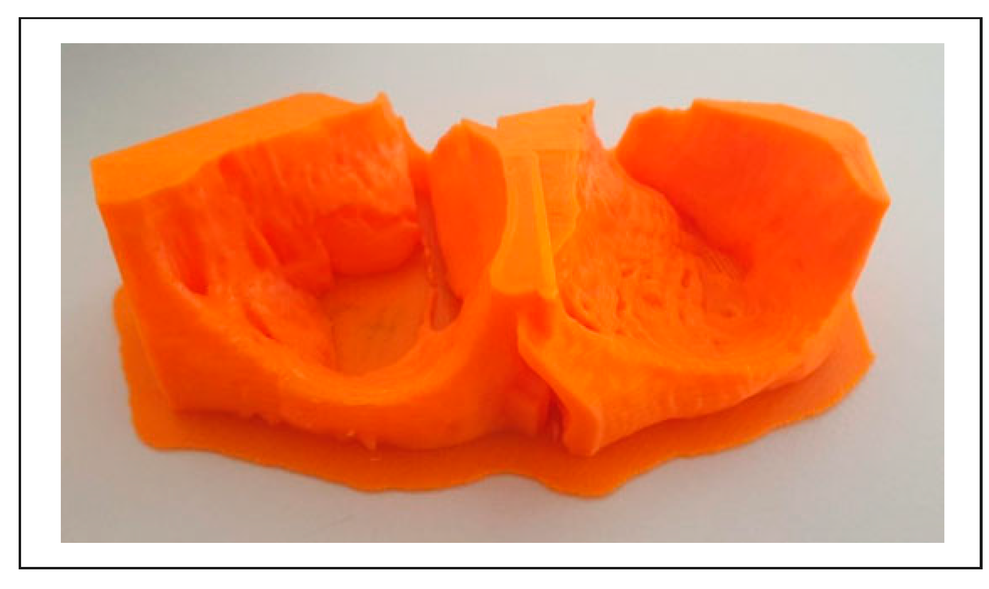
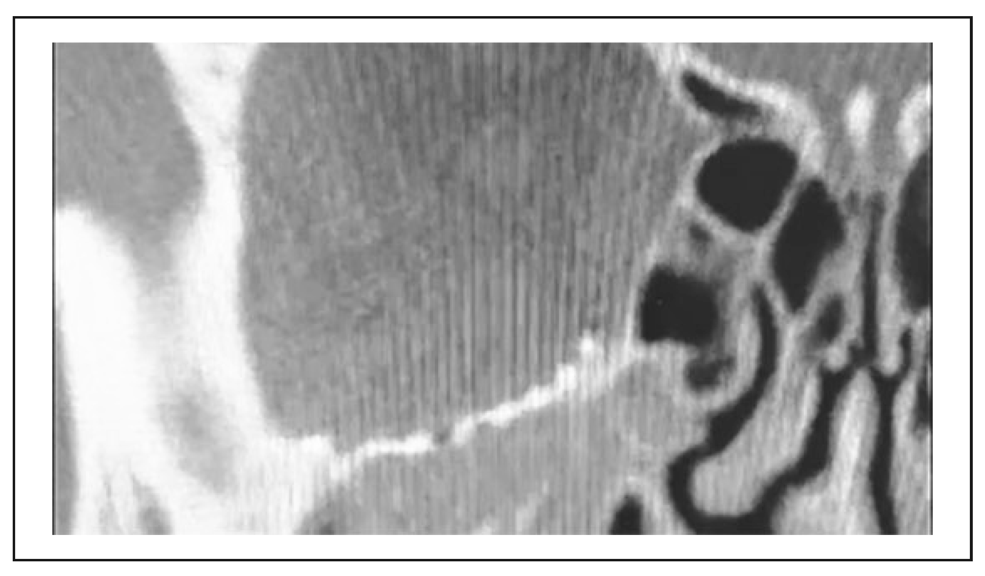
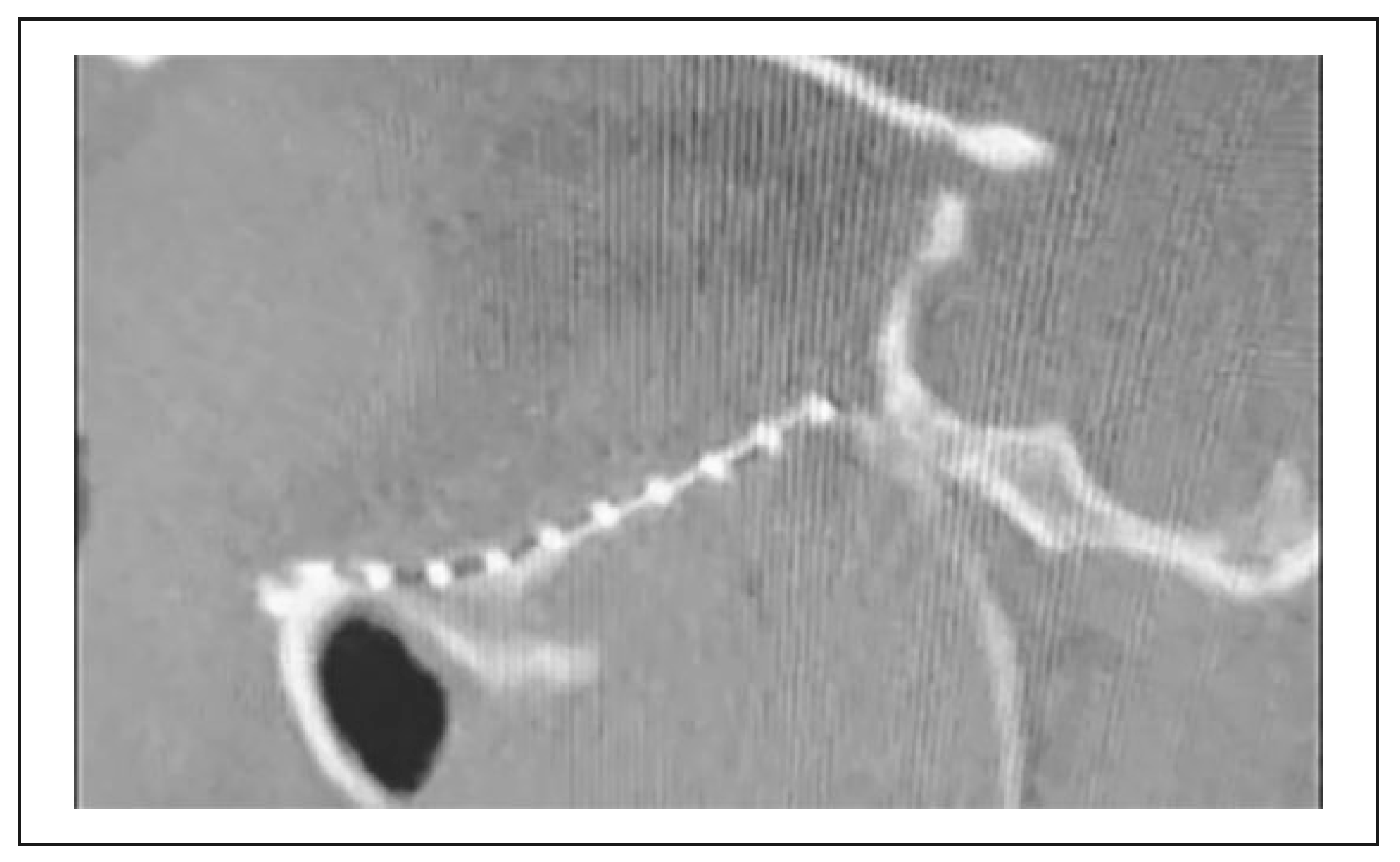
Figure 3 gives an example of a finished print. Figure 4 shows a bent titanium-mesh implant to reconstruct the defect zone anatomically.

3. Results

From May 2019 to March 2020, we treated 4 cases of severe orbital trauma with the approach mentioned above. Indication for performing orbital reconstruction with titanium-mesh implant was firm. All patients showed a disturbing esthetical enophthalmos of more than 2 mm and had persistent diplopia. All patients received an early reconstruction within 1 week from the onset. Every patient was seen by an ophthalmologist before and after surgery to document the clinical status. Informed consent has been obtained in every case emphasizing in detail the technique of 3D printing to best match the patient’s anatomy.
Within 48 h, we were able to perform the surgery. However, the prepared implant has to be handed over at least 24 h before surgery to complete the sterilization cycle. The time invested in segmentation varies significantly upon the defect size and artifacts. The segmentation process requires 30 min to 1 ½ h. Depending on the quality and the size, the print times varied between 2 ½ h and 6 h. Shaping and adapting the titanium-mesh takes between 10 and 20 min. The same process for bending a mandibular reconstruction plate takes approximately 45 to 60 min, depending on plate thickness and defect size.
The surgical approach for orbital reconstruction varied depending on the operator, 2 times subciliary vs. 2 times transconjunctival. In our hospital, KLS martin LevelOne is used as an osteosynthesis system. Orbital reconstruction was therefore performed with KLS martin LevelOne
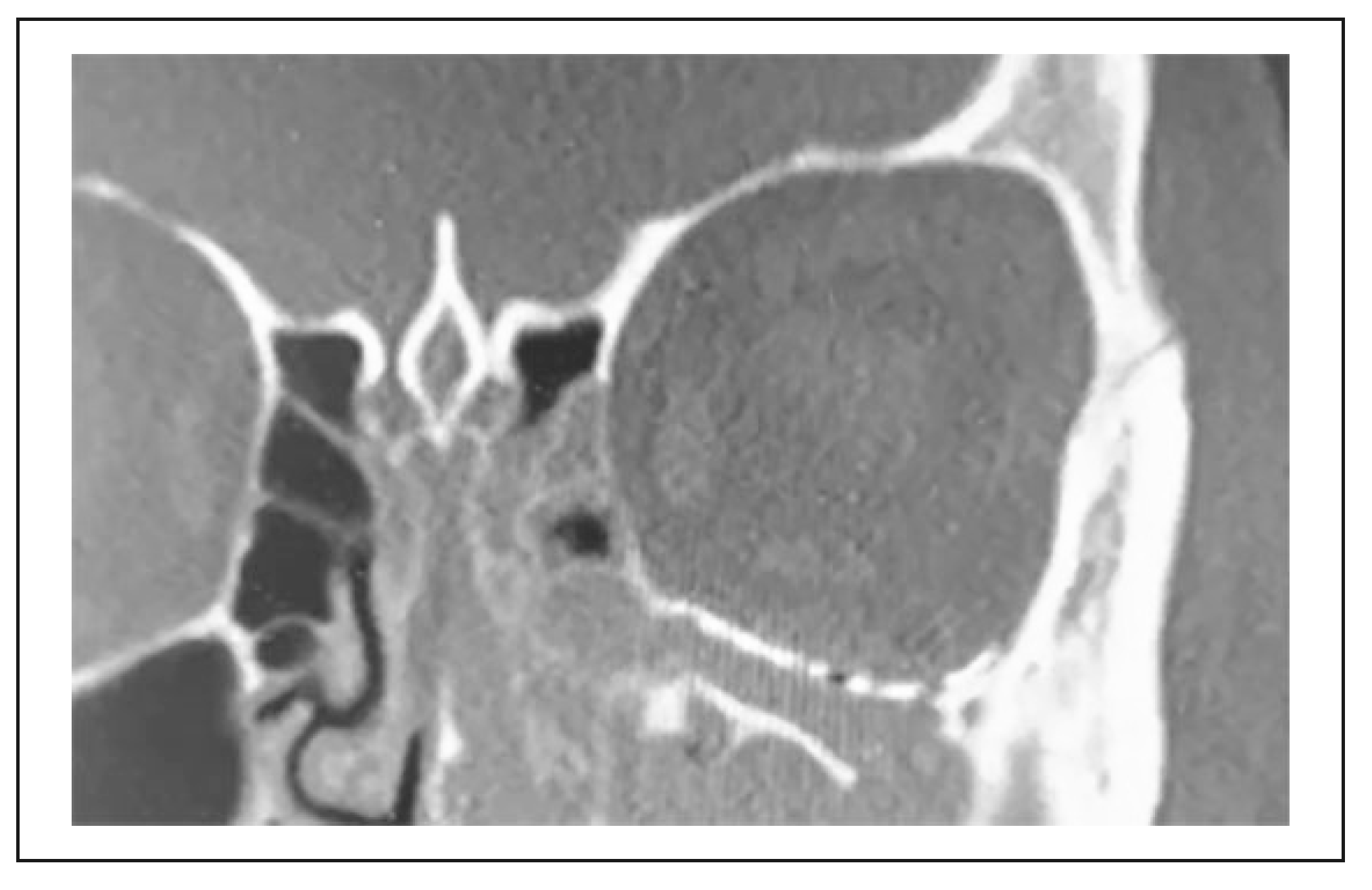
Midface 1.0 micro-mesh, as seen in Figure 4. We did not use intraoperative navigation during surgery to control the result. Hence a postoperative CT scan was performed as soon as possible to control the reconstruction radiologically. Figure 5 and Figure 6 show a successful reconstruction.
The CT scan displayed the correct anatomical position and regular reduction of the fracture. Vision and pupils were controlled every 15 min within the first 2 h and then extended to 30 min until 4 h postoperatively. After that, the interval was further stretched to an hourly control until 8 h postoperatively. After that, once per shift and stopped after 24 h. No loss of vision occurred, and no surgical revision was needed in any case. The postoperative ophthalmological examination showed no restriction in eye movement, no enophthalmos, and no diplopia. The same is true for freehand approach. Subjectively postoperative swelling varied depending on the surgical approach but was less than compared to conventional freehand open reduction and internal fixation. Reduction of postoperative swelling is most likely due to the decreased iterative manipulation and adaption processes resulting in shorterprocedure time, which varied from 45 min to 60 min with the pre-bent titanium mesh compared to 1 ½h to 2 h with freehand technique. Figure 7, Figure 8, Figure 9 and Figure 10, show another case treated with this technique.
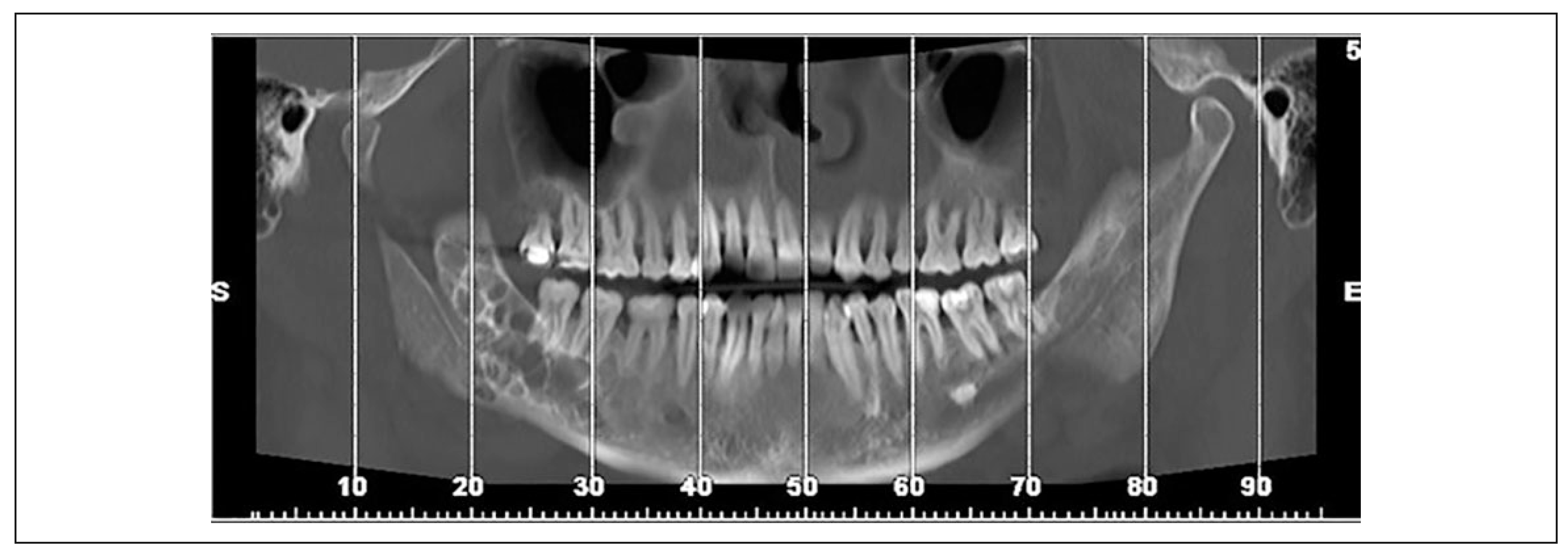
Besides the treatment of orbital trauma, we treated 1 case of severe odontogenic keratocyst (OKC) of the mandible. Figure 11 shows a reconstruction of the CT scan done to determine the extent of the cyst.
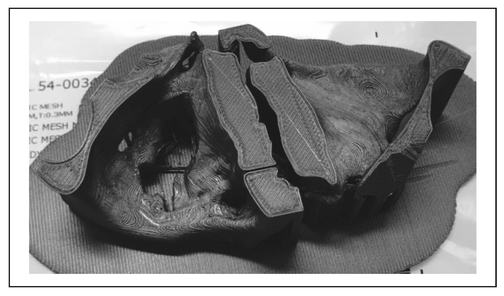
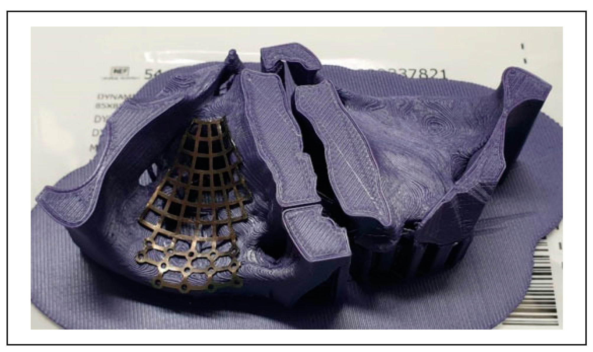
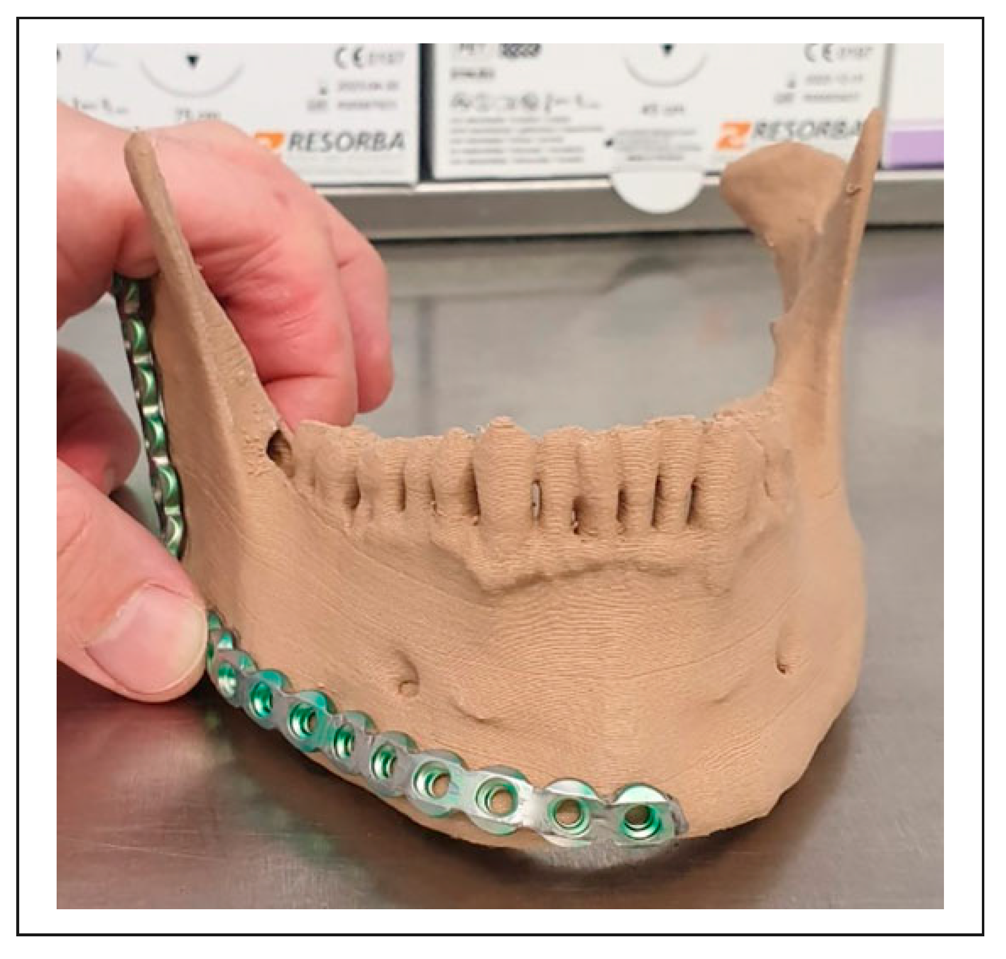
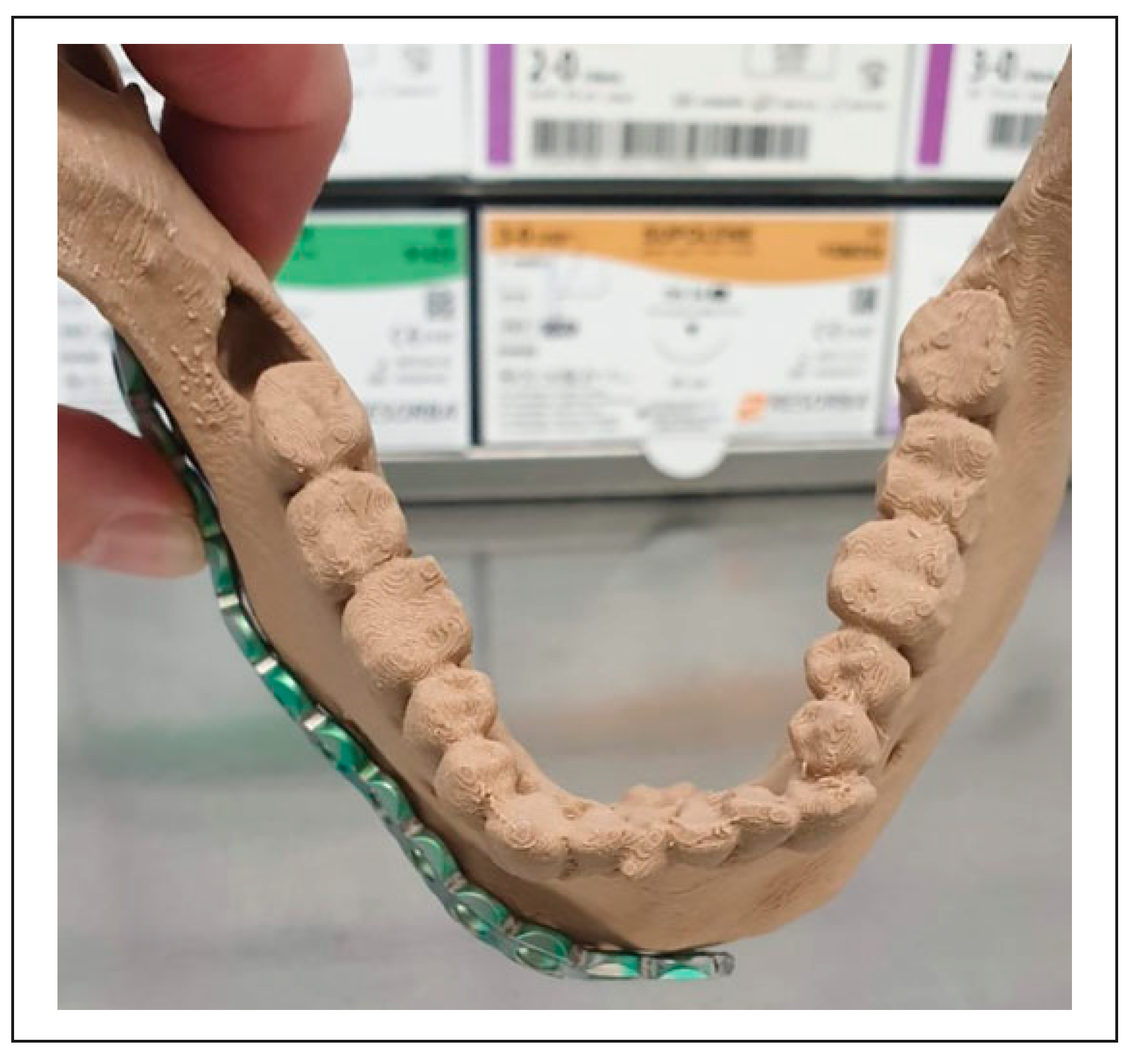
Because of the great extension in transversal, coronal, and sagittal planes, we decided on a continuity resection of the mandible, as the mandible was thinned out massively. We created a 3D specific model of the mandible and bent a reconstruction plate to fit perfectly. Figure 12, Figure 13 and Figure 14 show the printed model with the adapted plate.
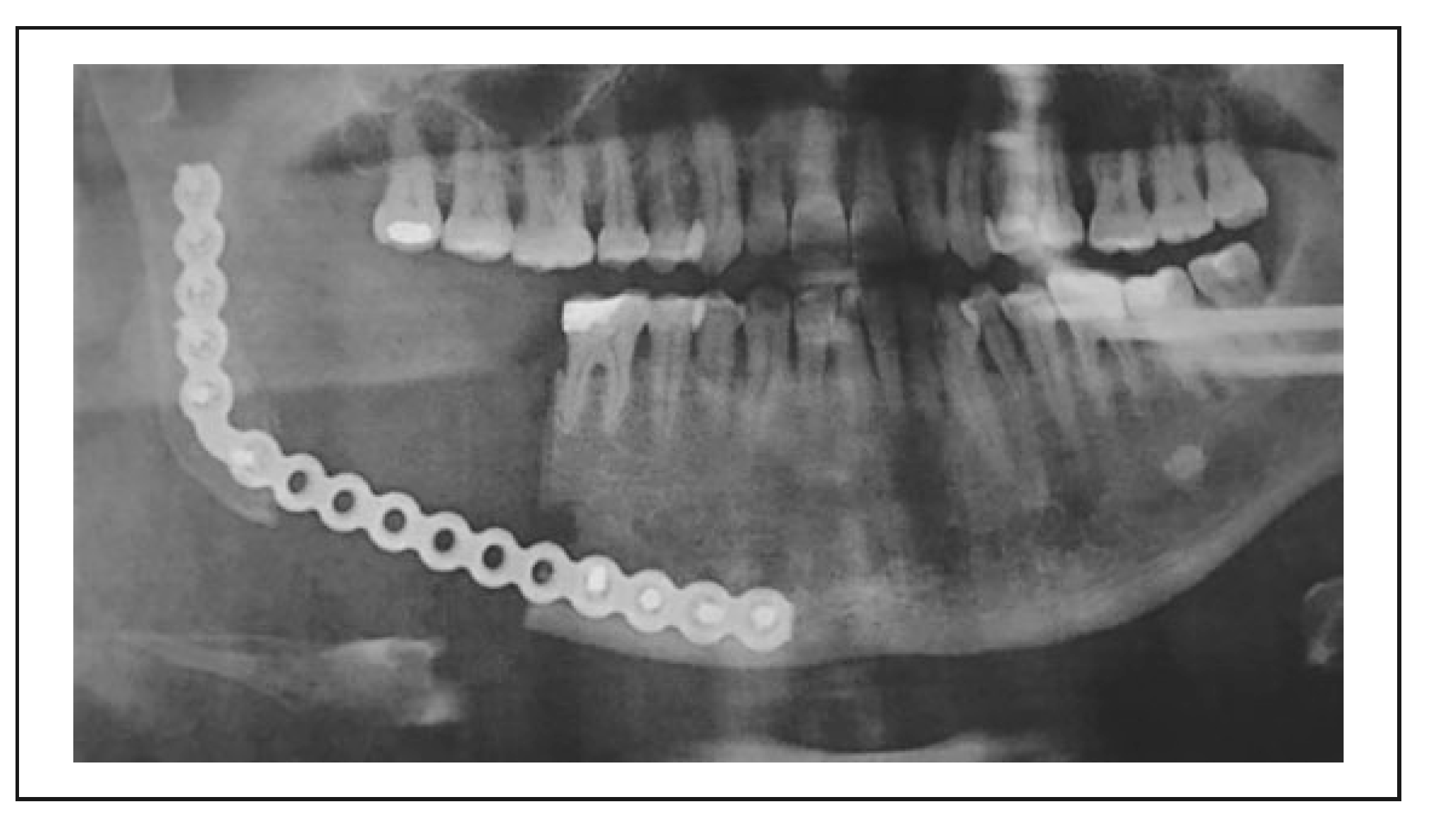
During surgery, the mandibular nerve was lateralized and preserved. Any excess plate material was cut off during operation. The postoperative X-rays show the plate in place (Figure 15) and after reconstruction with a free iliac crest graft (Figure 16).
This case showed likewise excellent functional and aesthetic outcome and significant reduction of operating time (2 ½ h compared to 3 ½ to 4 h with conventional technique). The treatment is still ongoing, as dental rehabilitation with implants is pending. Patient satisfaction was also high, although minor sensations of discomfort were noted by the patient on the operated side, especially when shaving.
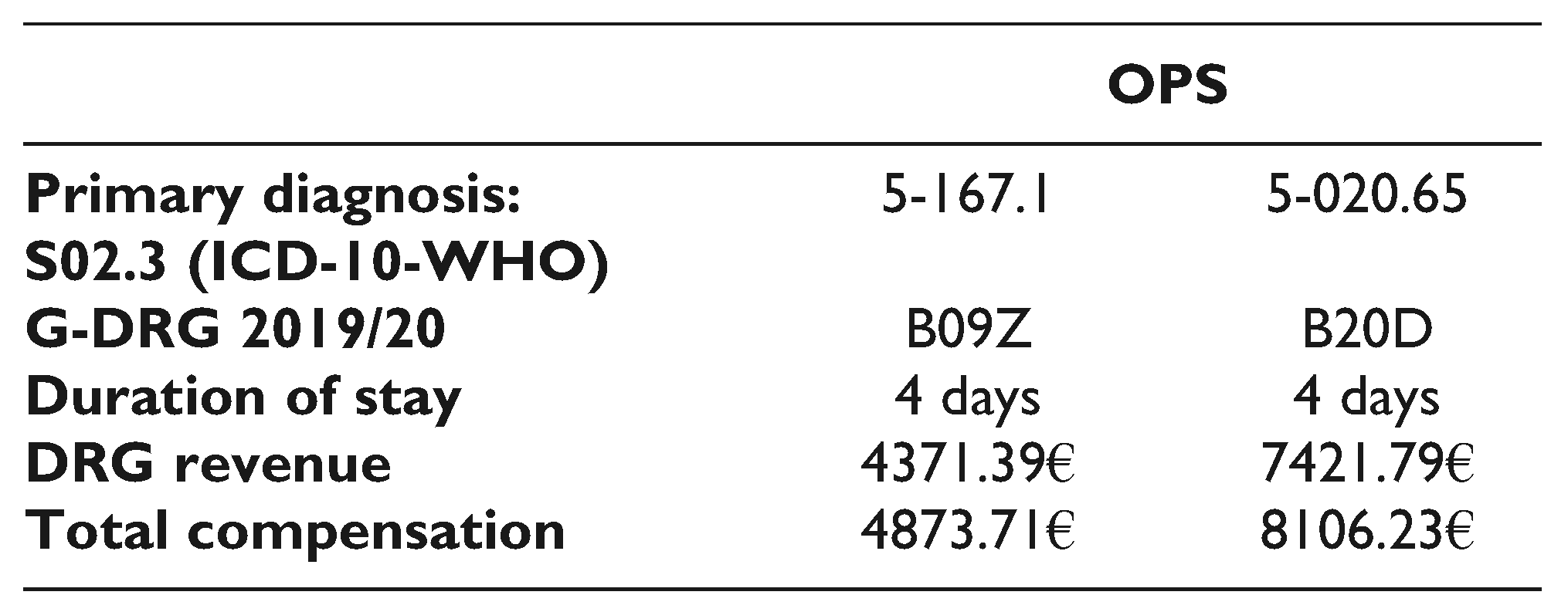
Finally, regarding DRG revenue, we applied the DRG Webgrouper algorithm of the DRG-Research-Group of the University of Mu¨nster, Germany. [46] Primary diagnosis in the case of orbital trauma was S02.3 fracture of the orbital floor (ICD-10-WHO). The DRG was selected automatically by the Webgrouper based on the OPS code used. Included procedures were 5-167.1 (reconstruction of the orbital wall: with metal plates or implants) and 5-020.65 (cranioplasty: reconstruction of the facial skull without involvement of the cerebral skull up to 2 regions with computer-assisted preformed implant [CAD-implant]). Duration of stay was 4 days. Total compensation accounted for 8106.23€ (G-DRG 2019/20) if OPS 5-20.65 was selected. If not, total compensation for OPS 5-167.1 was 4873.71€. So the total revenue with the use of a 3D printed patient-specific model was 3232.52€ higher. Therefore usage of 3D printing technologies not only offers better patient care but is also more attractive financially. Table 1 gives an overview of the revenues.
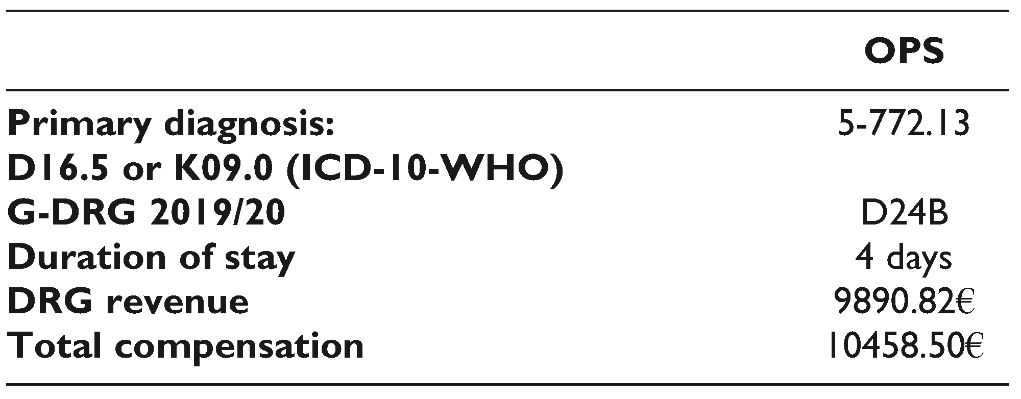
In contrast, the revenue for partial resection of the mandible was highest for OPS 5-772.13 (partial or total resection of the mandible: resection, partial with continuity cut: alloplastic reconstruction) and accounted for 10458.50€ (G-DRG 2019/20) total compensation. Resection and reconstruction of the mandible in the case presented in this study was a 2-stage approach. So, the initial resection was followed by a reconstruction with an autologous bone transplant, in particular a free iliac crest graft. The selected primary diagnosis in the case of OKC, which can be D16.5 (benign neoplasm of bone and articular cartilage: mandibular bone) or K09.0 (odontogenic cyst) made no difference in total compensation as the DRG remains the same. Here the provider of revenue was the selected OPS procedure. Table 2 shows the DRG revenue for odontogenic keratocyst (OKC).
The treatment of OKC presented here yielded no additional proceeds, and the benefit of a preformed reconstruction plate is the reduction of surgical time and less iterative manipulation of soft tissue. Furthermore, in the presented case, no plate loosening or plate fracture occurred and rendered it possible to perform the augmentation with free iliac crest graft without the need to exchange the plate.

4. Discussion

We showed that it is feasible to establish low-cost in-house 3D printing and adhere to legal requirements in Germany. Other studies already proposed 3D printing in a hospital environment and were adapted by us. [11,15,16,18] All cases of orbital floor reconstruction presented involved critical defect size and complex anatomical conditions. As mentioned earlier, a CT scan or CCT scan with 1 mm slices is obligatory. Without sound radiological data, the 3D model will have poor resolution and surface quality, thus rendering it unusable. In the case of external allocations, radiological imaging is, unfortunately, often inadequate or unsatisfactory. In orbital trauma treatment, high-quality imaging is key to successful reconstruction with a 3D printed model, which often leads to repeated CT scans and, thus, preventable radiation dose exposure. [47,48,49]
Nevertheless, orbital floor reconstruction and mandibular reconstruction without the aid of 3D printing would have been extremely challenging. Manually adapting a titanium-mesh or a reconstruction plate during surgery takes considerably more time and entails much manipulation, retractor dragging and pushing than our approach. However, we must mention that preoperative planning time is longer. Because of adapting and bending the titanium mesh/plate in advance, we were able to reduce operating time by at least 45 to 60 min. Likewise, the duration of general anesthesia decreased, too.
Thus reducing costs further and accounting for fewer comorbidities. [2,11,17,18,19,41] The total acquisition costs amounted to 383,42 Euros, which include the printer, and three 800 g filament spools, a smoke detector, and a fireproof base. As stated above, the material and energy cost for a 60 g 3D model are 1.58 Euros. Total expenditure is therefore significantly lower compared to known manufacturers 3D printing devices or outsourcing to a medical service provider. However, we note that 1 to 2 h of working time for the planning and shaping process are not included. Still, the cost can be roughly estimated, because the collective medical agreement for municipal hospitals in Germany is publicly available. For the lowest payment level and a 40 h work week, 1 h of working time accounts for approximately 26.86€. [50] So loosely speaking much less than an hour of surgical time. Also, the cost for the titanium-mesh, which accounts for a lower 3-digit€ number, are not included. The cost of the plates and meshes depends on the respective manufacturer and purchase price and can vary considerably.
The observed printing time 2 ½ h to 6 h depending on size and quality of post-processing, is similar to other studies. [11,41] However, we must say that the specific energy cost are highly likely to differ if tried to be reproduced. Ambient conditions and slicer settings inflict not only heat up times but also bed adhesion, viscosity, and cooling. As the 3D printer was not enclosed, the slightest change may affect the energy consumed.
An additional benefit was that the patients understood the planned treatment much better with the patient-specific model at hand. It helped the patients adhere to the treatment better compared to obtaining the informed consent with conventional methods such as radiological image demonstration and drawing a sketch of the surgical access way. [41] Having haptic feedback with the 3D model gives the surgeon a better sense of the fracture and the anatomical conditions, resulting in better performance in the operating theater. Equally to other studies, the educational aspects are also valuable for students and residents. [41,51,52,53]
One major limitation of our approach is that the surgery itself and the surgical dissection must be performed with great caution. If the prepared titanium-mesh implant is forced in place, it is highly likely to deform, thus rendering the whole previous work unusable. In order to overcome this limitation, plasma sterilization of the patient-specific 3D model for intraoperative use is necessary. Although we experimented with medical-grade I PLA filaments, the department for sterile goods processing did not grant the clearance for usage in the operating theater. Internal working instruction includes washing the sterile goods before sterilization. The manufacturer’s instruction sheet does not cover the solvents used, as already mentioned above.
In summary, we could show that low-cost 3D printing in a municipal hospital can be realized with little effort and monetary expenditures and achieve excellent results at the same time. It is worth noting that revenues are higher as well, at least in the case of orbital trauma. However, each case will be accurately assessed by the medical service of the health insurance, and the use of more profitable OPS codes may require additional justification. Also, we included only the primary diagnosis. Therefore, no casemix which may benefit to additional compensation can be calculated. Nevertheless, in our opinion, the reduction of operating time and the favorable outcome more than justifies the use of 3D printing techniques.
Looking ahead to the future rising expenditures in healthcare and the economic burden caused by the COVID-19 pandemic render it inevitable to search for cost savings, process optimization, timeand cost-efficient techniques to maintain high-quality patient care. Our approach offers the advantage of complete independence from medical service providers or other outsourced work like dental laboratories. Furthermore, our concept is adaptable to suit the needs of other specialties like neurosurgery or orthopedic surgery. [52,54,55] Our study aims to allow others to save time with the implementation process and address the challenging aspects right away. Moreover, we want to inspire more researchers to get past the obstacles we could not overcome and offer the best patient care in other hospitals with limited financial resources worldwide.

Author Contributions

All authors contributed to the study conception and design. The idea, material preparation, data collection, technical and practical realization were performed by Harald Gleissner. The first draft was written by Harald Gleissner, and all authors commented on previous versions of the manuscript. All authors read and approved the final manuscript.

Funding

The author(s) received no financial support for the research, authorship, and/or publication of this article.

Data Availability Statement

Anonymized data, all materials are available as over the counter products.

Acknowledgments

We acknowledge having conducted this study without any third party funding. The funding of this study is strictly private.

Conflicts of Interest

The author(s) declared no potential conflicts of interest with respect to the research, authorship, and/or publication of this article.

Ethics Approval

According to §15 BO (professional code of conduct) of BLAEK (regional medical association), no ethics committee consult is necessary. The study was performed in accordance with the ethical standards as laid down in the 1964 Declaration of Helsinki, its later amendments or comparable ethical standards.

Informed Consent to Participate

Informed consent was obtained from all individual participants included in the study.

Informed Consent for Publication

Patients signed informed consent regarding publishing their data and photographs.

References

  1. Banks, J. Adding value in additive manufacturing: Researchers in the United Kingdom and Europe look to 3D printing for customization. IEEE Pulse. 2013, 4, 22–26. [Google Scholar]
  2. Mehra, P.; Miner, J.; D’Innocenzo, R.; Nadershah, M. Use of 3-d stereolithographic models in oral and maxillofacial surgery. J. Maxillofac. Oral. Surg. 2011, 10, 6–13. [Google Scholar]
  3. Winder, J.; Bibb, R. Medical rapid prototyping technologies: State of the art and current limitations for application in oral and maxillofacial surgery. J. Oral. Maxillofac. Surg. Off. J. Am. Assoc. Oral. Maxillofac. Surg. 2005, 63, 1006–1015. [Google Scholar]
  4. Valding, B.; Zrounba, H.; Martinerie, S.; May, L.; Broome, M. Should you buy a three-dimensional printer? A study of an orbital fracture. J. Craniofac. Surg. 2018, 29, 1925–1927. [Google Scholar] [CrossRef]
  5. Sinha, P.; Skolnick, G.; Patel, K.B.; Branham, G.H.; Chi, J.J. A 3-dimensional-printed short-segment template prototype for mandibular fracture repair. JAMA Facial Plast. Surg. 2018, 20, 373–380. [Google Scholar]
  6. Ren, W.; Gao, L.; Li, S.; Chen, C.; Li, F.; Wang, Q.; Zhi, Y.; Song, J.; Dou, Z.; Xue, L.; et al. Virtual planning and 3D printing modeling for mandibular reconstruction with fibula free flap. Med. Oral. Patol. Oral. Cir. Bucal. 2018, 23, e359–e366. [Google Scholar]
  7. Fan, B.; Chen, H.; Sun, Y.-J.; Wang, B.-F.; Che, L.; Liu, S.-Y.; Li, G.-Y. Clinical effects of 3-D printingassisted personalized reconstructive surgery for blowout orbital fractures. Graefes Arch. Clin. Exp. Ophthalmol. [Albrecht Von Graefes Arch. Klin Exp. Ophthalmol.] 2017, 255, 2051–2057. [Google Scholar] [CrossRef]
  8. Chen, C.H.; Chen, C.T.; Wang, P.F.; Wang, Y.T.; Hsu, P.H.; Lin, C.L. A novel anatomical thin titanium mesh plate with patient-matched bending technique for orbital floor reconstruction. J. CranioMaxillofac. Surg. Off. Publ. Eur. Assoc. Cranio-Maxillofac. Surg. 2018, 46, 1526–1532. [Google Scholar] [CrossRef]
  9. Kozakiewicz, M.; Elgalal, M.; Loba, P.; Komuński, P.; Arkuszewski, P.; Broniarczyk-Loba, A.; Stefańczyk, L. Clinical application of 3D pre-bent titanium implants for orbital floor fractures. J. Cranio-Maxillofac. Surg. Off. Publ. Eur. Assoc. CranioMaxillofac. Surg. 2009, 37, 229–234. [Google Scholar] [CrossRef]
  10. Raisian, S.; Fallahi, H.R.; Khiabani, K.S.; Heidarizadeh, M.; Azdoo, S. Customized titanium mesh based on the 3D printed model vs. manual intraoperative bending of titanium mesh for reconstructing of orbital bone fracture: A randomized clinical trial. Rev. Recent. Clin. Trials. 2017, 12, 154–158. [Google Scholar] [CrossRef]
  11. Msallem, B.; Beiglboeck, F.; Honigmann, P.; Jaquie’ry, C.; Thieringer, F. Craniofacial reconstruction by a cost-efficient template-based process using 3D printing. Plast. Reconstr. Surg. Glob. Open 2017, 5, e1582. [Google Scholar]
  12. Yang, W.-F.; Choi, W.S.; Leung, Y.Y.; Curtin, J.P.; Du, R.; Zhang, C.-Y.; Chen, X.-S.; Su, Y.-X. Three-dimensional printing of patient-specific surgical plates in head and neck reconstruction: A prospective pilot study. Oral. Oncol. 2018, 78, 31–36. [Google Scholar] [PubMed]
  13. Batstone, M.D. Reconstruction of major defects of the jaws. Aust. Dent. J. 2018, 63 (Suppl 1), S108–S113. [Google Scholar] [PubMed]
  14. Orabona, G.D.; Abbate, V.; Maglitto, F.; Bonavolontà, P.; Salzano, G.; Romano, A.; Reccia, A.; Committeri, U.; Iaconetta, G.; Califano, L. Lowcost, self-made CAD/CAM-guiding system for mandibular reconstruction. Surg. Oncol. 2018, 27, 200–207. [Google Scholar]
  15. Elegbede, A.; Diaconu, S.C.; McNichols, C.H.; Seu, M.; Rasko, Y.M.; Grant, M.P.; Nam, A.J. Officebased three-dimensional printing workflow for craniomaxillofacial fracture repair. J. Craniofac. Surg. 2018, 29, e440–e444. [Google Scholar] [CrossRef] [PubMed]
  16. Mendez, B.M.; Chiodo, M.V.; Patel, P.A. Customized in-office three-dimensional printing for virtual surgical planning in craniofacial surgery. J. Craniofac. Surg. 2015, 26, 1584–1586. [Google Scholar]
  17. Salgueiro, M.I.; Stevens, M.R. Experience with the use of prebent plates for the reconstruction of mandibular defects. Craniomaxillofacial Trauma. Reconstr. 2010, 3, 201–208. [Google Scholar] [CrossRef]
  18. Smithers, F.A.E.; Cheng, K.; Jayaram, R.; Mukherjee, P.; Clark, J.R. Maxillofacial reconstruction using in-house virtual surgical planning. ANZ, J. Surg. 2018, 88, 907–912. [Google Scholar] [CrossRef]
  19. Abo Sharkh, H.; Makhoul, N. In-house surgeon-led virtual surgical planning for maxillofacial reconstruction. J. Oral. Maxillofac. Surg. Off. J. Am. Assoc. Oral. Maxillofac. Surg. 2020, 78, 651–660. [Google Scholar] [CrossRef]
  20. Kamio, T.; Hayashi, K.; Onda, T.; Takaki, T.; Shibahara, T.; Yakushiji, T.; Shibui, T.; Kato, H. Utilizing a low-cost desktop 3D printer to develop a “one-stop 3D printing lab” for oral and maxillofacial surgery and dentistry fields. 3D Print. Med. 2018, 4, 6. [Google Scholar] [CrossRef]
  21. Ghai, S.; Sharma, Y.; Jain, N.; Satpathy, M.; Pillai, A.K. Use of 3-D printing technologies in craniomaxillofacial surgery: A review. Oral. Maxillofac. Surg. 2018, 22, 249–259. [Google Scholar] [CrossRef] [PubMed]
  22. Pawlaczyk-Luszczynska, M.; Dudarewicz, A.; Szymczak, W.; Sliwinska-Kowalska, M. Evaluation of annoyance from low frequency noise under laboratory conditions. Noise Health 2010, 12, 166–181. [Google Scholar] [CrossRef] [PubMed]
  23. Yi, J.; LeBouf, R.F.; Duling, M.G.; Nurkiewicz, T.; Chen, B.T.; Schwegler-Berry, D.; Virji, M.A.; Stefaniak, A.B. Emission of particulate matter from a desktop three-dimensional (3D) printer. J. Toxicol. Environ. Health A 2016, 79, 453–465. [Google Scholar] [CrossRef]
  24. Vance, M.E.; Pegues, V.; van Montfrans, S.; Leng, W.; Marr, L.C. Aerosol emissions from fuse-deposition modeling 3d printers in a chamber and in real indoor environments. Environ. Sci. Technol. 2017, 51, 9516–9523. [Google Scholar] [CrossRef]
  25. Steinle, P. Characterization of emissions from a desktop 3D printer and indoor air measurements in office settings. J. Occup. Environ. Hyg. 2016, 13, 121–132. [Google Scholar] [CrossRef]
  26. Stefaniak, A.B.; Bowers, L.N.; Knepp, A.K.; Luxton, T.P.; Peloquin, D.M.; Baumann, E.J.; Ham, J.E.; Wells, J.R.; Johnson, A.R.; LeBouf, R.F.; et al. Particle and vapor emissions from vat polymerization desktop-scale 3-dimensional printers. J. Occup. Environ. Hyg. 2019, 16, 519–531. [Google Scholar] [CrossRef]
  27. Stefaniak, A.; Johnson, A.; du Preez, S.; Hammond, D.; Wells, J.; Ham, J.; LeBouf, R.; Menchaca, K.; Martin, S.; Duling, M.; et al. Evaluation of emissions and exposures at workplaces using desktop 3-dimensional printer. J. Chem. Health Saf. 2019, 26, 19–30. [Google Scholar] [CrossRef]
  28. Stefaniak, A.B.; LeBouf, R.F.; Yi, J.; Ham, J.; Nurkewicz, T.; Schwegler-Berry, D.E.; Chen, B.T.; Wells, J.R.; Duling, M.G.; Lawrence, R.B.; et al. Characterization of chemical contaminants generated by a desktop fused deposition modeling 3-dimensional printer. J. Occup. Environ. Hyg. 2017, 14, 540–550. [Google Scholar] [CrossRef] [PubMed]
  29. 3-D-Drucker Sicher Verwenden. Sichere Arbeitsplätze in Produktion und Industrie. Published 9 December 2019. Accessed 12 December 2019. Intranet. 9 December.
  30. Musterspule Splint Fill Filament, 2,85 mm—Shop 3D Agency. Available online: https://eshop-3d-agency.de/p/musterspule-splintfill-filament-2-85mm (accessed on 14 May 2020).
  31. Kunststoffverabeitung, B. Sicherheitsdatenblatt gemä ß 91/155/EWG. Available online: https://eshop-3dagency.de/WebRoot/Store16/Shops/e50a3f7b-8bd4-440da099-1dda18a98e9c/5CE2/94FF/438B/CCDF/18ED/0A48/3548/D079/SD-SplintFill-DE.pdf (accessed on 14 May 2020).
  32. Arfona LLC. Arfona 3D Printing Materials. Arfona. Available online: https://www.arfona.com/materials (accessed on 14 May 2020).
  33. DAS FILAMENT Inh. Roman Stieben AS 6 91448 Emskirchen. PLA Filament—1,75 mm—Neonorange. Published 14 May 2020. Available online: https://www.dasfilament.de/filament-spulen/pla-1-75-mm/96/pla-filament-1-75-mm-neonorange?c¼11 (accessed on 14 May 2020).
  34. BDEW. BDEW-Strompreisanalyse Januar 2020. Available online: http://www.bdew.de/service/daten-und-grafiken/bdew-strompreisanalyse/ (accessed on 29 May 2020).
  35. 20200107_BDEW-Strompreisanalyse_Januar_2020.pdf. Available online: https://www.bdew.de/media/documents/20200107_BDEW-Strompreisanalyse_Januar_2020.pdf (accessed on 29 May 2020).
  36. Slicer Wiki contributors. Documentation/4.x/Acknowledgments. Published 9 April 2020. Available online: https://www.slicer.org/w/index.php?title¼Documentation/4.x/Acknowledgments&oldid¼61143 (accessed on 22 September 2021).
  37. Jolesz, F.A. Intraoperative Imaging and Image-Guided Therapy; Springer: Berlin/Heidelberg, Germany, 2014. [Google Scholar]
  38. Kikinis, R.; Pieper, S.D.; Vosburgh, K.G. 3D slicer: A platform for subject-specific image analysis, visualization, and clinical support. In Intraoperative Imaging and Image-Guided Therapy; Jolesz, F.A., Ed.; Springer: New York, NY, USA, 2014; pp. 277–289. [Google Scholar]
  39. Kapur, T.; Pieper, S.; Fedorov, A.; Fillion-Robin, J.-C.; Halle, M.; O’Donnell, L.; Lasso, A.; Ungi, T.; Pinter, C.; Finet, J.; et al. Increasing the impact of medical image computing using community-based openaccess hackathons: The NA-MIC and 3D slicer experience. Med. Image Anal. 2016, 33, 176–180. [Google Scholar] [CrossRef]
  40. Fedorov, A.; Beichel, R.; Kalpathy-Cramer, J.; Finet, J.; Fillion-Robin, J.-C.; Pujol, S.; Bauer, C.; Jennings, D.; Fennessy, F.; Sonka, M.; et al. 3D Slicer as an image computing platform for the quantitative imaging network. Magn. Reson. Imaging. 2012, 30, 1323–1341. [Google Scholar] [PubMed]
  41. Velasco, I.; Vahdani, S.; Ramos, H. Low-cost method for obtaining medical rapid prototyping using desktop 3D printing: A novel technique for mandibular reconstruction planning. J. Clin. Exp. Dent. 2017, 9, e1103–e1108. [Google Scholar] [PubMed]
  42. Autodesk, Inc. Autodesk Meshmixer Free Software for Making Awesome Stuff. Available online: http://www.meshmixer.com/ (accessed on 14 May 2020).
  43. Foundation, B. blender.org—Home of the Blender Project—Free and Open 3D Creation Software. blender.org. Available online: https://www.blender.org/ (accessed on 14 May 2020).
  44. Ultimaker, B.V.; Ultimaker Cura: Leistungsstarke, Benutzerfreundliche 3D-Drucksoftware. ultimaker.com. Available online: https://ultimaker.com/de/software/ultimakercura (accessed on 14 May 2020).
  45. Hofmann, M. Formular Laufzettel/Implantat, M.K.G. Published online 27 May. 2019.
  46. DRG-Research Group Webgrouper. Available online: https://www.drg-research-group.de/index.php?option¼com_webgrouper&Itemid¼112&view¼webgrouperv (accessed on 31 July 2020).
  47. Hinzpeter, R.; Sprengel, K.; Wanner, G.A.; Mildenberger, P.; Alkadhi, H. Repeated CT scans in trauma transfers: An analysis of indications, radiation dose exposure, and costs. Eury, J. Radiol. 2017, 88, 135–140. [Google Scholar] [CrossRef]
  48. Bracco, D.; Deckelbaum, D.; Artho, G.; Khwaja, K.; Mulder, D.S.; Gruska, J.; Razek, T. Additional and repeated computed tomography in interfacility trauma transfers: Room for standardization. Surgery 2018, 164, 872–878. [Google Scholar] [CrossRef]
  49. Jones, A.C.; Woldemikael, D.; Fisher, T.; Hobbs, G.R.; Prud’homme, B.J.; Bal, G.K. Repeated computed tomographic scans in transferred trauma patients: Indications, costs, and radiation exposure. J. Trauma. Acute Care Surg. 2012, 73, 1564–1569. [Google Scholar] [CrossRef]
  50. Vereinigung der kommunalen Arbeitgeberverbände, M.B. Tarifvertrag für Ärztinnen und Ärzte an kommunalen Kranken-häusern im Bereich der kommunalen Arbeitgeberverbände (TV-Ärzte/VKA). Published 31 July 2020. Available online: https://bit.ly/316orRP (accessed on 31 July 2020).
  51. Werz, S.M.; Zeichner, S.J.; Berg, B.I.; Zeilhofer, H.F.; Thieringer, F. 3D Printed surgical simulation models as educational tool by maxillofacial surgeons. Eur. J. Dent. Educ. Off. J. Assoc. Dent. Educ. Eur. 2018, 22, e500–e505. [Google Scholar]
  52. Ploch, C.C.; Mansi, C.S.S.A.; Jayamohan, J.; Kuhl, E. Using 3D printing to create personalized brain models for neurosurgical training and preoperative planning. World Neurosurg. 2016, 90, 668–674. [Google Scholar]
  53. Nicot, R.; Druelle, C.; Schlund, M.; Roland-Billecart, T.; Gwénaël, R.; Ferri, J.; Gosset, D. Use of 3D printed models in student education of craniofacial traumas. Dent. Traumatol. Off. Publ. Int. Assoc. Dent. Traumatol. 2019, 35, 296–299. [Google Scholar]
  54. Hasan, O.; Atif, M.; Jessar, M.M.; Hashmi, P. Application of 3D printing in orthopaedic surgery. A new affordable horizon for cost-conscious care. JPMA, J. Pak. Med. Assoc. 2019, 69 (Suppl 1), S46–S50. [Google Scholar]
  55. Chen, K.; Yang, F.; Yao, S.; Xiong, Z.; Sun, T.; Zhu, F.; Telemacque, D.; Drepaul, D.; Ren, Z.; Guo, X. Application of computerassisted virtual surgical procedures and three-dimensional printing of patient-specific pre-contoured plates in bicolumnar acetabular fracture fixation. Orthop. Traumatol. Surg. Res. 2019, 105, 877–884. [Google Scholar] [PubMed]
Figure 1. DICOM anonymization sequence.
Cmtr 15 00049 g001
Figure 2. Workflow 3D to surgery, marked with (*) shows sterile processes.
Cmtr 15 00049 g002
Figure 3. Finished patient-specific 3D printed model.
Cmtr 15 00049 g003
Figure 4. Finished titanium-mesh implant for reconstruction.
Cmtr 15 00049 g004
Figure 5. Coronary plane showing reconstruction.
Cmtr 15 00049 g005
Figure 6. Sagittal plane showing reconstruction.
Cmtr 15 00049 g006
Figure 7. 3D printed model.
Cmtr 15 00049 g007
Figure 8. 3D model with bent titanium-mesh.
Cmtr 15 00049 g008
Figure 9. Coronary plane showing reconstruction.
Cmtr 15 00049 g009
Figure 10. Sagittal plane showing reconstruction.
Cmtr 15 00049 g010
Figure 11. Panoramic reconstruction of the CT scan.
Cmtr 15 00049 g011
Figure 12. Bent plate frontal view.
Cmtr 15 00049 g012
Figure 13. Bent plate upper view.
Cmtr 15 00049 g013
Figure 14. Bent plate bottom view.
Cmtr 15 00049 g014
Figure 15. Postoperative X-ray after continuity resection and mandible bridging plate in place.
Cmtr 15 00049 g015
Figure 16. Postoperative X-ray after free iliac crest graft transplant.
Cmtr 15 00049 g016
Table 1. DRG Revenue Orbital Floor Fracture S02.3 (ICD-10WHO).
Cmtr 15 00049 i001
Table 2. DRG Revenue Partial Mandibular Resection.
Cmtr 15 00049 i002