Next Article in Journal
Facial Trauma During the COVID-19 Pandemic
Previous Article in Journal
The Role of Bone Grafts in Preventing Medication-Related Osteonecrosis of the Jaw: Histomorphometric, Immunohistochemical, and Clinical Evaluation in Animal Model
 
 
Craniomaxillofacial Trauma & Reconstruction is published by MDPI from Volume 18 Issue 1 (2025). Previous articles were published by another publisher in Open Access under a CC-BY (or CC-BY-NC-ND) licence, and they are hosted by MDPI on mdpi.com as a courtesy and upon agreement with Sage.
Article

Intraoperative Positioning in Maxillofacial Trauma Patients with Cervical Spine Injury—Is It Safe? Radiological Simulation in a Healthy Volunteer

by
Thomas Pepper
1,
Harry Spiers
2,
Alex Weller
3 and
Clare Schilling
4,*
1
Institute of Naval Medicine, Defence Medical Services, UK
2
Department of Surgery, Addenbrooke’s Hospital, Cambridge, UK
3
Department of Head and Neck Radiology, Northwick Park Hospital, Watford Road, London, UK
4
Department of Head and Neck Surgery, University College Hospital, Academic Head and Neck Unit, University College London, 250 Euston Road, London NW1 2PG, UK
*
Author to whom correspondence should be addressed.
Craniomaxillofac. Trauma Reconstr. 2022, 15(4), 312-317; https://doi.org/10.1177/19433875211053091
Submission received: 1 November 2020 / Revised: 1 December 2020 / Accepted: 1 January 2021 / Published: 3 January 2022

Abstract

:
Study Design: Observational. Objective: To investigate the effects on the cervical spine of positioning patients for maxillofacial procedures by simulating intraoperative positions for common maxillofacial procedures. Methods: Magnetic resonance imaging was used to assess the effects of head position in common intraoperative configurations – neutral (anterior mandible position), extended (tracheostomy position) and laterally rotated (mandibular condyle position) on the C-spine of a healthy volunteer. Results: In the tracheostomy position, maximal movement occurred in the sagittal plane between the cervico-occipital junction and C4–C5, as well as at the cervico-thoracic junction. Minimal movement occurred at C2 (on C3), C5 (on C6) and C6 (on C7). In the mandibular condyle position, C-spine movements occurred in both rotational and sagittal planes. Maximal movement occurred above the level of C4, concentrated at atlanto-occipital and atlanto-axial (C1–2) joints. Conclusions: Neck extension is likely to be relatively safe in injuries that are stable in flexion and extension, such as odontoid peg fracture and fractures between C5 and C7. Head rotation is likely to be relatively safe in fractures below C4, as well as vertebral body fractures, and laminar fractures without disc disruption. Early dialogue with the neurosurgical team remains a central tenet of safe management of patients with combined maxillofacial and C-spine injuries.

Introduction

Cervical spine injury (CSI) is known to coexist in 1–10% of patients with maxillofacial fractures,[1,2,3,4,5,6,7] the presence of which has been reported to double the odds of CSI.[4]
The most commonly reported aetiology of combined maxillofacial fractures and CSI is high-energy trauma such as road traffic accidents.[1,4,5,8,9,10] Such trauma often results in mixed fractures involving two or more facial thirds. Mukherjee et al[4] reported that the incidence of CSI was eight times higher when associated with such a mixed maxillofacial fracture pattern, compared with an isolated mandibular fracture or non-mandibular facial fractures. A similar trend has been observed by other authors.[1,7]
This association has importance for the assessment of trauma patients, as well as implications for general anaesthesia and the operative management of maxillofacial injuries, both of which may necessitate manipulation of the head and neck.
Patients with proven or suspected cervical spine injuries are commonly immobilised in a semi-rigid collar, with the aim of preventing secondary injury resulting from movement. Definitive maxillofacial intervention may be delayed due to uncertainty over the status of the cervical spine[4,5,11] and whether removal of the collar and movement of the head is permitted. For the patient, the effects of a delay in treatment may lead to prolonged discomfort and return to oral nutrition, resulting in reduced quality of life. To date, there remains a lack of good-quality prospective data on the effect of deferred treatment on the risk of infection, wound dehiscence, and delayed-, non- or malunion.[12]
The aim of this study was to investigate the biomechanical effects on the cervical spine of positioning patients for maxillofacial procedures in theatre. This may in turn provide evidence regarding the safety of providing definitive maxillofacial intervention in patients with concomitant spinal injuries.

Methods

Magnetic resonance imaging (MRI) using a Siemens Aera 1.5T scanner was used to assess the effect of head movement on the cervical spine of a healthy volunteer. The volunteer was a 43-year-old female with no history of neck pain or injury and a normal range of neck movement. All image acquisitions were performed during free breathing using phased-array body coils (two anterior and two posterior elements). T2-weighted (T2W) axial and sagittal images comprised 2D spin-echo sequences with single-shot echo planar readout over a large field of view, with the whole C-spine imaged in a single scan volume. The fatsuppressed T2W sagittal images were acquired as 4-mm slices with 0.4 mm slice interspace, using a short-tau inversion recovery (STIR) fat suppression, repetition time (TR) = 667 ms, echo time (TE) = 24 ms and number of signal averages (NSA) = 2. Sagittal images were acquired without fat suppression at 3 mm slice thickness, 0.3 mm slice interspace, with TR = 3800 ms, TE = 84 ms and NSA = 2.
Axial and sagittal images were performed simulating three commonly used surgical positions (Table 1):
The effect of C-spine manipulation on different vertebral levels was evaluated by calculating the angle (in degrees) of the axes of each vertebra on the level below, in all three surgical positions. This was performed using an electronic protractor on the sagittal images for AP flexion/extension (Figure 1) and the axial images for lateral rotation. AP flexion/extension was calculated at each vertebral level, as the ‘sagittal intervertebral angle’ made between the inferior vertebral endplate and a similar line at the level below (Figure 2a–c). Rotation was calculated on axial images the ‘transverse intervertebral angle’, made by a line traversing the midpoint of the vertebral body plus spinous process at each level compared with the level below. At the skull base, these angles were defined by the line connecting basion to opisthion (anterior and posterior margins of foramen magnum) relative to the line traversing inferior margin of C1. All image analysis was performed in OsiriX (Pixmeo, Geneva, Switzerland).

Results

Images showing gross spinal alignment during intraoperative simulation are shown in Figure 1 and Figure 2.

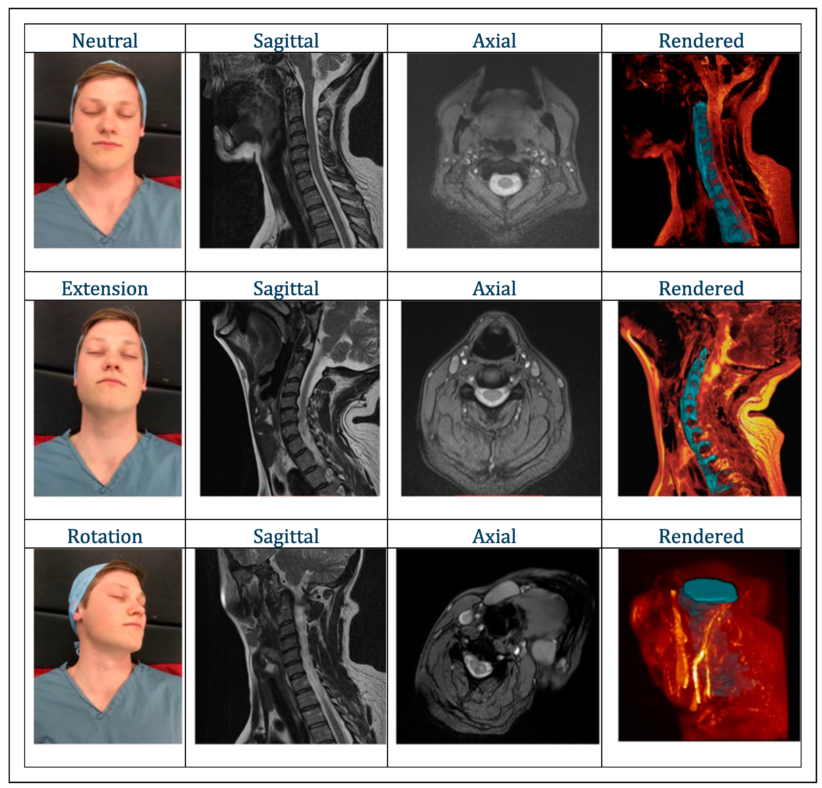
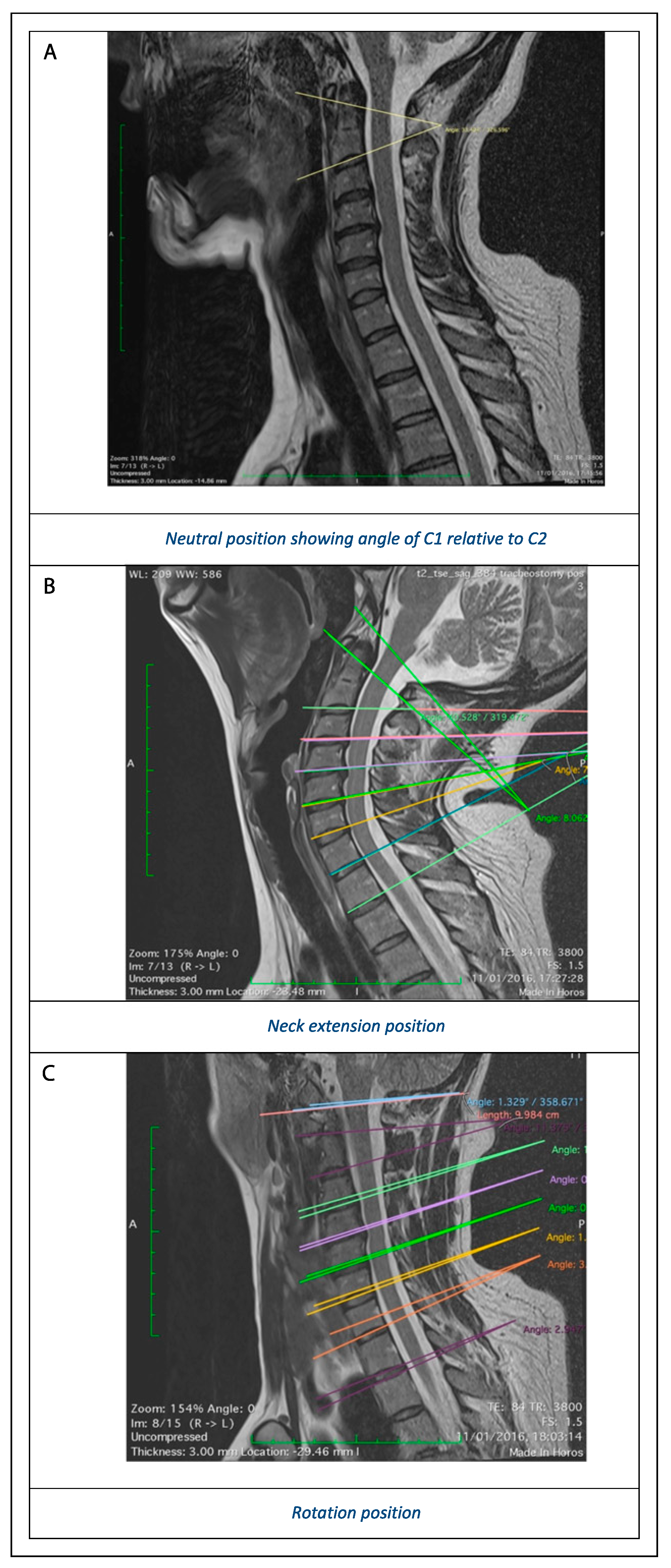
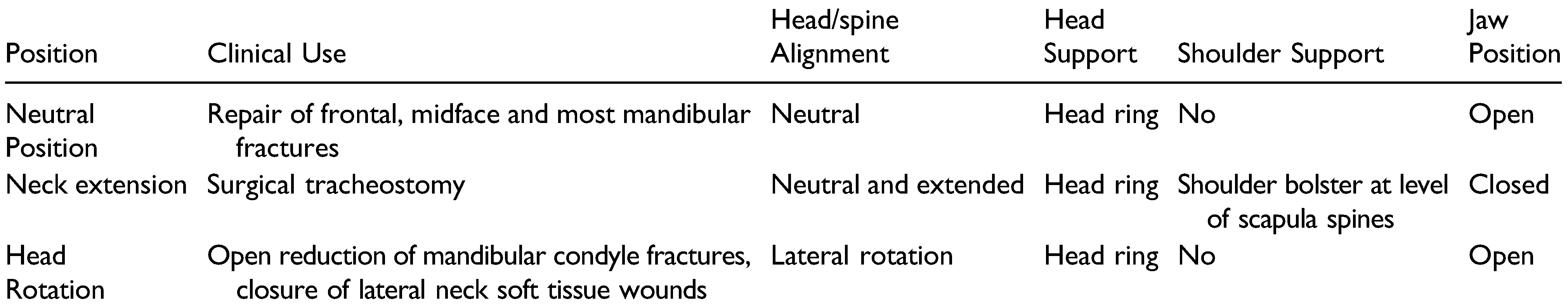
Effect of positioning on cervical spine alignment:

Intervertebral angles in both sagittal (for extension) and transverse (for rotation) planes are recorded in Table 2(a), alongside the change in these angles relative to the ‘anterior mandible’ position, which was taken as a clinically relevant position that was close to true neutral. Relative to this position, the effect of cervical manipulation on ’sagittal’ and ‘transverse’ intervertebral angles was calculated at each level, for both extension (on assuming the ‘tracheostomy’ position) and rotation (on assuming the ‘mandibular condyle’ position) (Table 2 (b–c)).

Effect of Extension

In the ‘tracheostomy’ position compared with ‘anterior mandible’, the vast majority of movement of each vertebra on the level below occurred in the sagittal plane. As expected, the intervertebral angle of extension increases at every level, as well as at the cervico-occipital and cervicothoracic junctions. Maximal movement (between 7–10°) was recorded between the cervico-occipital junction and C4–C5, plus at the cervico-thoracic junction, whereas minimal movement (1–3°) was observed at C2 (on C3), C5 (on C6) and C6 (on C7).

Effect of Rotation

In the ‘mandibular condyle’ position, compared with ‘anterior mandible’, movement of each vertebra on the level below occurred in both the transverse (rotational) and the sagittal (flexion/extension) planes. The majority of flexion (22°) occurred at the atlanto-axial joint (C1 on C2), with paradoxical extension of 9° at C3 on C4. The majority of rotation occurred at the atlanto-axial joint (32°), with lesser contribution from the atlanto-occipital joint (7°). Neck rotation was associated with very little intervertebral movement below the level of C4, including at the cervico-thoracic junction (6° or less).

Discussion

Until relatively recently, three-view (lateral, anteroposterior and odontoid peg) radiographs were the standard trauma imaging for the cervical spine. The sensitivity of these views for detecting CSI is reported to be 52%, compared with 98% for computed tomography (CT).[13] The latter is currently recommended as first-line imaging in selected adult trauma patients[14]; as a result, a greater number of CSIs are likely to be identified, which will increase the need for communication between the spinal and maxillofacial surgery teams. The data obtained in this study should help to inform such discussions. The most common types of hard-tissue CSIs reported in maxillofacial trauma patients are bony fractures, cervical subluxations and dislocations, while the most common softtissue CSIs are disc herniations and cord contusions.[1,4] The most common levels involved in hard tissue injury are C1–2 and C6–7.[1,4,5,6,8,15,16,17] Soft-tissue CSI is reported to incrementally increase in frequency, the more caudal the spinal level.[1,4] Some authors have reported an association between upper-level CSI and lower facial third (i.e. mandibular) fractures, and lower cervical spine injury and middle facial third fractures.[4,10] However, numbers in these samples were small and this finding has not been reproduced in other studies.[1]
The range of motion at each cervical spinal segment during different movements has been defined over a range of studies (Table 3).
This has been a problematic area of research because movement at each joint rarely occurs in isolation; axial rotation is always coupled with flexion-extension and lateral flexion.[22] Also, while cervical vertebral movements are reproducible within the same individual, they are not simple, and there may be variability of order of contribution between individuals, and this may be altered following injury. Despite these complexities, what can be deduced from the research to date is that axial rotation occurs primarily at the atlanto-axial joint (C1–C2), while lateral flexion occurs primarily in the subaxial cervical spine (C2– 7). Flexion and extension, in contrast, occur in a more equitable fashion along the length of the cervical spine.
Our images show that C-spine neutral alignment is maintained with a horseshoe head ring. In extension (the tracheostomy position), maximal movement occurred in the sagittal plane between the cervico-occipital junction and C4–C5, as well as at the cervico-thoracic junction. Minimal movement occurred at C2 (on C3), C5 (on C6) and C6 (on C7). This reflects that upper cervical, cervico-occipital and cervico-thoracic bony or soft-tissue injuries are likely to pose a higher risk of displacement during neck extension than isolated lower cervical (e.g. C5–6) injuries.
In lateral rotation (the mandibular condyle position), our images showed that cervical spine movements were more complex, occurring in both rotational and sagittal planes. Maximal movement occurred above the level of C4, meaning that as for neck extension, upper cervical injuries (above C4) are likely to pose a greater risk of displacement during rotational manipulation than those in lower cervical levels. However, unlike for extension, rotation is associated with movement that is concentrated at atlanto-occipital and atlanto-axial (C1–2) joints, suggesting caution during rotation in the setting of injuries at these levels.
Neck extension is likely to be safe in injuries that are stable in flexion and extension, such as odontoid peg fracture and fractures between C5 and C7. Head rotation is likely to be safe in fractures below C4, as well as vertebral body fractures, and laminar fractures without disc disruption. This is based on the premise that these segments move relatively little during the stated movements, but with the caveat that at present, there is a lack of data to describe exactly what magnitude of movement may result in secondary cord injury in a variety of CSIs.
The principal limitation of this study is that one healthy volunteer was used. Future studies should involve a greater number of participants, both male and female of a range of ages, in order to increase the applicability of findings. Although the biomechanics of spinal movement may differ in an injured neck, it would be unethical to conduct a similar investigation in such subjects.
We advocate seeking specialist spinal opinion prior to maxillofacial surgical intervention in patients with proven or suspected CSI in order to reduce the risk of secondary injury. Risk reduction may be further facilitated by further cross-sectional imaging in the theatre, if available, in order to assess stability. Once out of the collar on the operating table, the neck may be stabilised using blocks and tape in order to further reduce the risk of adverse movements. If there is any doubt about spinal safety, maxillofacial surgery should ideally be deferred.

Conclusions

We have demonstrated for the first time the effect of positioning for maxillofacial surgery on the cervical spine in a healthy volunteer. This information can inform interdisciplinary discussions about those patients in which it may be possible to safely remove the collar and alter the position of the head and neck. Early dialogue with the neurosurgical team remains a central tenet of safe management of patients with combined maxillofacial and cervical spine injuries.

Funding

This research received no external funding.

Acknowledgments

Many thanks to the Imaging Department of London Bridge Hospital for permitting the use of their MRI scanner for this study.

Conflicts of Interest

The authors declare no conflict of interest.

References

  1. Elahi, M.M.; Brar, M.S.; Ahmed, N.; Howley, D.B.; Nishtar, S.; Mahoney, J.L. Cervical spine injury in association with craniomaxillofacial fractures. Plast Reconstr Surg. 2008, 121, 201–208. [Google Scholar] [CrossRef] [PubMed]
  2. Mithani, S.K.; Mithani, S.K.; St-Hilaire, H.; et al. Predictable patterns of intracranial and cervical spine injury in craniomaxillofacial trauma: analysis of 4786 patients. Plast Reconstr Surg. 2009, 123, 1293–1301. [Google Scholar] [CrossRef] [PubMed]
  3. Chu, M.W.; Soleimani, T.; Evans, T.A.; et al. C-spine injury and mandibular fractures: lifesaver broken in two spots. J Surg Res. 2016, 206, 386–390. [Google Scholar] [CrossRef] [PubMed]
  4. Mukherjee, S.; Abhinav, K.; Revington, P.J. A review of cervical spine injury associated with maxillofacial trauma at a UK tertiary referral centre. Ann R Coll Surg Engl; 2015, 91, 66–72 101308/003588414X14055925059633. [Google Scholar]
  5. Roccia, F.; Cassarino, E.; Boccaletti, R.; Stura, G. Cervical spine fractures associated with maxillofacial trauma: An 11-year review. J Craniofac Surg. 2007, 18, 1259–1263. [Google Scholar] [CrossRef] [PubMed]
  6. Jamal, B.T.; Diecidue, R.; Qutub, A.; Cohen, M. The Pattern of Combined Maxillofacial and Cervical Spine Fractures. J Oral and Maxillofacial Surg. 2009, 67, 559–562. [Google Scholar] [CrossRef]
  7. Mulligan, R.P.; Mahabir, R.C. The prevalence of cervical spine injury, head injury, or both with isolated and multiple craniomaxillofacial fractures. Plast Reconstr Surg. 2010, 125, 1647–1651. [Google Scholar] [CrossRef] [PubMed]
  8. Mourouzis C, Schoinohoriti O, Krasadakis C, Rallis G, Cervical spine fractures associated with maxillofacial trauma: a 3-year-long study in the Greek population. J cranio-maxillofacial surg. 2018, 46, 1712–1718. [CrossRef] [PubMed]
  9. Beirne, J.C.; Butler, P.E.; Brady, F.A. Cervical spine injuries in patients with facial fractures: a 1-year prospective study. International journal of oral and maxillofacial surgery. 1995, 24, 26–29. [Google Scholar] [CrossRef] [PubMed]
  10. Lalani, Z.; Bonanthaya, K.M. Cervical spine injury in maxillofacial trauma. Br J Oral and Maxillofac Surg. 1997, 35, 243–245. [Google Scholar] [CrossRef]
  11. Schilling, C. Audit of Time from Admission to Open Reduction and Internal Fixation of Mandibular Fractures. Internal audit; 2016 (unpublished).
  12. Stone, N.; Corneman, A.; Sandre, A.R.; Farrokhyar, F.; Thoma, A.; Cooper, M.J. Treatment delay impact on open reduction internal fixation of mandibular fractures: a systematic review. Plasti reconstr surgery. Glob open. 2018, 6, e1829. [Google Scholar] [CrossRef] [PubMed]
  13. Holmes, J.F.; Akkinepalli, R. Computed tomography versus plain radiography to screen for cervical spine injury: a metaanalysis. J trauma. 2005, 58, 902–905. [Google Scholar]
  14. National Institute for Health and Care Excellence. Spinal Injury: Assessment and Initial Management. NICE Guideline NG41; National Clinical Guideline Centre: London, UK, 2016. [Google Scholar]
  15. Reich, W.; Surov, A.; Eckert, A.W. Maxillofacial trauma – Underestimation of cervical spine injury. J Cranio-Maxillofacial Surg. 2016, 44, 1469–1478. [Google Scholar] [CrossRef] [PubMed]
  16. Hackl, W.; Hausberger, K.; Sailer, R.; Ulmer, H.; Gassner, R. Prevalence of cervical spine injuries in patients with facial trauma. Oral Surg, Oral Med, Oral Pathol, Oral Radiol, and Endod. 2001, 92, 370–376. [Google Scholar] [CrossRef]
  17. Davidson, J.S.; Birdsell, D.C. Cervical spine injury in patients with facial skeletal trauma. The Journal of trauma. 1989, 29, 1276–1278. [Google Scholar] [PubMed]
  18. Dvorak, J.; Froehlich, D.; Penning, L.; Baumgartner, H.; Panjabi, M.M. Functional radiographic diagnosis of the cervical spine: flexion/extension. Spine. 1988, 13, 748–755. [Google Scholar] [PubMed]
  19. Penning, L.; Wilmink, J.T. Rotation of the cervical spine. A CT study in normal subjects. Spine. 1987, 12, 732–738. [Google Scholar] [PubMed]
  20. Bogduk, N.; Mercer, S. Biomechanics of the cervical spine. I: normal kinematics. Clinical biomechanics (Bristol, Avon). 2000, 15, 633–648. [Google Scholar] [PubMed]
  21. Dankmeijer, J.; Rethmeier, B.J. The lateral movement in the atlanto-axial joints and its clinical significance. Acta Radiologica. 1943, 24, 55–66. [Google Scholar]
  22. Mimura, M.; Moriya, H.; Watanabe, T.; Takahashi, K.; Yamagata, M.; Tamaki, T. Three-dimensional motion analysis of the cervical spine with special reference to the axial rotation. Spine. 1989, 14, 1135–1139. [Google Scholar] [PubMed]
Figure 1. Effect of simulated intraoperative position on gross spinal alignment.
Cmtr 15 00042 g001
Figure 2. (a) Neutral position showing angle of C1 relative to C2. (b) Neck extension position. (c) Rotation position.
Cmtr 15 00042 g002
Table 1. Intraoperative positions recreated in magnetic resonance imaging scans.
Cmtr 15 00042 i001
Table 2. Effect of positioning on cervical spine alignment. (a) Anterior mandible position (‘Neutral’). (b) Tracheostomy position (extension). (c) Mandibular condyle position (lateral rotation).
(a) Anterior Mandible Position (‘Neutral’)
AP Flexion (Sagittal Plane)Change in Extension Angle Relative to NeutralRotation (Trans plane)Change in Rotation Angle Relative to Neutral
SB – C1—1-0-
C1–C233-0-
C2–C31-0-
C3–C4—8-0-
C4–C5—1-0-
C5–C65-0-
C6–C76-0-
C7–T1—4-0-
(b) Tracheostomy position (extension)
AP flexion (Sagittal plane) Change in extension angle relative to neutral Rotation (Trans plane) Change in rotation angle relative to neutral
SB – C18900
C1–C241800
C2–C32100
C3–C421000
C4–C56711
C5–C68311
C6–C793—1—1
C7–T13700
(c) Mandibular Condyle Position (Lateral rotation)
AP flexion (Sagittal plane) Change in extension angle relative to neutral Rotation (Trans plane) Change in rotation angle relative to neutral
SB – C1—1077
C1–C211—223232
C2–C32133
C3–C41922
C4–C51266
C5–C61—411
C6–C74—200
C7–T1—3111
Table 3. Range of motion at each cervical spinal segment, in degrees.
JointFlexion-Extension [18]Lateral FlexionAxial Rotation [19] (Each side)
C0–1 (OA)18.6 [20]3.9 [20]1.0
C1–2 (AA)125 [21]40.5
C2–31070 [19]3.0
C3–415 6.5
C4–519 6.8
C5–620 6.9
C6–719 5.4

