Next Article in Journal
Management of Maxillofacial Gunshot Injuries with Emphasis on Damage Control Surgery During the Yemen Civil War. Review of 173 Victims from a Level 1 Trauma Hospital in Najran, Kingdom of Saudi Arabia
Previous Article in Journal
Cycling-Related Injuries During COVID-19 Lockdown: A North London Experience
 
 
Craniomaxillofacial Trauma & Reconstruction is published by MDPI from Volume 18 Issue 1 (2025). Previous articles were published by another publisher in Open Access under a CC-BY (or CC-BY-NC-ND) licence, and they are hosted by MDPI on mdpi.com as a courtesy and upon agreement with Sage.
Editorial

“6 Anatomical Landmarks” Technique for Satisfactory Free-Hand Orbital Reconstruction with Standard Preformed Titanium Mesh

by
Gabriele Canzi
1,*,
Federica Corradi
2,
Giorgio Novelli
2,
Alberto Bozzetti
2 and
Davide Sozzi
2
1
Maxillofacial Surgery Unit, Emergency Department, ASST-GOM Niguarda, Niguarda Hospital, Milan, Italy
2
O.U. Maxillofacial Surgery, Department of Medicine and Surgery, School of Medicine, ASST-Monza, S. Gerardo Hospital, University of Milano-Bicocca, Monza, Italy
*
Author to whom correspondence should be addressed.
Craniomaxillofac. Trauma Reconstr. 2022, 15(1), 51-57; https://doi.org/10.1177/19433875211007614
Submission received: 1 November 2020 / Revised: 1 December 2020 / Accepted: 1 January 2021 / Published: 6 April 2021

Abstract

:
Study Design: Retrospective study. Objective: Resolution of clinical signs and symptoms following orbital fractures depends on the accurate restoration of the orbital volume. Computer-Assisted procedures and Patient Specific Implants represent modern solutions, but they require additional resources. A more reproducible option is the use of standard preformed titanium meshes, widely available and cheaper; with their use quality of results is proportional to the accuracy with which they are positioned. This work identifies 6 reproducible and constant anatomical landmarks, as an intraoperative guide for the precise positioning of titanium preformed meshes. Methods: 90 patients treated at the Maxillofacial Surgery Department, Niguarda Trauma Center, Milan, for unilateral orbital reconstruction (January 2012 to December 2018), were studied. In all cases reconstruction was performed respecting the 6 proposed anatomical landmarks. The outcomes analyzed are: post-operative CT adherence to the 6 anatomical markers and symmetry achieved respect to controlateral orbit; number/year of re-interventions and duration of surgery; resolution of clinical defects (at least 12-months follow-up); incidence of complications. Results: Satisfactory results were obtained in terms of restoration of orbital size, shape and volume. Clinical defects early recovered with a low incidence of complications and re-interventions. Operating times and radiological accuracy have shown a progressive improvement during years of application of this technique. Conclusions: The proposed “6 anatomical landmarks” is an easy free-hand technique that allows everyone to obtain high levels of reconstructive accuracy and it should be a skill of all surgeons who deal with orbital reconstruction in daily clinical activity.

Introduction

Orbital fractures are reported in up to 40% of facial trauma and significant clinical defects, including enophthalmos, restriction of gaze, diplopia, eyeball dystopia and loss of vision may occur.[1,2]
Orbital reconstruction can be difficult due to the pecu- liar and sophisticated anatomy of hard and soft tissues of this region, the limited visibility of the operating field and the extreme precision required for surgery. Any imperfec- tion can bring to uncorrected defects and persisting func- tional disorders.[3,4]
A satisfactory reconstruction requires the restoration of the normal volume and shape of orbital walls, with careful manipulation of its content and soft tissues involved in the surgical approach.[4,5]
Identification of ideal reconstructive materials and surgical strategy is a crucial aspect.[6] For primary and secondary orbital reconstruction current literature recom- mends Computer-Assisted Surgery (CAS), with preopera- tive virtual planning and intraoperative navigation.[2,7,8,9] Selected and difficult cases could be better managed with CAD/CAM techniques and the use of Patient Specific Implant (PSI).[3,7,9,10,11,12]
Titanium has been demonstrated to be the most reliable material thanks to its malleability, biocompatibility, stabi- lity over time, safety for low susceptibility to infections, availability and costs. Its radiopacity allows post-operative check of the results.[2,5,9,13,14,15,16]
In daily routine, many cases with 1 or 2 walls involved can still be properly reconstructed using standard pre- formed titanium meshes, which offer good results and are nowadays widely available and at low cost compared to PSI.[2,8,9,16,17] The use of these implants still requires an adequate surgical technique and a meticulous positioning of the orbital plate.
Preformed meshes have the right shape, but only 1 allowed position.[9,18] Any exception to this rule leads to errors in reconstruction with possible clinical sequelae and necessity of surgical revision.[8]
Many surgeons need a solution to achieve the highest accuracy in the reconstruction with preformed meshes avoiding the reliance on intraoperative navigation.[18] In this way navigation, if available, can be used just for the check- out of the result, reducing the overall operating time.
The aim of this study is to propose a free-hand recon- struction technique, for orbital floor with or without medial wall fractures, using 6 intraoperative pre-established and reproducible anatomical markers as a guide to get the cor- rect positioning of preformed titanium meshes.
The retrospective evaluation of the results, obtained by the prospective intraoperative application of this technique over a period of 7 years in a single-center and for a single- operator case series, was analyzed.

Materials and Methods

Orbital reconstructions performed by a single-operator (GC1) working at Maxillo-Facial Surgery Department of Niguarda Trauma Centre, Milan, Italy, between January 2012 and December 2018, were retrospectively considered.
Inclusion’s criteria were:
  • ⁻ unilateral orbital fracture ≥2 cm2 involving orbital floor, with or without medial wall fracture;
  • ⁻ primary reconstruction using standard preformed titanium meshes;
  • ⁻ availability of full clinical documentation, with pre- and post-operative high-quality CT scans (slices thickness ≤1 mm) with coronal, sagittal and 3D reconstructions;
  • ⁻ follow-up of at least 12 months. Exclusion’s criteria were:
  • ⁻ craniofacial malformation, especially those with orbital asymmetries;
  • ⁻ history of previous orbital trauma or surgery;
  • ⁻ pre-existing ocular functional defects;
  • ⁻ bilateral orbital walls fractures;
  • ⁻ reconstruction performed with other solutions (resorbable plates, traditional non-preformed tita- nium plates, Patient Specific Implant or reposition- ing of bony wall fragments);
  • ⁻ incomplete pre- and post-operative clinical and radi- ological documentation;
  • ⁻ less than 12 months of follow-up.
The study included 90 patients. Not only pure blow out fractures, but also orbito-zygomatic fractures, in which orbital floor with or without medial wall displacement required reconstruction using titanium meshes, were considered.
Simple orbito-zygomatic fractures have been distin- guished from complex and multifragmentary ones by the CFI score attributed[19] (equal to or greater than CFI 9 respectively).
Surgical procedures were always performed under general balanced anesthesia. Transconjunctival- preseptal approach, associated with retrocaruncular extension in case of medial wall fractures, was used. Adjunctive lateral canthotomy with inferior cantholysis (“swinging eyelid approach”) was necessary in only 2 patients because of limited lower lid eversion at the “pinch or snap-back test.”[20] Herniated orbital soft tis- sues were carefully repositioned and almost 2 mm of healthy bone margins were circumferentially exposed. All the plates used were preformed Large, 0.4 mm thick-ness, titanium mesh (Matrix Orbital®, DePuy Synthes, J&J, West Chester, PA).
Six anatomical markers were identified and used as an intraoperative guide for mesh positioning. Among these, 3 were identified as principal markers and the others as sec- ondary markers.
The principal markers are:
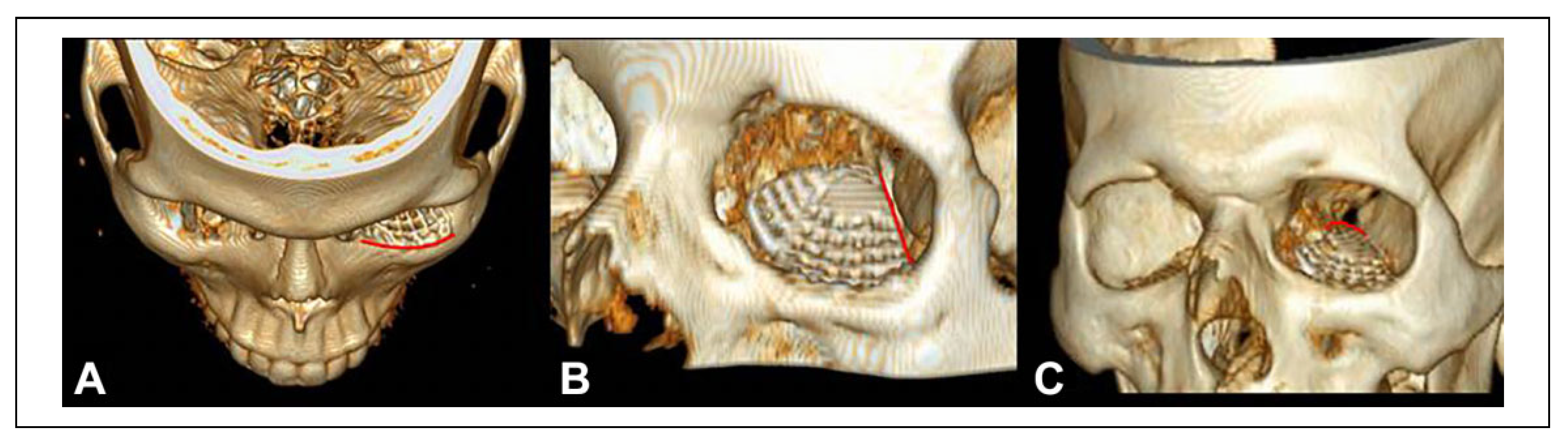
  • The inferior orbital rim, which must correspond with the position of the screw holes of the mesh (Figure 1A).
  • The inferior orbital fissure, which determines the position of the lateral edge of the mesh (Figure 1B).
  • The orbital process of the palatine bone, known as “posterior bony ledge,” as the support structure for the apex of the mesh (Figure 1C).
The secondary markers are:
1.
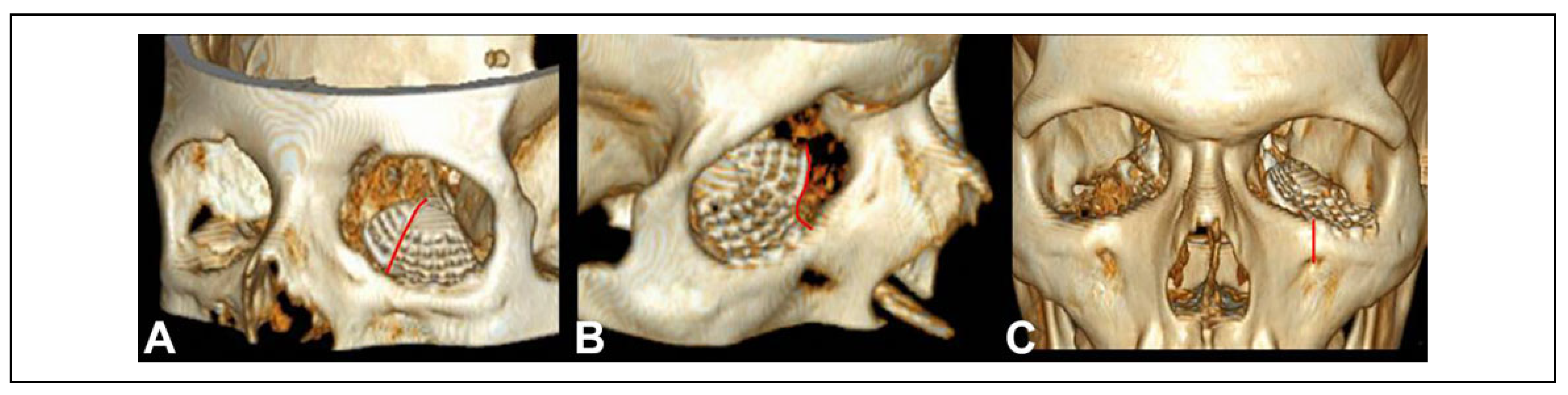
The transition zone represented by the inferome- dial bony strut, for the position of the inferomedial part of the mesh (Figure 2A).
2.
The lacrimal sac, the posterior lacrimal crest and the origin of the inferior oblique muscle, for the posi- tion of the antero-medial notch of the mesh (Figure 2B).
x
The emergence of the infraorbital nerve, which has to be vertically aligned with the first medial screw hole (Figure 2C).
In order to facilitate its placement, the redundant parts of the mesh were previously cut according to the size of the defect measured on the preoperative CT. Plate insertion was achieved with a slight lateral-to-medial rotation and an antero-posterior sliding movement, avoiding forced insertion. In this way the plate reaches a spontaneous posi- tion, perceived by the operator as the “best fit.”
In all cases, the implant position was verified checking the adherence with each of the “6 anatomical landmarks” moving from 1 to 6. Once the optimal position was guar- anteed, stabilization of the mesh was obtained with 1 or 2 monocortical screws at the orbital rim; in many cases the spontaneous stability was sufficient.
At the end of the surgical procedure clinical ocular pro- jection and forced duction test were checked.
Conjunctival approach was always sutured with 7/0 resorbable polyfilament suture.
The radiological accuracy of the reconstruction was assessed by evaluating the orbital symmetry and the match- ing with the 6 anatomical markers on immediate post- operative CT scan.
In order to compare the symmetry of the reconstructed orbit with the mirrored image of the contralateral healthy one, the analysis was conducted using I plan 3.0 CMF software (Brainlab, Munich, Germany). Reconstruction was considered accurate when discrepancy was �1 mm.[5]
In a similar way previously described by Ellis and Tan,[21] a 1 to 3 scale of radiological accuracy was used:
1.
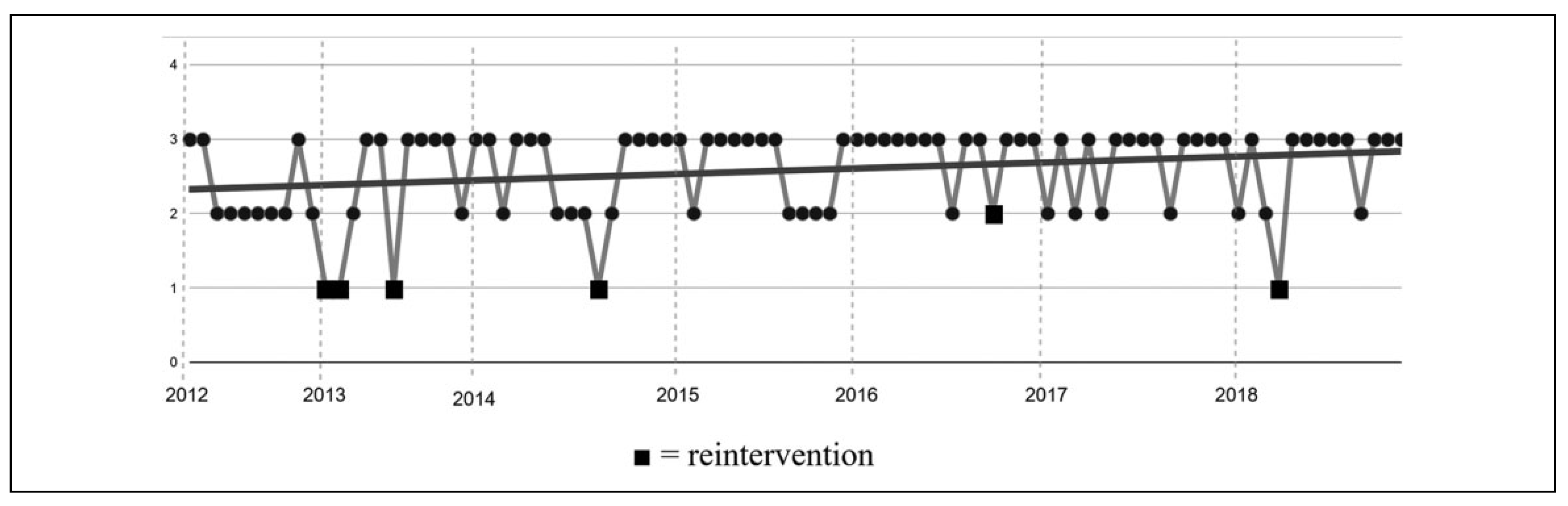
¼ Not accurate radiological result (grossly mesh malposition).
2.
¼Partial accurate radiological result (discrepancy >1 mm compared to controlateral mirrored orbit and/or adherence to less than 6/6 anatomical markers).
3.
¼ Accurate radiological result (good adherence to controlateral mirrored orbit and/or total adherence to the anatomical markers).
The mean accuracy (SD) rated for every year and its trend along the entire period examined were calculated.
The number of re-interventions and intra/post-operative complications was also registered, as well as the mean (SD) duration of surgery.
Subjective diplopia confirmed by Hess-Lancaster Test and the amount, if present, of clinical enophthalmos were assessed preoperative and at 3, 6 and 12 months after surgery.
This study was performed according to Helsinki declara- tion and local ethical rules. Anonymous and retrospective radiologic and clinical data analysis required neither pre- vious informed consent acquisition from patients nor the ethical committee approval.

Results

The study included 90 patients, 70 were males (78% of the sample) and 20 were females (22%). The average age was 41 (SD15) years (range 18-75 years). Right orbit was involved in 41 cases (46%), left orbit in 49 (54%). Fracture patterns are reported in Table 1.
The post-operative evaluation of radiological accuracy showed that level 1 of accuracy (Not accurate results) was obtained in 5/90 patients (5.6%), level 2 of accuracy (Partial accurate results) was reached in 28/90 patients (31.1%) and level 3 of accuracy (Accurate result) in 57/90 (63.3%).
All patients rated with level 1 of accuracy underwent a re-intervention, after which in 3 cases a level 2 of accuracy was reached and 2 cases obtained level 3 of accuracy.
Only 1 patient, rated with level 2 of accuracy, needed a re-intervention because of clinical symptoms, due to mesh’s interference with the medial rectus muscle; after revision surgery a level 3 of accuracy was assessed and functional defect totally recovered.
Mean accuracy for each year is presented in Table 2. Figure 3 shows the trend in accuracy along the entire period considered.
Total revision rate was 6/90 (6.7%). 4/36 (11.1%) revi- sions occurred between 2012 and 2014 and 2/54 (3.6%)
between 2015 and 2018.
3/6 surgical reoperations were needed for orbital recon- structions associated to complex orbito-zygomatic (CFI score >9) fractures, in which 1 or more anatomical land- marks were themselves disrupted (2 cases in 2013 and 1 case in 2016).
No major complications occurred during surgery. One patient developed a mild post-operative scleral show and 3 patients had post-traumatic sequelae (1 dacryocystitis and 2 ipsilateral hypovirus).
The mean duration of surgery was 120 minutes (SD56), blow out floor fractures required a mean surgical time of 74 (SD24) minutes. The trend of duration of surgery is shown in Figure 4.
68/90 (76%) patients had clinical evident enophthalmos (>2 mm) immediately after trauma while 65/90 patients (72%) had diplopia.
All of the patients who needed surgical revision had total recovery from symptoms presented after first surgical reconstruction. Three months after surgery mild enophthal- mos (<2 mm) persisted in 11% of patients and diplopia in 30%. Twelve months after surgery 10% of patients had still a mild enophthalmos (none of them had level 3 of accu- racy) while diplopia was recovered in all patients who reached level 2 or 3 of radiological accuracy.
Discussion
Orbital reconstruction is one of the most frequent surgical procedures in facial traumatology.[1]
The use of Computer-Assisted Surgery (CAS) with intraoperative navigation[2,6,8,9] or Patient Specific Implant (PSI)[6,7,9,10] are certainly excellent resources, especially in complex Orbito-zygomatic fractures.
Their availability is not as widespread as the clinical request to perform this type of procedure.[2,8]
Reconstruction with titanium mesh is an ideal recon- structive solution and the availability of standard pre- formed meshes has certainly allowed a significant improvement in surgical results.[2,5,9,13,14,15,16]
Unfortunately, this material requires high precision in its positioning, leading frequently to reported poor plant inser- tion (up to 23% of cases[8,22,23]) and high rate of revision surgery (from 3.6% to 17% of cases[8,14,16,22,23)).
The intraoperative use of “6 anatomical landmarks” is a very efficient technique for a proper preformed mesh posi- tioning, improving the accuracy of free-hand orbital recon- struction and reducing overall surgical time and the number of surgical revisions. This is confirmed in the 2015-2018 period of our single-operator and single-center sample in which, after an adequate learning curve, the percentage of surgical revisions further drops to 3.7%.
Only 5/90 (5.6%) cases in our sample were considered the lowest level (Level 1) of accuracy and therefore requested re-intervention. Three of them were complex Orbito-zygomatic (CFI score >9) fractures in which 1 or more anatomical landmarks were themselves disrupted.
None of the Level 3 of accuracy patients required revi- sion surgery; only 1 patient with Level 2 accuracy needed reoperation because of diplopia for horizontal ipsilateral ocular movement restriction.
The average duration of surgery is 120 (SD56) minutes. In our sample pure blow-out fractures are reported in 56.7% of cases. The remaining 43.3% are orbito-zygomatic frac- tures with different degrees of surgical difficulty expressed by proportional CFI scores.[24]
Preformed mesh positioning guided by anatomical land- marks even needs meticulous reconstruction of the associ- ated zygomatic fractures. This is a fundamental requirement to avoid imperfections that can be emphasized during orbital reconstruction, but obviously leads to longer procedures. The entire defect and all the anatomical land- marks should be exposed during preparation.[2,14,18]
According to the literature, our blow out floor fracture reconstructions required a mean surgical time of 70 min- utes.[9,25] Nevertheless, there is a constant improvement in surgical time during the years: this is the second advantage from using this technique.
The 6 landmarks proposed have a sort of hierarchy. The most important are: the inferior orbital rim, which prevents from antero-posterior mesh dislocation toward orbital apex and implant yaw; the medial margin of the inferior orbital fissure, which prevents from medial or lateral dislocation, yaw of the mesh and ethmoidal displacement of its apex; the posterior bony ledge, which prevents from implant pitch and falling of the apex into maxillary sinus. If we ensure the match with the leading landmark 2,[2,18] the others can generally be simply verified.
All the plates used in our study were preformed Large, 0.4 mm thickness, titanium mesh (Matrix Orbital®, DePuy Synthes, J&J, West Chester, PA). Small preformed meshes are also available, however they are not only smaller in size but different in shape. This makes them unusable for the leading landmark (inferior orbital fis-sure). We always prefer to use a Large mesh trimming its size as needed, cutting the exceeding parts at intersection bars, but keeping a suitable shape for at least the 3 prin- cipal landmarks.[9,18] Generally, we recommend to keep 1 to 2 millimeters of the medial wall portion of the mesh to be able to rely also on landmark 4 (inferomedial bony strut) and to obtain a 360-degree control of the correct positioning. This does not necessarily require a retrocar- uncular extension but can be done with a standard trans- conjunctival incision.
Post-operative point to point radiological verification of the adherence to the 6 landmarks allows to early identify correctable errors and to perform targeted procedures, in order to avoid ocular disfunction and ocular position defects caused by less effective late surgery.
Complications following orbital reconstruction are not so rarely described, with a range between 3% and 85.5%.[25] The whole “6 anatomical landmark” procedure turned out to be safe with only minor and transient complications.
A 12-months follow-up revealed a complete correction of diplopia in every patient who reached at least level 2 of radiological accuracy (98.9% of patients) and an acceptable correction of enophthalmos (90% of patients).
The main limits to our findings are the retrospective nature of the study and lack of a control group.

Conclusion

The “6 anatomical landmarks” technique for the recon- struction of orbital floor and medial wall fractures is an effective and easy procedure that allows to obtain accurate surgical results, even when computer-assisted surgery is not available. No adjunctive costs are requested.
The high level of confidence in local anatomy acquired by its systematic use supports the surgeons during the inter- pretation of preoperative imaging, intraoperative choices and post-operative evaluation of results.
This means that everyone could achieve faster proce- dures and less need for surgical revisions.
Even when revision surgery is necessary it can be focused, faster and earlier improving the final outcome.
Surgeons who have easy access to surgical navigation could use this free-hand technique and rely on technologi- cal support only as final intraoperative control.
The 1 reported is a single-center and single-operator experience, the involvement of other centers will confirm its validity.

Funding

The author(s) received no financial support for the research, authorship, and/or publication of this article.

Conflicts of Interest

The author(s) declared no potential conflicts of interest with respect to the research, authorship, and/or publication of this article.

References

  1. Ellis E, III, el-Attar A, Moos KF. An analysis of 2,067 cases of zygomatico-orbital fracture. J Oral Maxillofac Surg, 4: 43(6). [CrossRef]
  2. Bittermann G, Metzger MC, Schlager S, et al. Orbital recon- struction: prefabricated implants, data transfer, and revision surgery. Facial Plast Surg. [CrossRef] [PubMed]
  3. Gander T, Essig H, Metzler P, et al. Patient specific implants (PSI) in reconstruction of orbital floor and wall fractures. J Craniomaxillofac Surg. [CrossRef]
  4. Zavattero E, Ramieri G, Roccia F, Gerbino G.Comparison of the outcomes of complex orbital fracture repair with and without a surgical navigation system: a prospective cohort study with historical controls. Plast Reconstr Surg, 9: 139(4). [CrossRef]
  5. Sozzi D, Gibelli D, Canzi G, et al. Assessing the precision of posttraumatic orbital reconstruction through “mirror” orbital superimposition: a novel approach for testing the anatomical accuracy. J Craniomaxillofac Surg, 1258. [CrossRef]
  6. Dubois L, Steenen SA, Gooris PJ, Bos RR, Becking AG. Controversies in orbital reconstruction-III. Biomaterials for orbital reconstruction: a review with clinical recommenda- tions. Int J Oral Maxillofac Surg. [CrossRef]
  7. Schramm A, Suarez-Cunqueiro MM, Ru¨ cker M, et al. Computer-assisted therapy in orbital and mid-facial recon- structions. Int J Med Robot. [CrossRef]
  8. Schlittler F, Schmidli A, Wagner F, Michel C, Mottini M, Lieger O. What is the incidence of implant malpositioning and revision surgery after orbital repair? J Oral Maxillofac Surg. [CrossRef]
  9. Zimmerer RM, Ellis E, 3rd, Aniceto GS, et al. A prospective multicenter study to compare the precision of posttraumatic internal orbital reconstruction with standard preformed and individualized orbital implants. J Craniomaxillofac Surg, 1485. [CrossRef]
  10. Rana M, Chui CH, Wagner M, Zimmerer R, Rana M, Gellrich NC. Increasing the accuracy of orbital reconstruction with selective laser-melted patient-specific implants combined with intraoperative navigation. J Oral Maxillofac Surg, 1113. [CrossRef] [PubMed]
  11. Kim YC, Jeong WS, Park TK, Choi JW, Koh KS, Oh TS. The accuracy of patient specific implant prebented with 3D- printed rapid prototype model for orbital wall reconstruction. J Craniomaxillofac Surg. [CrossRef]
  12. Huempfner-Hierl H, Doerfler HM, Kruber D, Hierl T. Mor- phologic comparison of preformed orbital meshes. J Oral Maxillofac Surg, 1119. [CrossRef]
  13. Bourry M, Hardouin JB, Fauvel F, Corre P, Lebranchu P, Bertin H.Clinical evaluation of the efficacy of materials used for primary reconstruction of orbital floor defects: meta- analysis [published online ahead of print, 2020 Nov 4]. Head Neck. [CrossRef]
  14. Scolozzi P, Momjian A, Heuberger J, et al. Accuracy and predictability in use of AO three-dimensionally preformed titanium mesh plates for posttraumatic orbital reconstruction: a pilot study. J Craniofac Surg, 1108. [CrossRef] [PubMed]
  15. Potter JK, Malmquist M, Ellis E, 3rd. Biomaterials for recon- struction of the internal orbit. Oral Maxillofac Surg Clin North Am. [CrossRef]
  16. Peng MY, Merbs SL, Grant MP, Mahoney NR. Orbital frac- ture repair outcomes with preformed titanium mesh implants and comparison to porous polyethylene coated titanium sheets. J Craniomaxillofac Surg. [CrossRef] [PubMed]
  17. Strong EB, Fuller SC, Wiley DF, Zumbansen J, Wilson MD, Metzger MC. Preformed vs intraoperative bending of titanium mesh for orbital reconstruction. Otolaryngol Head Neck Surg. [CrossRef]
  18. Purnell CA, Vaca EE, Ellis MF. Orbital fracture reconstruc- tion using prebent, anatomic titanium plates: technical tips to avoid complications. J Craniofac Surg. [CrossRef]
  19. Canzi G, De Ponti E, Novelli G, et al. The CFI score: valida- tion of a new comprehensive severity scoring system for facial injuries. J Craniomaxillofac Surg. [CrossRef]
  20. Novelli G, Ferrari L, Sozzi D, Mazzoleni F, Bozzetti A. Transconjunctival approach in orbital traumatology: a review of 56 cases. J Craniomaxillofac Surg. [CrossRef]
  21. Ellis E 3rd, Tan Y. Assessment of internal orbital reconstruc- tions for pure blowout fractures: cranial bone grafts versus titanium mesh. J Oral Maxillofac Surg. [CrossRef] [PubMed]
  22. Schlittler F, Vig N, Burkhard JP, Lieger O, Michel C, Holmes, S. What are the limitations of the non-patient-specific implant in titanium reconstruction of the orbit? Br J Oral Maxillofac Surg. [CrossRef]
  23. Shaye DA, Tollefson TT, Strong EB. Use of intraoperative computed tomography for maxillofacial reconstructive sur- gery. JAMA Facial Plast Surg. [CrossRef]
  24. Canzi G, De Ponti E, Fossati C, et al. Understanding the relevance of comprehensive facial injury (CFI) score: statis- tical analysis of overall surgical time and length of stay out- comes. J Craniomaxillofac Surg, 1456. [CrossRef]
  25. Reich W, Seidel D, Bredehorn-Mayr T, Eckert AW. Recon- struction of isolated orbital floor fractures with a prefabri- cated titanium mesh. Klin Monbl Augenheilkd. [CrossRef]
Figure 1. The 3 principal landmarks for free-hand standard preformed mesh positioning: the inferior orbital rim (A), the inferior orbital fissure (B) and the posterior bony ledge (C).
Cmtr 15 00009 g001
Figure 2. The secondary landmarks for standard preformed mesh positioning: the inferomedial bony strut (A); the lacrimal sac– posterior lacrimal crest–origin of the inferior oblique muscle (B); the emergence of the infraorbital nerve (C).
Cmtr 15 00009 g002
Figure 3. Specific level of accuracy reached for each surgical reconstruction (first intervention only, no surgical revision level of accuracy was considered). Number and year of surgical revision (&) and linear trend of accuracy during the entire period are highlighted.
Cmtr 15 00009 g003
Figure 4. Specific duration of surgery and linear trend during the entire period.
Cmtr 15 00009 g004
Table 1. Sample Size for Each Type of Fracture Reconstructed Using Standard Preformed Titanium Meshes: Isolate Blow Out Orbital Floor Fractures (A) or Associated With Blow Out Medial Wall Fractures (B). Simple Zygomatic Fractures With Orbital Floor Displacement Which Need Reconstruction (C) or With Orbital Floor and Medial Wall Fractures (D). Complex and Multifragmentary Zygomatic Fractures With Orbital Floor Displacement Which Need Reconstruction (E) or With Orbital Floor and Medial Wall Fractures (F).
Type of fractureNumber of fractures%
A. Orbital floor4550%
B. Orbital floor + medial wall910%
C. Orbito-zygomatic (CFI = 9)1719%
D. Orbito-zygomatic (CFI = 9) + medial wall56%
E. Orbito-zygomatic (CFI >9)1213%
F. Orbito-zygomatic (CFI >9) + medial wall22%
Table 2. Mean Accuracy (SD) Per Year and Number of Surgical Revisions Needed Per Year.
2012201320142015201620172018
Mean accuracy (SD)2.30 (0.48)2.27 (0.90)2.53 (0.64)2.62 (0.51)2.80 (0.41)2.71 (0.47)2.67 (0.65)
Surgical revisions0310101
Disclaimer/Publisher’s Note: The statements, opinions and data contained in all publications are solely those of the individual author(s) and contributor(s) and not of MDPI and/or the editor(s). MDPI and/or the editor(s) disclaim responsibility for any injury to people or property resulting from any ideas, methods, instructions or products referred to in the content.

