Next Article in Journal
A Literature Review of Rapid Prototyping and Patient Specific Implants for the Treatment of Orbital Fractures
Previous Article in Journal
Complications of the Transmasseteric Anteroparotid Approach for Subcondylar Fractures: A Retrospective Study
 
 
Craniomaxillofacial Trauma & Reconstruction is published by MDPI from Volume 18 Issue 1 (2025). Previous articles were published by another publisher in Open Access under a CC-BY (or CC-BY-NC-ND) licence, and they are hosted by MDPI on mdpi.com as a courtesy and upon agreement with Sage.
Review

Pneumomediastinum as a Complication of Oral and Maxillofacial Injuries: Report of 3 Cases and a 50-Year Systematic Review of Case Reports

by
Ioannis (Yiannis) Papadiochos
1,2,*,
Stavros-Evangelos Sarivalasis
2,3,
Meg Chen
4,
Lampros Goutzanis
2,5 and
Aristotelis Kalyvas
2,6
1
«Attikon» University General Hospital of Athens, Haidari, Greece
2
Evaggelismos General Hospital of Athens, Athens, Greece
3
Department of Plastic Surgery, Hygeia Hospital, Athens, Greece
4
Department of Oral & Maxillofacial Surgery, National Cheng Kung University Hospital, Tainan, Taiwan
5
Dental School of Athens, Evaggelismos General Hospital of Athens, Athens, Greece
6
Neuro-Oncology and Skull Base Surgery, University of Toronto, Toronto, ON, Canada
*
Author to whom correspondence should be addressed.
Craniomaxillofac. Trauma Reconstr. 2022, 15(1), 72-82; https://doi.org/10.1177/1943387521997236
Submission received: 1 November 2020 / Revised: 1 December 2020 / Accepted: 1 January 2021 / Published: 4 March 2021

Abstract

:
Objectives: Pneumomediastinum (PM) secondary to oromaxillofacial trauma (OMF) is a rare but well-described com- plication/pathologic finding. The aim of this study was twofold: first, to report our experience in treatment of maxillofacial trauma patients with PM, and second, to review the literature regarding the clinical features, severity, course, and management of the aforementioned complication. Material and methods: We retrospectively reviewed the medical records and charts of patients who suffered from maxillofacial trauma and treated in our hospital between September 1, 2013 and September 31, 2017. The inclusion criteria were patients with radiologically confirmed PM. In addition, the electronic databases PubMed, Scopus, and Science Direct were queried for articles reporting PM cases secondary to OMF injuries and published in English, French, and German language. Results: Three cases of PM out of 3,514 cases of craniomaxillofacial trauma were found; there were 3 male patients who presented in our emergency department with the chief complaint of cervicofacial swelling. Literature search isolated 58 selected articles and 63 cases were assessed in total; posttraumatic repeated blowing of nose was proved as most frequent triggering factor among them. Furthermore, the outcomes of review showed that thoracic pain, respiratory distress, and swallowing difficulties were not frequently reported in patients with ME due to facial trauma. Conclusions: Both our experience and the results of systematic literature review indicated that patients with PM due to OMF injuries present mild clinical course. If properly managed, this specific pathologic condition may have no further complications or relative comorbidities. The exact etiology and mechanism of PM in the context of maxillofacial injuries always needs to be identified. Radiographic, laboratory, and endoscopic examinations should be applied to rule out the more serious and frequently diagnosed aerodigestive, thoracic, and abdominal causes of PM.

Introduction

Pneumomediastinum (PM) or mediastinal emphysema (ME) is a pathologic clinical finding that denotes abnormal collection of air or gas within the mediastinal cavity. Its occurrence may be spontaneous in otherwise healthy indi- viduals or may involve various causes of either intra- or extrathoracic origin. PM can be referred as ether sponta- neous (primary) or secondary (related injury or underlying disease), depending the variety of its causes. In 1827, Laenek made the first clinical description of PM,[1] while Hamman published a case of post-partum PM combined with subcutaneous emphysema that is currently termed as Hamman’s syndrome.[2,3]
PM in the context of oromaxillofacial (OMF) trauma is a rare but well-known complication; several case reports have been published due to the involvement of facial fractures and/or orofacial lacerations in PM formation.[4,5] However, the literature currently lacks adequate informa- tion on the prevalence of PM as result of OMF injuries, as well as on frequency and severity of its presenting signs and symptoms, accompanying comorbidities, complica- tions, clinical course, and prognosis.
The purpose of this article was twofold; first, to retro- spectively report our clinical experience in management of patients with various OMF injuries that resulted in forma- tion of PM, and second, to systematically review the liter- ature regarding the clinic-epidemiological aspects of the aforementioned complication.

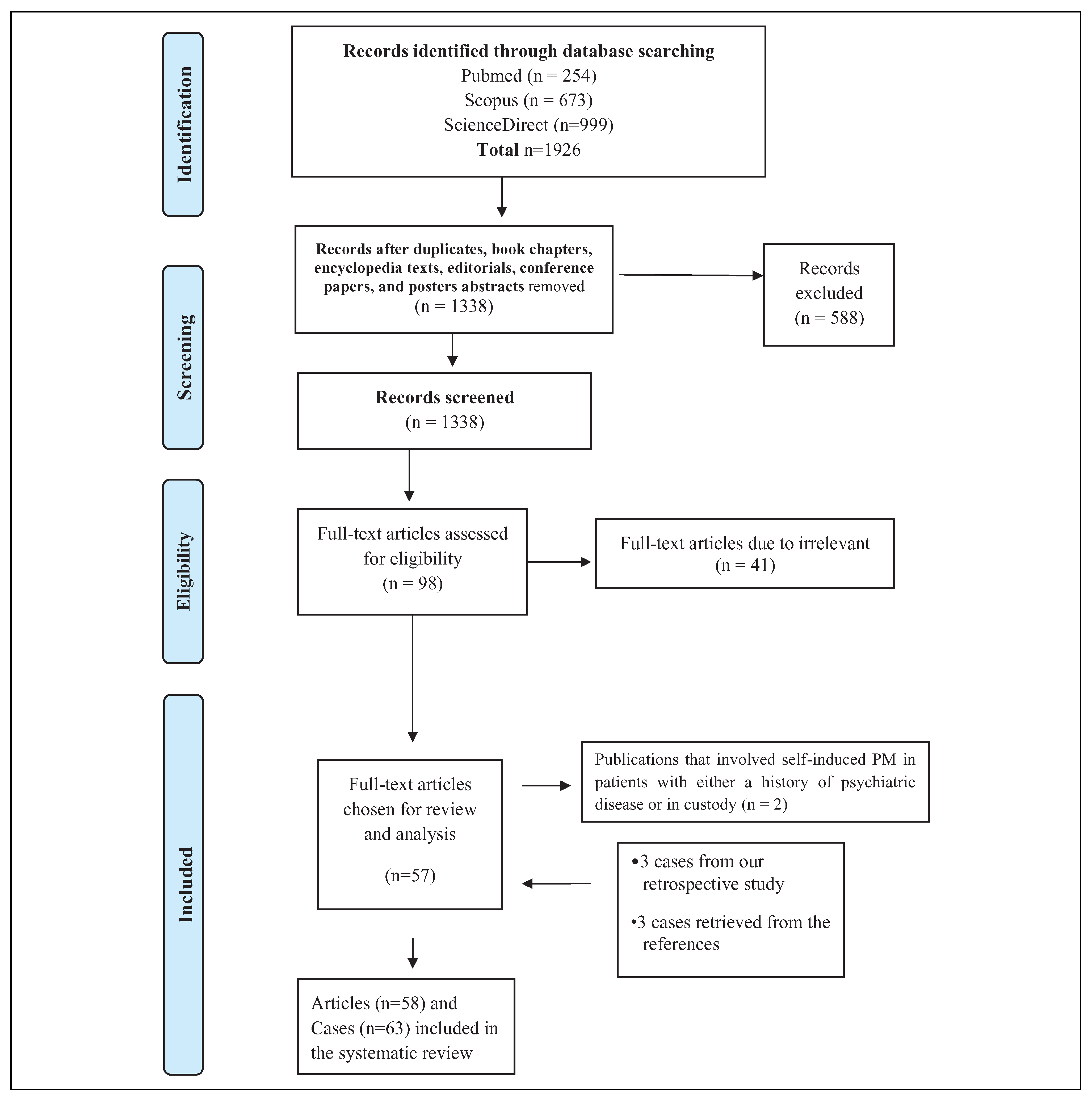
Material and Methods
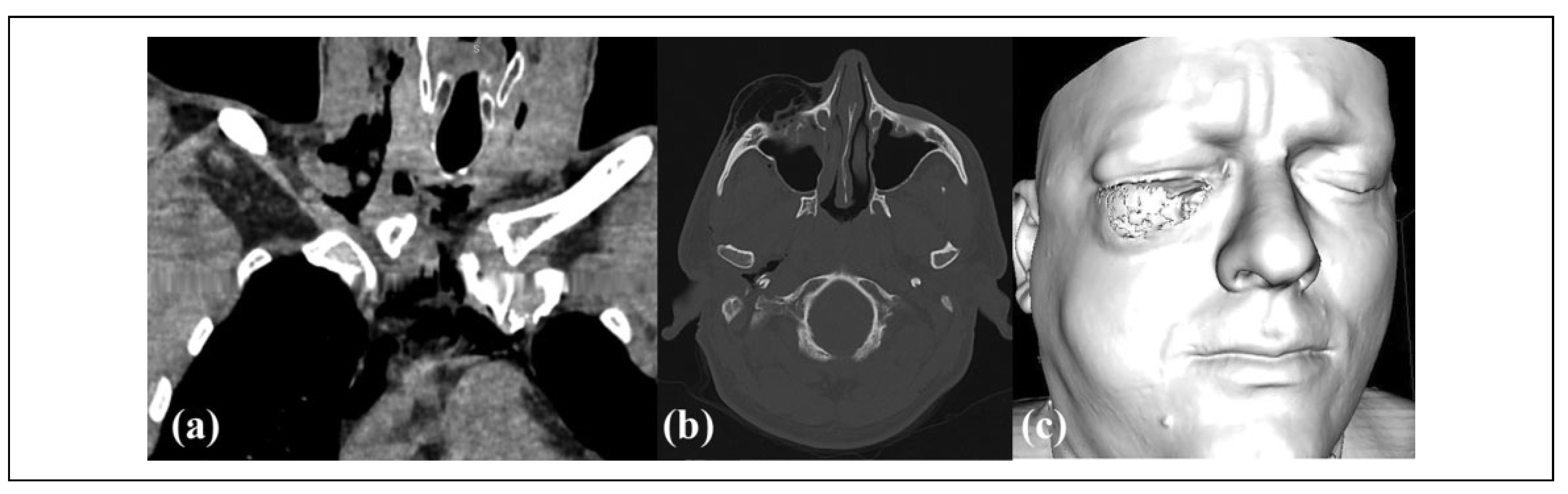
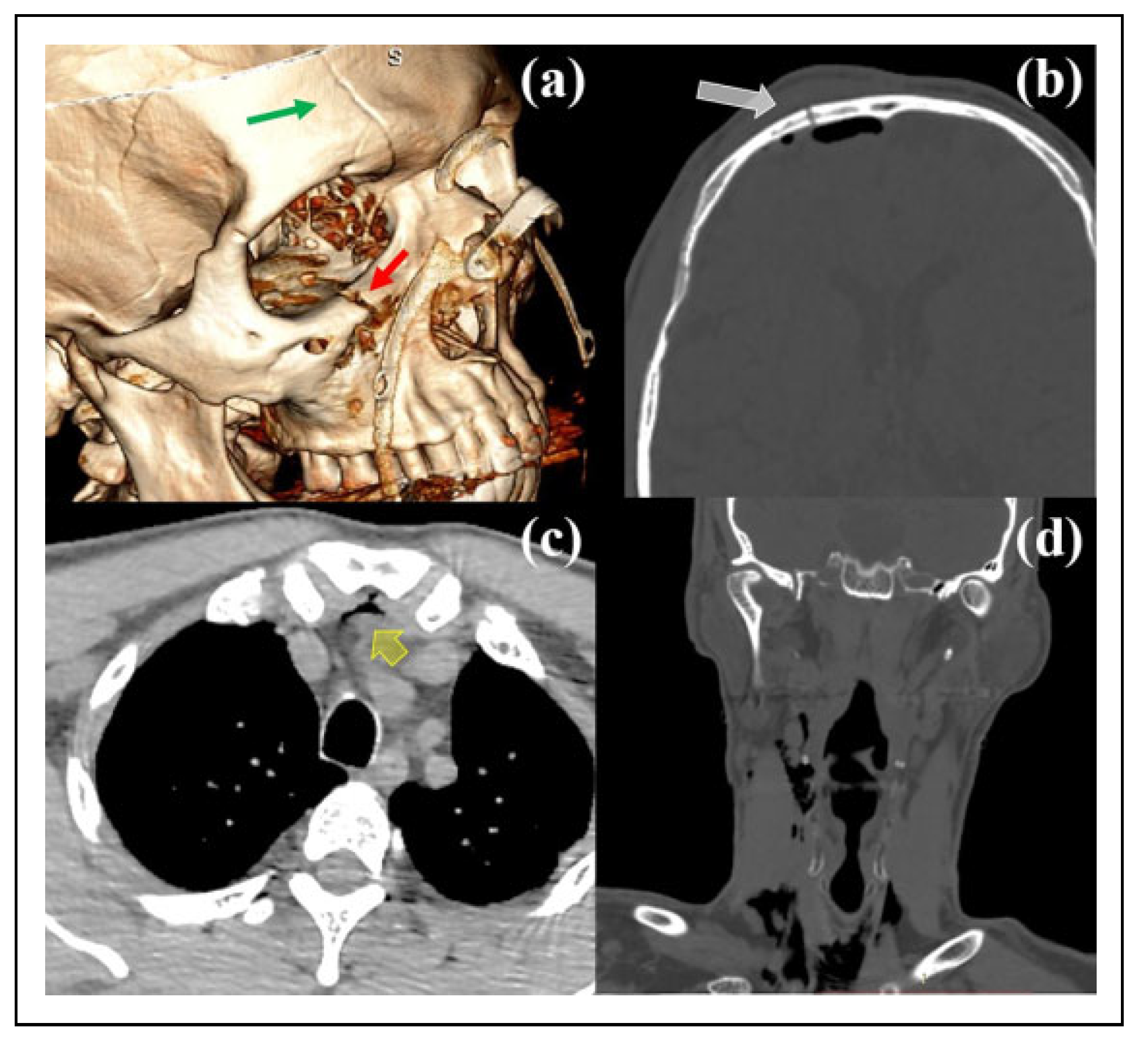
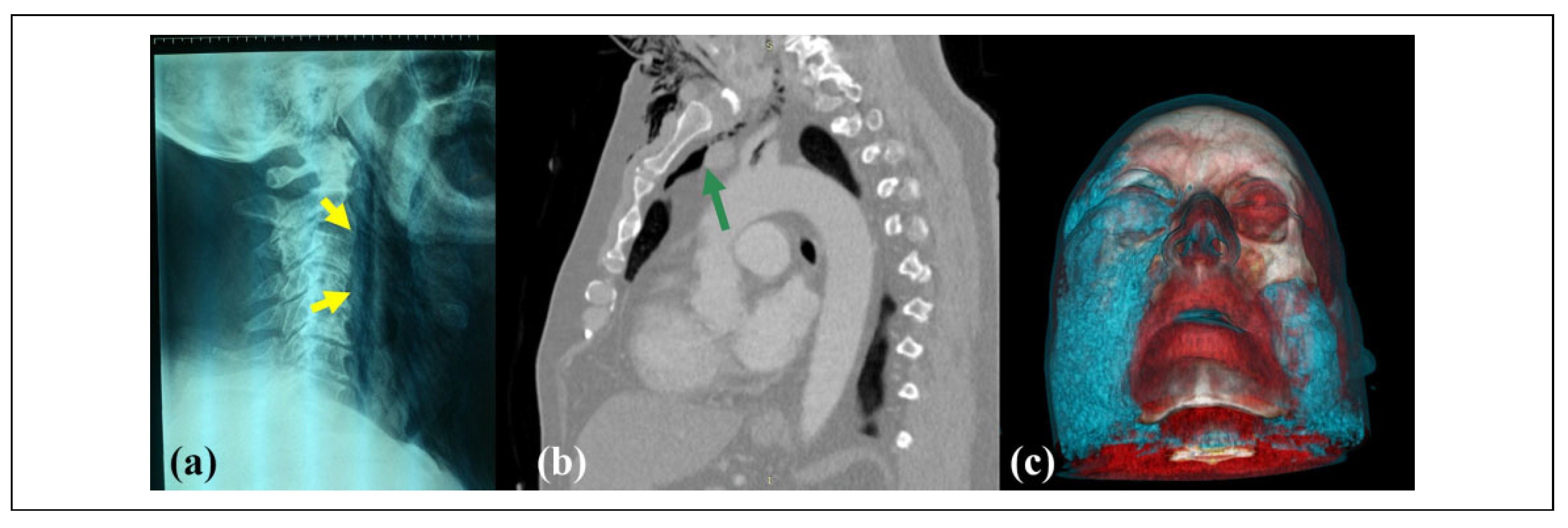
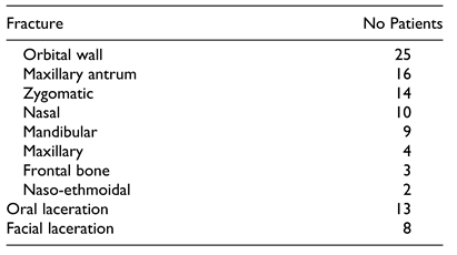
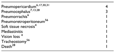
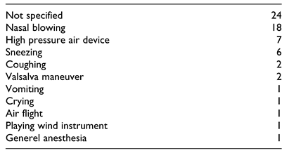
An electronic search of PubMed, Scopus, and ScienceDirect was carried out for identifying case reports and short case series of PM or ME secondary to craniomaxillo- facial (CMF) trauma that had been published in English, German, and French from January 1, 1970 to January 1, 2020. The search included combination of the follo- wing terms “pneumomediastinum,” “mediastinal emphysema,” “cervicofacial emphysema,” “retropharyngeal emphysema,” “massive emphysema,” “facial fracture,” “orbital fracture,” “orbital trauma,” “mandibular fracture,” “zygomatic fracture,” “maxillofacial fracture,” “maxillofacial trauma,” “facial trauma,” and “facial injury.”
The study was made in accordance with Preferred Reporting Items for Systematic Reviews and Meta- Analyses (PRISMA) statement (Figure 1). First, there was removal of the duplicates, book chapters, encyclopedia texts, editorials, conference papers, and posters abstracts. Afterwards, all the titles and abstracts of the remaining 1338 results were reviewed. Publications were only included if they reported patients with a history of an intraoral and/or CMF injury that provoked emphysema in at least one of the following anatomic sites: orbit, face, neck, thorax, and mediastinum. We excluded cases of PM that either was induced spontaneously or air leakage arose from an anatomical site other than CMF complex. More- over, non-traumatic cases of PM/ME such as those resulted from tracheostomy, tonsillectomy, dental/intraoral proce- dures, orthognathic surgery, and other iatrogenic interven- tions were also not included.
Of 98 full-text articles assessed for eligibility, 57 were chosen for review and analysis. To increase the validity of clinical information derived from patients (chief complaint, symptoms, history of injury etc.), we did not also encom- pass 2 publications that involved self-induced PM in patients with either a history of psychiatric disease or in custody. References of the selected articles were checked to consider other eligible publications and ultimately 3 arti- cles were integrated for analysis too. Finally, we isolated 58 articles.
Eligible articles were evaluated independently by 2 investigators (IP and SES). Disagreements were resolved by consensus and, as necessary, involvement of a 3rd reviewer (LG). Data extracted included author and year of publication, demographics (gender, age), injury mechan- ism and pattern, presenting signs and symptoms e.g. crepi- tus, respiratory distress, chest pain, Hamman’s sign (a crunch-like sound that is best audible over the left side of the patient’s chest or retrosternum during the systole and expiration),[2] dysphagia, voice alteration, concurrent comorbidities and complications (e.g. pneumocephalus, mediastinitis, death), and therapeutic outcomes.
In addition, a retrospective search of the comprehensive clinical database of Evaggelismos General Hospital of Athens was applied to isolate patients who treated for var- ious types of oromaxillofacial injuries in our clinic; the hard-copy records of our emergency department were scru- tinized too. Special focus was laid on those patients who were diagnosed with cervicofacial emphysema and under- went radiologic chest examinations. This study was approved by the institutional review board of Evaggelismos General Hospital of Athens (protocol number 380/20-11- 2018)

Results

Concerning to our clinic’s experience, we managed between September 1, 2013 and September 31, 2017 over- all 3,514 patients who presented with different types of OMF injuries in ED. Among them, we found 13 cases (0.37%) that had been clinically diagnosed) with cervicofacial emphysema (puffy facial/ periocular swelling, crepitation on palpation). Three males of this subgroup (0.09%) were admitted or referred to our clinic due OMS injuries, and a PM was documented after clinical and radiographic exams (Figure 2, Figure 3 and Figure 4). All the patients were conservatively treated with administration of antibio- tics, oxygen administration (with a non-rebreather mask at a rate of 10-15 lt/min), corticosteroids, and analgesics. Vision abnormalities did not found, while endoscopic evaluation of esophagus, larynx, and trachea did not reveal other sources of air leakage.
In addition, the selected publications contained 61 patients (Table 1).[4,5,6,7,8,9,10,11,12,13,14,15,16,17,18,19,20,21,22,23,24,25,26,27,28,29,30,31,32,33,34,35,36,37,38,39,40,41,42,43,44,45,46,47,48,49,50,51,52,53,54,55,56,57,58,59,60,61] One of the selected articles com- prised one which was among the 3 aforementioned identi- fied patients of our retrospective search. The overwhelming majority of them were males (87.1%, n ¼ 56) with mean age 34.9 years (median 34.5; 2 months-82 years).
The most commonly reported sign was crepitation or SE (81%), while Hamman’s sign was recorded in 14.3% of cases (Table 2). Respiratory distress was reported in only 12.7%% of cases, whereas dysphagia and voice alteration (rhinolalia, hoarseness) was cited in 6 and 4 cases respec- tively. Among OMF injuries, bone fractures were princi- pally diagnosed in 74.6% (n ¼ 47) of cases. Within this category, maxillary sinus fractures and orbital floor and inner wall fractures were the most frequently seen pattern (Table 3). A single case of mediastinitis was recorded by Shuker et al [49] who cited a case of deep cervical infection being extended to the upper anterior mediastinum (Table 4.). Besides, there was 1 death reported by Gouda et al[28] and it was attributed to both blood aspiration and PM caused by isolated facial fractures. Furthermore, nasal blowing was recognized as the most common mechanism responsible for cervicofacial emphysema and PM (Table 5).

Discussion

PM formation secondary to OMF injuries implies the pres- ence of cervicofacial emphysema. The responsible mechanism involves either forceful entrance of air/gas through skin or mucosal lacerations in orofacial region or exit of air via mucoperiostal tears on the fractured bones making up the walls of paranasal sinuses. From an epide- miological perspective, among 390 patients who were diag- nosed with 458 paranasal sinus fractures, traumatic surgical emphysema was identified in 29 patients (7.43%) patients[62]; however, there was no information in this pro- spective study about the prevalence of PM in this group. SE is most commonly localized in orbital cavity, whereas an orbital emphysema more frequently occurs whenever med- ial orbital wall is fractured (isolated or combined).[63]
Various factors such as vigorous nose blowing, sneeze, or cough as well as fierce blast from a compressed air- device, Valsalva maneuver, drugs inhalation, crying, vomiting, asthma crisis, and barotrauma have been found responsible to cause this mechanism.[38,64] Depending on the intensity and/or repeatability of the triggering factor, the entrapped air accumulations within cervicofacial soft tis- sues may propagate downwards to mediastinal cavity by dissecting neck fascial spaces the route of the least resis- tance. The anatomical communications of parapharyngeal space with the carotid sheath and retropharyngeal space enable this emphysema propagation .Except for OMF inju- ries, another type of head injury, i.e. comminuted mastoid process fracture, may inflict PM with the triggering mechanism of Valsalva maneuver.[65] PM is considered an acute medical condition given that patients are almost always used to visit hospital less than 24 hours from the onset of symptoms.[66] SE of the face has been observed by Von Arx et al[67] even 7 years after an orbital fracture.
Taking into account the results of this review, crepita- tion is the predominant sign among patients with PM secondary to OMS injuries. Nevertheless, clinical manifes- tations seem to differ from those of spontaneous PM. In the event of latter, the symptom of retrosternal thoracic pain is predominately seen in various studies (60-100%),[68] and is characterized by radiation to the dorsum, neck, and shoulder.[22] It may be aggravated by swallowing, coughing, deep inspiration, and clinostatism.[30] Similarly to pain, other more commonly observed symptoms may include dyspnea (up to 75%), coughing spells (up to 80%), and neck pain (up to 44%).[68] The positivity of Hamman’s sign varies among different studies of PM other than OMF inju- ries (0%-100%)[66]; this percentage was over 50% in older studies,[44] while in more recent the sign was found positive in approximately 1 out 5 patients.[69]
Voice alteration, dysphonia, aphonia, dysphagia, odyno-phagia, and rhinolalia may also be associated with PM. In some retrospective studies the prevalence of these clinical findings exceeds 20% of cases with spontaneous PM,[66,70,71] while hoarseness was present in 8% of 410 PM cases due to blunt neck or chest trauma in a systematic review.[72] Hoover et al have postulated that closed rhinolalia due to PM may be not duly appreciated by thoracic surgeons.[73] Closed rhinola- lia in the context of cervicofacial emphysema is likely to signify pneumonasopharynx and air within the retropharyn- geal space. The latter represents a potential risk of respiratory obstruction. Closed rhinolalia or “rhinolalia clausa” is char- acterized by hyponasal voice quality, which entails reduction or absence of normal resonance of nasal the nasal “m,” “n,” “ng” to be heard as their plosives “b,” “d,” and “g,” respec- tively.[74,75] Examiners should never neglect the clinical sce- nario where a subcutaneous neck emphysema combined with dysphonia, neck asymmetry, and pain upon deglutition may be result from a laryngeal fracture.[76] Five to 6 cm3 have been enough to lead to mechanical obstruction of the larynx and emergency tracheotomy or decompression.[77]
Although a CT scan is the imaging modality of choice for PM diagnosis, simple radiographs may also provide reliable evidence for entrapped air collections. A poster- oanterior chest radiography is able to suggest the diagnosis of PM providing whether an air line is seen parallel to the left hilus.[27] Other diagnostic radiological signs for PM are the “ring-around-the-artery sign” (well- defined lucency along or surrounding the right pulmonary artery on lateral chest radiographs), “Naclerio’s V sign” (V-shaped air lucency in the left lower mediastinal area), and “continuous diaphragm sign” (lucency is above the diaphragm).[64] Noteworthy, Kaneki et al[77] demonstrated that a normal radiograph is seen in up to 30%.
Management of PM is very similar to subcutaneous emphysema and is mainly conservative; surgical decom- pression may be reserved only for those rare cases where there is compression of mediastinal structures (respiratory or vascular compromise) due to large quantities of entrapped gas.[46] Bronchodilators, steroids, cough suppres- sants, decongestants, stool softeners, oxygen, and antibio- tics are used to be administrated.[78] Patients with PM should refrain from any strenuous physical activity and bed rest is warranted too. Strong recommendations are given to them to avoid coughing, nose blowing, arrest of sneezing, or performing any other maneuver that may potentially pro- duce increased air pressure within the paranasal sinuses. Hospitalization is advised for cases presented with respira- tory distress, visual disturbances, generalized facial edema and emphysema, fever, infection development, need for fracture reconstruction, overanxiety, malaise, and discom- fort. Anterior nasal packing is also suggested as a
Administration of 100% oxygen is recommended because it can accelerate gas absorption by six fold.[68] Oxy- gen induces decrease of partial pressure of blood nitrogen and, consequently, rapid resorption of subcutaneous (nitro- gen-containing) air is facilitated by creating a nitrogen incline between the enclosed air and arterial blood.[80] On the other hand, nitrous oxide during general anesthesia and devices of high pressure oxygen strongly need to be avoided.[46,81,82] Moreover, clinicians should pay attention to not perform vigorous palpation of head and neck SCEs, since such a maneuver could move the air into a deeper location such as the mediastinum.[81]
Provided that emphysematous air collections are derived from upper aerodigestive tract, pathogenic bacteria may be transferred into mediastinal cavity. The vast majority of the authors endorsed the administration of prophylactic anti- microbials to eliminate the risk of locoregional infection. Only Alix et al[25] did not prescribe antibiotics in their patient with maxillary sinus fracture and PM and ques- tioned their prophylactic use too.
Many publications underlined that PM may potentially provoke life-threatening complications such as pneu- mothorax, pneumopericardium, and mediastinitis.[21] Though rare, when a large amount of air is entrapped within the cervical fascial planes and mediastinal cavity, airway obstruction may occur due to trachea compression. Further- more, a serious subtype of PM is defined as malignant as long as it impairs venous return to the heart resulting in cardiac tamponade and cardiopulmonary collapse.[78] Severe dyspnea, dysphagia, dysphonia, retrosternal chest pain irra- diating to the arms, neck, and back, cyanosis, distended nonpulsatile jugular veins, tachycardia, hypotension, and collapse are emergent manifestations that require emergent intervention.[22] If cardiac tamponade occurs secondary to pneumopericardium, pericardial needle aspiration must be accurately applied.[83] Relevant clinical signs and symptoms hypotension may comprise distended neck veins, Hamman crunch, and muffled heart sounds. To prevent irreversible loss of vision, Linberg first proposed decompression of orbital emphysema with a syringe.[84] Accurate penetration of an air pocket can be confirmed where a blunt-tipped Atkinson needle (or sharp 22-25 gauge) joined with a syr- inge filled with normal saline (without its plunger) is inserted and bubbles are appeared within the fluid.[85]
The entrapped air is progressively absorbed and PM typically is anticipated to resolve within over a 2-to 14 day period[11]; yet, Carmicael et al[39] cited that both swelling and crepitus in a patient lasted for several weeks.
A very special form of PM occurs as a result of self- induced facial emphysema and is related to medico-legal issues. In 1950, Reading used the term “autemphysesis” to describe self-induced facial emphysema in Second World War captives who attempted to alter their facial features and their identity.[86] Indeed, they made an incision with a safety-razor blade either in the mucosal side of the cheek or in interdental gingiva of maxillary molars and, afterwards they applied the Valsalva maneuver to provoke it. Mart’ınez-Carpio et al[87] cited the forensic term “camouflaging facial emphysema” to report six cases of intimates who had carried out punctures on their upper lips by using either a sewing needle or fishbone. To achieve facial camouflaging deformation, they repeated intensively the Valsalva maneuver many times over approximately 10–15 min. Similar cases (factitious subcutaneous emphy- sema) have been documented among psychiatric patients and some of them were diagnosed with PM during radiolo- gic evaluation; consequently, clinicians should methodi- cally investigate the presence of mucosal puncture marks whether there is a suspicious case of self-infliction.[88]
Current literature does not provide complete and extended documentation about the exact period of absti- nence from air flights in case of facial fractures. According to Boyle’s law,[89] the volume and pressure of a gas are inversely proportional considering that temperature remains stable. Therefore, a potential risk arises for patients with facial emphysema or fractured walls of paranasal sinuses due to air expansion as long as the airplane ascends and cabin’s pressure decreases. A study from UK had eval- uated the recommendations of 184 oral and maxillofacial surgeons to patients with zygomatic complex fractures regarding the required post-injury period of avoidance air travels.[90] The outcomes of study did not provide evidence- based recommendations, with a significant percentage (40%) of participants did not indicate a specific time- frame.[91] Some authors have suggested 4 weeks of avoiding air for small orbital fractures with minimal displacement.[90] Due to the fact that bone callus is typically formed approx- imately 2 to 3 weeks after fracture, it can be hypothesized that structural integrity is achieved.[89]
Taking into account that the atmospheric pressure at sea level is approximately 760 mm Hg, or 1 atm absolute (ATA), Seiff mentioned that ocular problems because of orbital emphysema probably take place if there is rapid unpressur- ized ascent to 18,000 feet where the ambient pressure decreases to 0.5 ATA.[91] Yet, commercial aircraft cabins are pressurized to between 5000 and 10,000 feet. Tan-Gore et al[92] drawn the conclusion that air transports should be not consid- ered as contraindication for patients with midfacial fractures. Their retrospective study did not report orbital complications in that category of patients (n ¼ 48) who transferred to hos- pitals via aircrafts. Remarkably, Ye et al[93] published a case of a fatal exacerbation of dermatomyositis-related PM after a flight and this complication was not attributed to rapid disease progression. Even though commercial airplane cabins are pressurized at the flight altitude of 8000 ft (2438 m) or less, the authors pointed out that entrapped air/gas maintains its ability to enlarge within lung parenchyma or mediastinal cav- ity by approximately 38%. Furthermore, they advocated that PM must be an absolute contraindication for air travel simi- larly to pneumothorax

Conclusions

PM is considered as a multi-origin disorder and may con- cern several specialties. Patients, who present OMF injuries and in addition have experienced a recent episode of intense or repeated nasal blowing or vigorous sneeze, should be examined for the development of widespread emphysema extending to the mediastinal cavity. Crepitation on palpa- tion of cervicofacial region, closed rhinolalia, or symptoms such as sudden onset facial swelling, sensation of fullness of the face may represent the first clinical sign of a widespread SE. It is imperative for examiners not only to detect PM and but to accurately determine its etiology. Taking into con- sideration the history and mechanism of injury, patients with PM should undergo thorough diagnostic evaluation and be guided with appropriate instructions to eliminate further complications. Meticulous laboratory and endo- scopic examinations contribute to accurate differential diagnosis especially in patients with multiple injuries and/ or suffered from high-energy trauma. Notwithstanding that a traumatic source of air leakage in CMF region may be easily assumed during clinical examination, the likelihood of additional latent injuries in larynx, tracheobronchial tree, oesophagus, abdomen, or chest should be not disregarded. The outcomes of our experience have limitations due to the retrospective nature of our study, while many different clinicians/authors were involved in examination methods and collection of medical data for the other included cases. Within the limits of this systematic review of case reports, PM arisen from OMF injuries has very good prognosis if properly treated. It can be hypothesized that PM secondary OMF injuries have milder clinical manifestations and lower rate of complications than spontaneous PM. When the origin of the air entrance/leakage is located in OMF region, the air collections may perhaps have to “travel a long dis- tance” to produce similar clinical findings to those of PM of different etiologies.

Funding

The author(s) received no financial support for the research, authorship, and/or publication of this article.

Conflicts of Interest
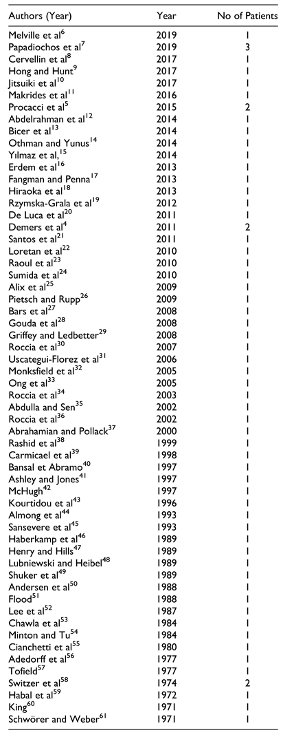
The author(s) declared no potential conflicts of interest with respect to the research, authorship, and/or publication of this article.

References

  1. Lae¨nnec RTH. De l’auscultation mediate ou Traite du Diag- nostic des Maladies des Poumon et du Coeur, 1819.
  2. Baumann MH, Sahn SA. Hamman’s sign revisited. Pneu- mothorax or pneumomediastinum? Chest, 1281.
  3. Bickley, J. Hamman syndrome. Br J Hosp Med (Lond), 1: 77(03).
  4. Demers G, Camp JL, Bennett D. Pneumomediastinum caused by isolated oral-facial trauma. Am J Emerg Med, 8: 29(07).
  5. Procacci P, Zanette G, Nocini PF. Blunt maxillary fracture and cheek bite: two rare causes of traumatic pneumomedias- tinum. Oral Maxillofac Surg.
  6. Melville JC, Balandran SS, Blackburn CP, Hanna IA. Massive self-induced subcutaneous cervicofacial, pneumo- mediastinum, and pneumopericardium emphysema sequelae to a nondisplaced maxillary wall fracture: a case report and literature review. J Oral Maxillofac Surg, 1867. [CrossRef]
  7. Papadiochos I, Sarivalasis SE, Papadogeorgakis N. Acute hyponasality (closed rhinolalia) and craniomaxillofacial frac- ture suggest the coexistence of retropharyngeal emphysema and pneumomediastinum. Craniomaxillofac Trauma Reconstr. [CrossRef]
  8. Cervellin G, Bellini C, Tarasconi S, Bresciani P, Lippi G. Massive pneumomediastinum following orbital fracture. Am J Emerg Med, 1585. [CrossRef]
  9. Hong B, Hunt P. Pneumomediastinum secondary to facial trauma. Am J Emerg Med. [CrossRef]
  10. Jitsuiki K, Hashimoto A, Yoshizawa T, Yanagawa Y. An infant case of intraoral penetrating injury with a toothbrush causing retropharyngeal and upper mediastinal emphysema. J Indian Soc Pedod Prev Dent. [CrossRef]
  11. Makrides H, Lawton LD. Don’t blow it! extensive subcuta- neous emphysema of the neck caused by isolated facial inju- ries: a case report and review of the literature. J Emerg Med. [CrossRef]
  12. Abdelrahman H, Shunni A, El-Menyar A, et al. Mediastinal emphysema following fracture of the orbital floor. J Surg Case Rep. 2014; 2014(5): rju032. Published May 2, 2014. [Google Scholar] [CrossRef]
  13. Bic¸er YO¨, Kesgin S, Tezcan E, Ko¨ybas¸ı S. Facial, cervical, and mediastinal emphysema of the clarinet player: case report. Balkan Med J. [CrossRef]
  14. Othman MN, Yunus MRM. Cervicofacial emphysema: a rare presentation of a nasal bone fracture. Brunei Int Med J, 1: 10(3).
  15. Yılmaz F, C¸ iftc¸i O, O¨ zlem M, Komut E, Altunbilek E. Sub-cutaneous emphysema, pneumo-orbita and pneumomediasti- num following a facial trauma caused by a high-pressure car washer. Ulus Travma Acil Cerrahi Derg. [CrossRef]
  16. Erdem Y, Karatay M, Sertbas¸ I_, Celik H, Bayar MA. Air compressor-induced pneumocephalus, pneumomediastinum, and extensive emphysema. Pediatr Neurosurg.
  17. Fangman B, Penna KJ. Pneumomediastinum, pneumopericar- dium, orbital subcutaneous emphysema as consequence of low energy impact facial trauma. N Y State Dent J, 2: 78(6).
  18. Hiraoka T, Ogami T, Okamoto F, Oshika T. Compressed air blast injury with palpebral, orbital, facial, cervical, and mediastinal emphysema through an eyelid laceration: a case report and review of literature. BMC Ophthalmol. 2013; 13: 68. Published November 7, 2013. [Google Scholar] [CrossRef]
  19. Rzymska-Grala I, Palczewski P, Błaz_ M, Zmorzyn’ski M, Gołe˛biowski M, Wanyura H. A peculiar blow-out fracture of the inferior orbital wall complicated by extensive subcu- taneous emphysema: a case report and review of the litera- ture. Pol J Radiol. [CrossRef]
  20. De Luca G, Petteruti F, Tanga M, Luciano A, Lerro A. Pneu- momediastinum and subcutaneous emphysema unusual com- plications of blunt facial trauma. Indian J Surg. [CrossRef]
  21. Santos SE, Sawazaki R, Asprino L, de Moraes M, Fernandes Moreira RW. A rare case of mediastinal and cervical emphy- sema secondary mandibular angle fracture: a case report. J Oral Maxillofac Surg, 2626. [CrossRef]
  22. Loretan S, Scolozzi P. Pneumomediastinum secondary to iso- lated orbital floor fracture. J Craniofac Surg, 1502. [CrossRef]
  23. Raoul G, Vanlergerghe B, Ferri J. Massive pneumomediasti- num and subcutaneous emphysema secondary to isolated zygomaticomaxillary complex fracture. J Craniofac Surg, 1316.
  24. Sumida T, Kobayashi A, Oka R, Yorozuya T, Nagaro T, Hamakawa H. Massive subcutaneous emphysema developing before surgery for mandibular fracture: a case report. Dent Traumatol.
  25. Alix T, Boutet C, Caquant L, Parrau G, Carlot B, Seguin P. Le pneumome’diastin, une complication rare d’une fracture mineure du maxillaire [Pneumomediastinum, a rare compli- cation secondary to a minor maxillary fracture]. Rev Stomatol Chir Maxillofac. [CrossRef]
  26. Pietsch C, Rupp P. Mediastinal emphysema after a simple maxillofacial fracture. Unfallchirurg.
  27. Bars N, Atlay Y, Tu¨ lay E, Tanju G. Extensive subcuta- neous emphysema and pneumomediastinum associated with blowout fracture of the medial orbital wall. J Trauma, 1366. [CrossRef]
  28. Gouda HS, Shashidhar D, Mestri C. Mediastinal emphysema due to an isolated facial trauma: a case report. Med Sci Law. [CrossRef]
  29. Griffey RT, Ledbetter S. Pneumomediastinum and subcuta- neous emphysema after isolated blunt facial trauma. J Trauma, 1201. [CrossRef]
  30. Roccia F, Diaspro A, Pecorari GC, Bosco G. Pneumomedias- tinum and cervical emphysema associated with mandibular fracture. J Trauma. [CrossRef]
  31. Uscategui-Florez T, Martinez-Devesa P, Gupta D. Mucosal tear in the oropharynx leading to pneumopericardium and pneumomediastinum: an unusual complication of blunt trauma to the face and neck. Surgeon. [CrossRef]
  32. Monksfield P, Whiteside O, Jaffe’ S, Steventon N, Milford C. Pneumomediastinum, an unusual complication of facial trauma. Ear Nose Throat J.
  33. Ong WC, Lim TC, Lim J, Sundar G. Cervicofacial, retro- pharyngeal and mediastinal emphysema: a complication of orbital fracture. Asian J Surg. [CrossRef]
  34. Roccia F, Griffa A, Nasi A, Baragiotta N. Severe subcuta- neous emphysema and pneumomediastinum associated with minor maxillofacial trauma. J Craniofac Surg. [CrossRef]
  35. Abdulla SR, Sen A. Mediastinal emphysema after a minor oral laceration. Emerg Med J. [CrossRef]
  36. Roccia F, Griffa A, Berardino M. Pneumomediastinum asso- ciated with cranio-maxillo-facial trauma. a case report. Riv Ital Chir Maxillo-Facciale.
  37. Abrahamian FM, Pollack CV. Traumatic pneumomediasti- num caused by isolated blunt facial trauma: a case report. J Emerg Med. [CrossRef]
  38. Rashid MA, Wikstro¨m T, Ortenwall P. Pneumomediastinum after nasal fracture. Eur J Surg.
  39. Carmichael F, Ward-Booth RP, Banks JM. Pneumomediasti- num after facial trauma. Oral Surg Oral Med Oral Pathol. [CrossRef]
  40. Bansal BC, Abramo TJ. Subcutaneous emphysema as an uncommon presentation of child abuse. Am J Emerg Med.
  41. Ashley M, Jones C. Pneumomediastinum: an unusual radio- graphic finding following mid-facial trauma injury. Injury. [CrossRef]
  42. McHugh, TP. Pneumomediastinum following penetrating oral trauma. Pediatr Emerg Care. [CrossRef]
  43. Kourtidou-Papadeli C, Paspatis A, Mohler S. Pneumomedias- tinum during flight secondary to facial fractures—a case report. Aviat Space Environ Med, 1201.
  44. Almog Y, Mayron Y, Weiss J, Lazar M, Avrahami E. Pneu- momediastinum following blowout fracture of the medial orbital wall: a case report. Ophthalmic Plast Reconstr Surg.
  45. Sansevere JJ, Badwal RS, Najjar TA. Cervical and mediast- inal emphysema secondary to mandible fracture: case report and review of the literature. Int J Oral Maxillofac Surg, 2: 22(5). [CrossRef]
  46. Haberkamp TJ, Levine HL, O’Brien G. Pneumomediastinum secondary to a mandible fracture. Otolaryngol Head Neck Surg. [CrossRef]
  47. Henry CH, Hills EC. Traumatic emphysema of the head, neck, and mediastinum associated with maxillofacial trauma:case report and review. J Oral Maxillofac Surg.
  48. Lubniewski AJ, Feibel RM. Traumatic air blast injury with intracranial, bilateral orbital, and mediastinal air. Ophthalmic, 1989.
  49. Shuker S, Hirmiz NH, Abd al-Sada RS. Pneumomediastinum and cervical emphysema subsequent to mandibular injury associated with a flare pistol shot. J Oral Maxillofac Surg. [CrossRef]
  50. Andersen C, Andersen C, 2ndRasmussen F.Pneumomedias- tinum associated with orbital fracture. Case report. Scand J Plast Reconstr Surg Hand Surg. [CrossRef]
  51. Flood, TR. Mediastinal emphysema complicating a zygomatic fracture: a case report and review of the literature. Br J Oral Maxillofac Surg. [CrossRef]
  52. Lee HY, Samit A, Mashberg A. Extensive post-traumatic subcutaneous emphysema and pneumomediastinum follow- ing a minor facial injury. J Oral Maxillofac Surg. [CrossRef]
  53. Chawla K, Steinbaum S, Alexander LL. Pneumomediastinum occurring in association with facial trauma. N Y State J Med.
  54. Minton G, Tu HK. Pneumomediastinum, pneumothorax, and cervical emphysema following mandibular fractures. Oral Surg Oral Med Oral Pathol. [CrossRef]
  55. Cianchetti JA, Carroll GF. Traumatic pneumomediastinum resulting from facial trauma. Ann Emerg Med.
  56. Adendorff D, Malherbe WD, Grotepass F. Generalized surgical emphysema as an early complication of facial frac- ture: a case report. S Afr Med J.
  57. Tofield, JJ. Pneumomediastinum following fracture of the maxillary antrum. Br J Plast Surg.
  58. Switzer P, Pitman RG, Fleming JP. Pneumomediastinum associated with zygomatico-maxillary fracture. J Can Assoc Radiol.
  59. Habal MB, Beart R, Murray JE. Mediastinal emphysema sec- ondary to fracture of orbital floor. Am J Surg.
  60. King, YY. Ocular changes following air-blast injury. Arch Ophthalmol.
  61. Schwo¨ rer J, Weber P. Mediastinalemphysem nach Fraktur im Bereich der Nasennebenho¨hlen [Mediastinal emphysema following fracture in the region of the paranasal sinuses]. Fortschr Geb Rontgenstr Nuklearmed.
  62. Brasileiro BF, Cortez AL, Asprino L, Passeri LA, De Moraes M, Mazzonetto R,... Moreira RW. Traumatic subcutaneous emphysema of the face associated with paranasal sinus frac- tures: a prospective study. J Oral Maxillofac Surg, 1: 63(8), 1080.
  63. van Issum C, Courvoisier DS, Scolozzi P. Posttraumatic orbi- tal emphysema: incidence, topographic classification and possible pathophysiologic mechanisms. A retrospective study of 137 patients. Oral Surg Oral Med Oral Pathol Oral Radiol.
  64. Bejvan SM, Godwin JD. Pneumomediastinum: old signs and new signs. AJR Am J Roentgenol, 1041.
  65. Ahmed Y, Ng M. Extensive Cervicomediastinal emphysema from mastoid injury. Craniomaxillofac Trauma Reconstr. [CrossRef]
  66. Macia I, Moya J, Ramos R, et al. Spontaneous pneumome- diastinum: 41 cases. Eur J Cardiothorac Surg, 1110.
  67. Von Arx DP, Gilhooly M. An unusual presentation of orbital floor fracture. Br J Oral Surg.
  68. Kouritas VK, Papagiannopoulos K, Lazaridis G, et al. Pneu- momediastinum. J Thorac Dis, S: 1).
  69. Sahni S, Verma S, Grullon J, Esquire A, Patel P, Talwar A. Spontaneous pneumomediastinum: time for consensus. N Am J Med Sci.
  70. Gerazounis M, Athanassiadi K, Kalantzi N, Moustardas M. Spontaneous pneumomediastinum: a rare benign entity. J Thorac Cardiovasc Surg.
  71. Perna V, Vila` E, Guelbenzu JJ, Amat I. Pneumomediastinum: is this really a benign entity? When it can be considered as spontaneous? Our experience in 47 adult patients. Eur J Cardiothorac Surg.
  72. von Bergen V, Dissanaike S, Jurkovich GJ. Pneumomedias- tinum in blunt trauma: a review. Trauma.
  73. Hoover LR, Febinger DL, Tripp HF. Rhinolalia: an under- appreciated sign of pneumomediastinum. Ann Thorac Surg.
  74. Dalston RM, Warren DW, Dalston ET. The identification of nasal obstruction through clinical judgments of hyponasality and nasometric assessment of speech acoustics. Am J Orthod Dentofacial Orthop.
  75. Clarke R, McMahon SD. Disorders of speech. In: Gleeson M, Clarke RW, ed. Scott-Brown’s Otolaryngology, 2: 2008; 99, 2008.
  76. Kirchner JA: Cervical mediastinal emphysema. Arch Otolar- yngol.
  77. Kaneki T, Kubo K, Kawashima A, Koizumi T, Sekiguchi M, Sone S. Spontaneous pneumomediastinum in 33 patients: yield of chest computed tomography for the diagnosis of the mild type. Respiration.
  78. Oliver AJ, Diaz EM, JrHelfrick JF. Air emphysema secondary to mandibular fracture: case report. J Oral Maxillofac Surg, 1143.
  79. Demas PN, Braun TW. Infection associated with orbital sub- cutaneous emphysema. J Oral Maxillofac Surg, 1239.
  80. Munsell, WP. Pneumomediastinum: a report of 28 cases and review of the literature. JAMA.
  81. Anderson JA, Tucker MR, Foley WL, Pillsbury HC, 3rd Norfleet EA. Subcutaneous emphysema producing airway compromise after anesthesia for reduction of a mandibular fracture. A case report and review of the literature. Oral Surg Oral Med Oral Pathol.
  82. Kramer NR, Fine MD, McRae RG, Millman RP. Unusual complication of nasal CPAP: subcutaneous emphysema fol- lowing facial trauma. Sleep.
  83. Beltran GW, Lopez MD. Fifty-year-old female with facial subcutaneous emphysema: a case report. Am J Emerg Med.
  84. Linberg, JV. Orbital emphysema complicated by acute central retinal artery occlusion: case report and treatment. Ann Ophthalmol.
  85. Hunts JH, Patrinely JR, Holds JB, Anderson RL. Orbital emphysema. Staging and acute management. Ophthalmol- ogy.
  86. Reading, P. Autemphysesis. Br Med J. 1950;1(4645):105. [CrossRef]
  87. Mart’ınez-Carpio PA, del Campillo A’ F, Leal MJ, Lleopart N, Marro’n MT, Trelles MA. Camouflaging facial emphysema: a new syndrome. Forensic Sci Int. [CrossRef]
  88. Lo’pez-Pela’ez MF, Rolda’n J, Mateo S.Cervical emphysema, pneumomediastinum, and pneumothorax following self-induced oral injury. Report of four cases and review of the literature. Chest.
  89. Holland TE, Smith DM, Gibson GN, Brinkerhoff JG. Water balloon-induced orbital fracture in an aviator. Mil Med Res.
  90. Mahmood S, Keith DJW, Lello GE. When can patients blow their nose and fly after treatment for fractures of zygomatic complex: the need for a consensus. Injury.
  91. Seiff, SR. Perspective: atmospheric pressure changes and the orbit: recommendations for patients after orbital trauma or surgery. Ophthalmic Plast Reconstr Surg.
  92. Tan-Gore E, Thanigaivel R, Wilson B, Thomas A, Thomas ME. Flying and midface fractures: the truth is out there. Int J Oral Maxillofac Surg, 1506.
  93. Ye Q, Hang LZ, Ian XT, Hi JS. Exacerbation of pneumome- diastinum after air travel in a patient with dermatomyositis. Aviat Space Environ Med.
Figure 1. Evidence search and selection.
Cmtr 15 00012 g001
Figure 2. A 38-year-old male patient presented to ED because of an assault-related injury. He chiefly complained for inability to open his right eye owing to periorbital ecchymosis and oedema. The patient was hemodynamically stable, fully orientated without alteration of mental status, and denied any chest pain, respiratory distress, or visual disturbances. Crepitation was apparent on palpation of right periorbital and midcheek area. Emergency computed tomography (CT) scan of head, neck, and thorax was conducted. CT findings evidenced a facial emphysema extended along the neck and up to superior mediastinum and a non-displaced fracture of the right zygomatico-maxillary complex (Figure 2a and b). Air also found in the right orbital cavity and periorbital region (Figure 2c). He was discharged 5 days later without evidence of residual PM in follow-up CT.
Cmtr 15 00012 g002
Figure 3. A 45-year-old male patient was admitted on our emergency department 2 hours after accidental fall from a height of 2 m. He presented with right periorbital edema and ecchy- mosis, and complained of unilateral moderate facial and chest pain, after forceful nose blowing and multiple episodes of sneeze. Full- body CT scan revealed linear undisplaced fractures involving the right: anterior sinus wall and orbital floor (Figure 3a, red arrow), anterior and posterior tables of frontal sinus (Figure 3a and b, green and white arrows), and roof and posterior wall of sphenoid sinus. These findings were accompanied by the presence of PM (Figure 3c, yellow arrow), cervicofacial emphysema (Figure 3d), orbital emphy- sema and pneumocephalus (Figure 3b). The patient was discharged 9 days later and both PM and pneumocephalus had completely resolved in follow-up CT.
Cmtr 15 00012 g003
Figure 4. A 63-year-old male patient with the presenting sign of closed rhinolalia who diagnosed with retropharyngeal emphysema (Figure 4a, yellow arrows) and PM (Figure 4b, green arrow) due to a linear and nondisplaced fracture of right anterior sinus wall extending to the base of frontal process of ipsilateral maxillary bone (Figure 4c). He mentioned multiple attempts of forceful nasal blowing shortly after a motorcycle accident. He was admitted in our clinic, and interruption in use of his face mask for continuous positive airway pressure due to history of sleep apnea was applied. Patient’s course was uncomplicated and he was discharged 5 days later, with almost complete resolution of cervicofacial emphysema and absence of residual PM in follow-up imaging tests.
Cmtr 15 00012 g004
Table 1. List of Cases.
Cmtr 15 00012 i001
Table 2. Signs and Symptoms.
Cmtr 15 00012 i002
Table 3. Injuries.
Table 3. Injuries.
Cmtr 15 00012 i003
Table 4. Complications and Comorbidities.
Cmtr 15 00012 i004
Table 5. Triggering Factor.
Cmtr 15 00012 i005
Disclaimer/Publisher’s Note: The statements, opinions and data contained in all publications are solely those of the individual author(s) and contributor(s) and not of MDPI and/or the editor(s). MDPI and/or the editor(s) disclaim responsibility for any injury to people or property resulting from any ideas, methods, instructions or products referred to in the content.

