Computer-Assisted Secondary Orbital Reconstruction
 ,
, 
Abstract
:Introduction
Materials and Methods
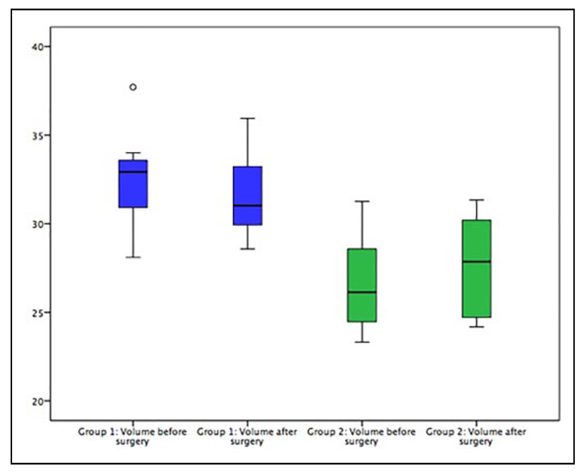
- Orbital Volume: Pre- and postoperative orbital CT volumes were measured (iPlan CMF 3.0.5, Brain-Lab).[14] The preoperative volume of the affected exe was compared to the volume of the unaffected side and to the postoperative volume. Analysis was performed by the primary surgeon (M.R.) to assure intra-observer repeatability. To calculate volumes, the orbital cavities were segmented automatically and in case of remaining defects or reduction of the orbital cavity due to titanium meshes, extensive scaring, bone fragments, and so on were manually adjusted using the smart shaper tool.
- Intraorbital angles (anterior, medial, and posterior angles between the medial orbital wall and the orbital floor): Intraorbital angles were compared at three points (anterior, middle, and posterior) in both the virtual plan and postoperative CT scan. They were measured in the coronal layers. The measurements were made using the anterior orbital rim, the posterior ledge and the exact in-between as fixed reference points in sagittal layers.
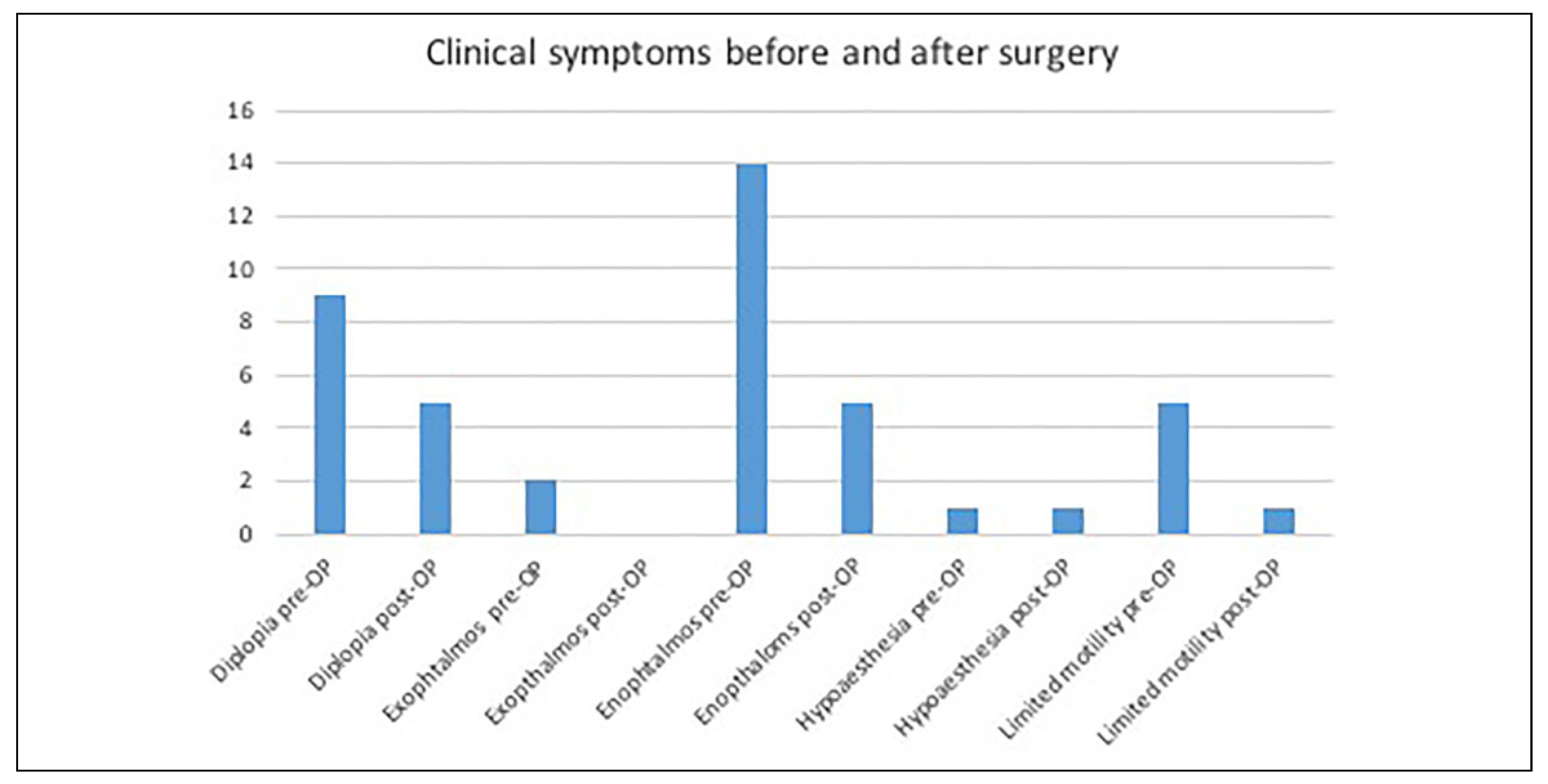
- Diplopia: Diplopia was documented as either negative or positive. If positive, it was documented as being either in primary gaze or with globe excursion (including the direction of excursion that resulted in diplopia).
- Visual acuity: Visual acuity was graded as either normal (0) or reduced (1). It was determined via finger perimetry.
- Motility: Extraocular movement was checked having the patient follow the examiner’s finger moving across their full range of horizontal and vertical eye movement.
- Hypesthesia: Reduced sensitivity in V2 areas was checked by clinical evaluation (sharp–dull, hot– cold, and 2-point discrimination using the Nerve Evaluation Protocol of 2014 by the California Association of Oral and Maxillofacial Surgeons, classified by the MRC Scale).
3. Results
4. Discussion
- Secondary orbital reconstruction can be performed using PSIs.
- In two-thirds of all cases, clinical symptoms can be eliminated or reduced when treated with customized intraorbital volume changes through the use of PSIs and titanium spacers.
Funding
Authors’ Note
Data Availability Statement
Acknowledgments
Conflicts of Interest
Ethical Approval
References
- Gellrich, N.C.; Schramm, A.; Hammer, B.; et al. Computerassisted secondary reconstruction of unilateral posttraumatic orbital deformity. Plast Reconstr Surg. 2002, 110, 1417–1429. [Google Scholar] [PubMed]
- Rana, M.; Chui, C.H.K.; Wagner, M.; Zimmerer, R.; Rana, M.; Gellrich, N.-C. Increasing the accuracy of orbital reconstruction with selective laser-melted patient-specific implants combined with intraoperative navigation. J Oral Maxillofac Surg. 2015, 73, 1113–1118. [Google Scholar] [CrossRef] [PubMed]
- Manson, P.N.; Markowitz, B.; Mirvis, S.; Dunham, M.; Yaremchuk, M. Toward CT-based facial fracture treatment. Plast Reconstr Surg. 1990, 85, 202–212, discussion 213–214. [Google Scholar] [CrossRef] [PubMed]
- Biesman, B.S.; Hornblass, A.; Lisman, R.; Kazlas, M. Diplopia after surgical repair of orbital floor fractures. Ophthal Plast Reconstr Surg. 1996, 12, 9–16, discussion 17. [Google Scholar] [CrossRef]
- Burnstine, M.A. Clinical recommendations for repair of isolated orbital floor fractures: an evidence-based analysis. Ophthalmology. 2002, 109, 1207–1210, discussion 1210–1211; quiz 1212–1213. [Google Scholar] [CrossRef]
- Bell, R.B.; Markiewicz, M.R. Computer-assisted planning, stereolithographic modeling, and intraoperative navigation for complex orbital reconstruction: a descriptive study in a preliminary cohort. J Oral Maxillofac Surg. 2009, 67, 2559–2570. [Google Scholar] [CrossRef]
- Whitaker, L.A.; Yaremchuk, M.J. Secondary reconstruction of posttraumatic orbital deformities. Ann Plast Surg. 1990, 25, 440–449. [Google Scholar] [CrossRef]
- Villarreal, P.M.; Monje, F.; Morillo, A.J.; Junquera, L.M.; Gonza´lez, C.; Barbo´n, J.J. Porous polyethylene implants in orbital floor reconstruction. Plast Reconstr Surg. 2002, 109, 877–885, discussion 886–887. [Google Scholar] [CrossRef]
- Rubin, P.A.; Bilyk, J.R.; Shore, J.W. Orbital reconstruction using porous polyethylene sheets. Ophthalmology. 1994, 101, 1697–1708. [Google Scholar] [CrossRef]
- Schittkowski, M.P.; Gundlach, K.K.; Guthoff, R.F. Treatment of congenital clinical anophthalmos with high hydrophilic hydrogel expanders. Ophthalmologe. 2003, 100, 525–534. [Google Scholar] [CrossRef]
- Nkenke, E.; Vairaktaris, E.; Spitzer, M.; et al. Secondary reconstruction of posttraumatic enophthalmos: prefabricated implants vs. titanium mesh. Arch Facial Plast Surg. 2011, 13, 271–277. [Google Scholar] [CrossRef] [PubMed]
- Baum, S.H.; Schmeling, C.; Pfo¨rtner, R.; Mohr, C. Autologous dermis—fat grafts as primary and secondary orbital transplants before rehabilitation with artificial eyes. J Craniomaxillofac Surg. 2018, 46, 90–97. [Google Scholar]
- Mantripragada, V.P.; Lecka-Czernik, B.; Ebraheim, N.A.; Jayasuriya, A.C. An overview of recent advances in designing orthopedic and craniofacial implants. J Biomed Mater Res A. 2013, 101, 3349–3364. [Google Scholar]
- Rana, M.; Essig, H.; Ru¨cker, M.; Gellrich, N.-C. Development and demonstration of a novel computer planning solution for predefined correction of enophthalmos in anophthalmic patients using prebended 3D titanium-meshes—a technical note. J Oral Maxillofac Surg. 2012, 70, e631–e638. [Google Scholar] [PubMed]
- Ramli, N.; Kala, S.; Samsudin, A.; Rahmat, K.; Abidin, Z.Z. Proptosis—correlation and agreement between Hertel exophthalmometry and computed tomography. Orbit. 2015, 34, 257–262. [Google Scholar] [CrossRef] [PubMed]
- Jeon, H.B.; Kang, D.H.; Oh, S.A.; Gu, J.H. Comparative study of Naugle and Hertel exophthalmometry in orbitozygomatic fracture. J Craniofac Surg. 2016, 27, 142–144. [Google Scholar]
- Wolfe, S.A.; Ghurani, R.; Podda, S.; Ward, J. An examination of posttraumatic, postsurgical orbital deformities: conclusions drawn for improvement of primary treatment. Plast Reconstr Surg. 2008, 122, 1870–1881. [Google Scholar] [CrossRef]
- Hammer, B.; Kunz, C.; Schramm, A.; DeRoche, R.; Prein, J. Repair of complex orbital fractures: technical problems, state-of-theart solutions and future perspectives. Ann Acad Med Singapore. 1999, 28, 687–691. [Google Scholar]
- Bartling, S.H.; Leinung, M.; Graute, J.; et al. Increase of accuracy in intraoperative navigation through high-resolution flatpanel volume computed tomography: experimental comparison with multislice computed tomography-based navigation. Otol Neurotol. 2007, 28, 129–134. [Google Scholar] [CrossRef]
- Rana, M.; Essig, H.; Eckardt, A.M.; et al. Advances and innovations in computer-assisted head and neck oncologic surgery. J Craniofac Surg. 2012, 23, 272–278. [Google Scholar]
- Stoetzer, M.; Rana, M.; von See, C.; Eckardt, A.M.; Gellrich, N.-C. Reconstruction of defects of maxillary sinus wall after removal of a huge odontogenic lesion using prebended 3D titanium-mesh and CAD/CAM technique. Head Face Med 2011, 7, 21. [Google Scholar] [PubMed]
- Bly, R.A.; Chang, S.-H.; Cudejkova, M.; Liu, J.J.; Moe, K.S. Computer-guided orbital reconstruction to improve outcomes. JAMA Facial Plast Surg. 2013, 15, 113–120. [Google Scholar] [PubMed]
- Metzger, M.C.; Scho¨n, R.; Zizelmann, C.; Weyer, N.; Gutwald, R.; Schmelzeisen, R. Semiautomatic procedure for individual preforming of titanium meshes for orbital fractures. Plast Reconstr Surg. 2007, 119, 969–976. [Google Scholar] [CrossRef]
- Tabakovic, S.Z.; Konstantinović, V.S.; Radosavljević, R.; Movrin, D.; Hadžistević, M.; Hatab, N. Application of computer-aided designing and rapid prototyping technologies in reconstruction of blowout fractures of the orbital floor. J Craniofac Surg. 2015, 26, 1558–1563. [Google Scholar] [PubMed]
- Baumann, A.; Sinko, K.; Dorner, G. Late reconstruction of the orbit with patient-specific implants using computer-aided planning and navigation. J Oral Maxillofac Surg 2015, 73 Suppl. S12, S101–106. [Google Scholar] [CrossRef]
- Chen, C.T.; Pan, C.-H.; Chen, C.-H.; Shyu, V.; et al. Clinical outcomes for minimally invasive primary and secondary orbital reconstruction using an advanced synergistic combination of navigation and endoscopy. J Plast Reconstr Aesthet Surg. 2018, 71, 90–100. [Google Scholar] [CrossRef]
- Gaffre´e, G.; Santos, R.; Cupello, V.; Polinati, J.M.; Paredes, L. Secondary reconstruction of posttraumatic enophthalmos with titanium mesh and buccal fat pad graft: case report. Surg J 2017, 3, e101–e106. [Google Scholar]
- Pan, H.; Zhang, Z.; Tang, W.; Li, W.; Deng, Y. Bioresorbable material in secondary orbital reconstruction surgery. J Ophthalmol. 2019, 2019, 8715314. [Google Scholar] [CrossRef]

© 2020 by the author. The Author(s) 2020.
Share and Cite
Singh, D.D.; Schorn, L.; Strong, E.B.; Grant, M.; Schramm, A.; Hufendiek, K.; Gellrich, N.-C.; Rana, M. Computer-Assisted Secondary Orbital Reconstruction. Craniomaxillofac. Trauma Reconstr. 2021, 14, 29-35. https://doi.org/10.1177/1943387520935004
Singh DD, Schorn L, Strong EB, Grant M, Schramm A, Hufendiek K, Gellrich N-C, Rana M. Computer-Assisted Secondary Orbital Reconstruction. Craniomaxillofacial Trauma & Reconstruction. 2021; 14(1):29-35. https://doi.org/10.1177/1943387520935004
Chicago/Turabian StyleSingh, Daman D., Lara Schorn, E. Bradley Strong, Michael Grant, Alexander Schramm, Karsten Hufendiek, Nils-Claudius Gellrich, and Majeed Rana. 2021. "Computer-Assisted Secondary Orbital Reconstruction" Craniomaxillofacial Trauma & Reconstruction 14, no. 1: 29-35. https://doi.org/10.1177/1943387520935004
APA StyleSingh, D. D., Schorn, L., Strong, E. B., Grant, M., Schramm, A., Hufendiek, K., Gellrich, N.-C., & Rana, M. (2021). Computer-Assisted Secondary Orbital Reconstruction. Craniomaxillofacial Trauma & Reconstruction, 14(1), 29-35. https://doi.org/10.1177/1943387520935004