Next Article in Journal
Traumatic Facial Tattoo Injuries From Gunpowder and Ammunition: A Case Series
Previous Article in Journal
Skeletal Deformity in Patients With Unilateral Coronal Craniosynostosis: Perceptions of the General Public
 
 
Craniomaxillofacial Trauma & Reconstruction is published by MDPI from Volume 18 Issue 1 (2025). Previous articles were published by another publisher in Open Access under a CC-BY (or CC-BY-NC-ND) licence, and they are hosted by MDPI on mdpi.com as a courtesy and upon agreement with Sage.
Short Note

Management of Zygomatic Arch Fractures by Intraoral Open Reduction and Transbuccal Fixation: A Technical Note

by
Elavenil Panneerselvam
,
Sasikala Balasubramanian
*,
Jaghandeep Kempraj
,
Vijitha Ravindira Babu
and
V. B. Krishna Kumar Raja
Department of Oral and Maxillofacial Surgery, SRM Dental College and Hospital, Ramapuram Campus, Ramapuram, Chennai 600089, Tamil Nadu, India
*
Author to whom correspondence should be addressed.
Craniomaxillofac. Trauma Reconstr. 2020, 13(2), 130-132; https://doi.org/10.1177/1943387520911866
Submission received: 1 December 2019 / Revised: 31 December 2019 / Accepted: 1 February 2020 / Published: 17 March 2020

Abstract

:
Fractures of the zygomatic arch are common due to its anatomical prominence. The post-traumatic restoration of the arch form is important to maintain the midfacial symmetry and anteroposterior projection of the face. Open reduction and internal fixation (ORIF) of fractured arch is indicated in specific clinical presentations. The traditional methods of ORIF of zygomatic arch fractures require cutaneous incisions, which are associated with complications such as scarring and facial nerve injury. This article presents a simple technique of “intraoral reduction and transbuccal fixation” of the arch that negates the problems associated with the conventional approaches to ORIF.

Introduction

Zygomatic arch fractures in isolation or associated with fractures of other bones are common. The arch is prone to fracture due to its long and thin structural framework which projects outside the facial skeleton.[1] Restoration of the arch form is important to maintain the symmetry of midface and its anteroposterior projection. Most often, they are managed by closed reduction without any fixation because of the thick periosteal envelope which holds the fragments together.
However, open reduction and internal fixation (ORIF) of the fractured arch is indicated for the following:[1] (1) comminuted fractures which are unstable after reduc- tion, (2) malunited arch fractures, and (3) failed closed reduction. The traditional methods of ORIF of zygo-matic arch fractures require either a cutaneous incision, such as a preauricular incision, or an extensive approach using a coronal incision, both of which are associated with potential complications.[1] This article presents a simple technique that negates the problems associated with the conventional approaches to ORIF of the zygo- matic arch.

Technique

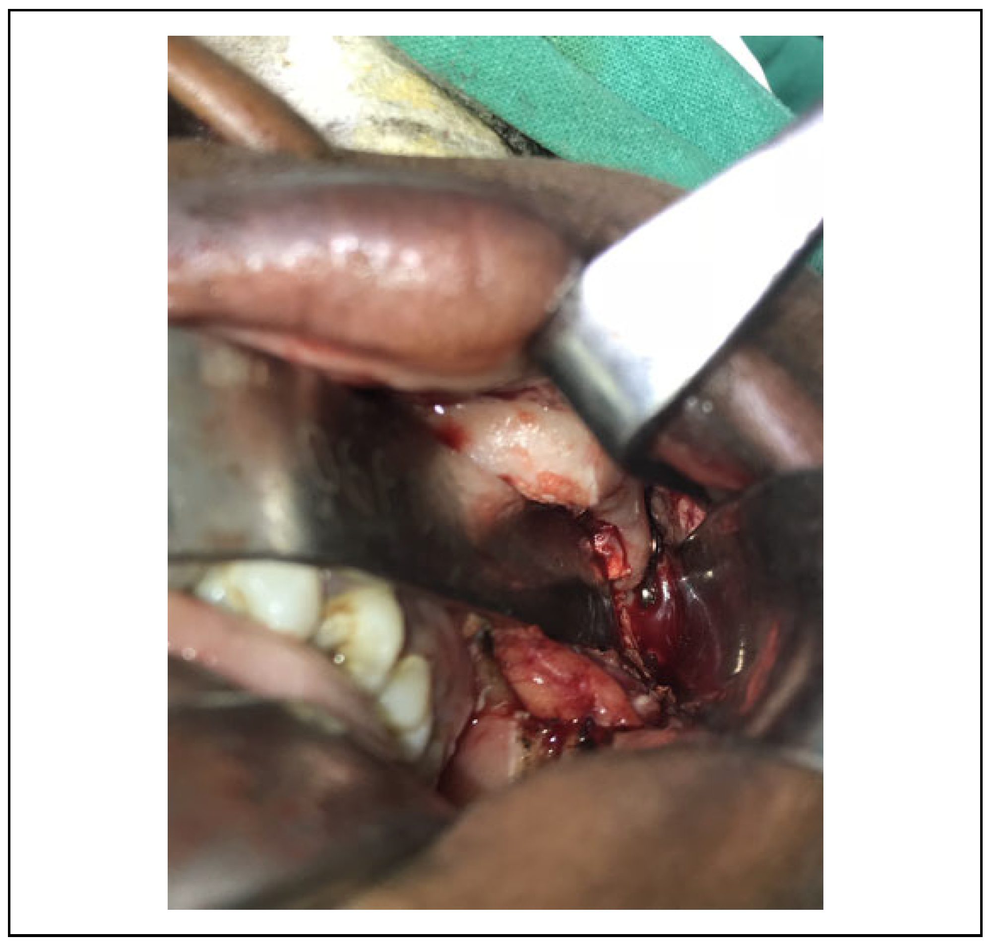
The procedure was planned for a 26-year-old male patient who presented with zygomatic arch fracture (Figure 1) fol- lowing road traffic accident. The patient had reported to our unit 2 weeks after trauma and required surgical intervention to treat complaints of facial asymmetry and restricted mouth opening. Under general anesthesia, the fracture was exposed using an intraoral vestibular incision which extended from 13 to 17 regions. Transoral reduction of the fracture was achieved with a zygomatic elevator. A 2-hole, 1.5-mm titanium miniplate was adapted intraorally across the reduced zygomatic arch fracture. A stab incision was placed on the facial skin corresponding to the arch through which a trocar was introduced to facilitate insertion of a transbuccal cannula (Figure 2). The miniplate was fixed transbuccally by inserting 6-mm screws through the can- nula (Figure 3).
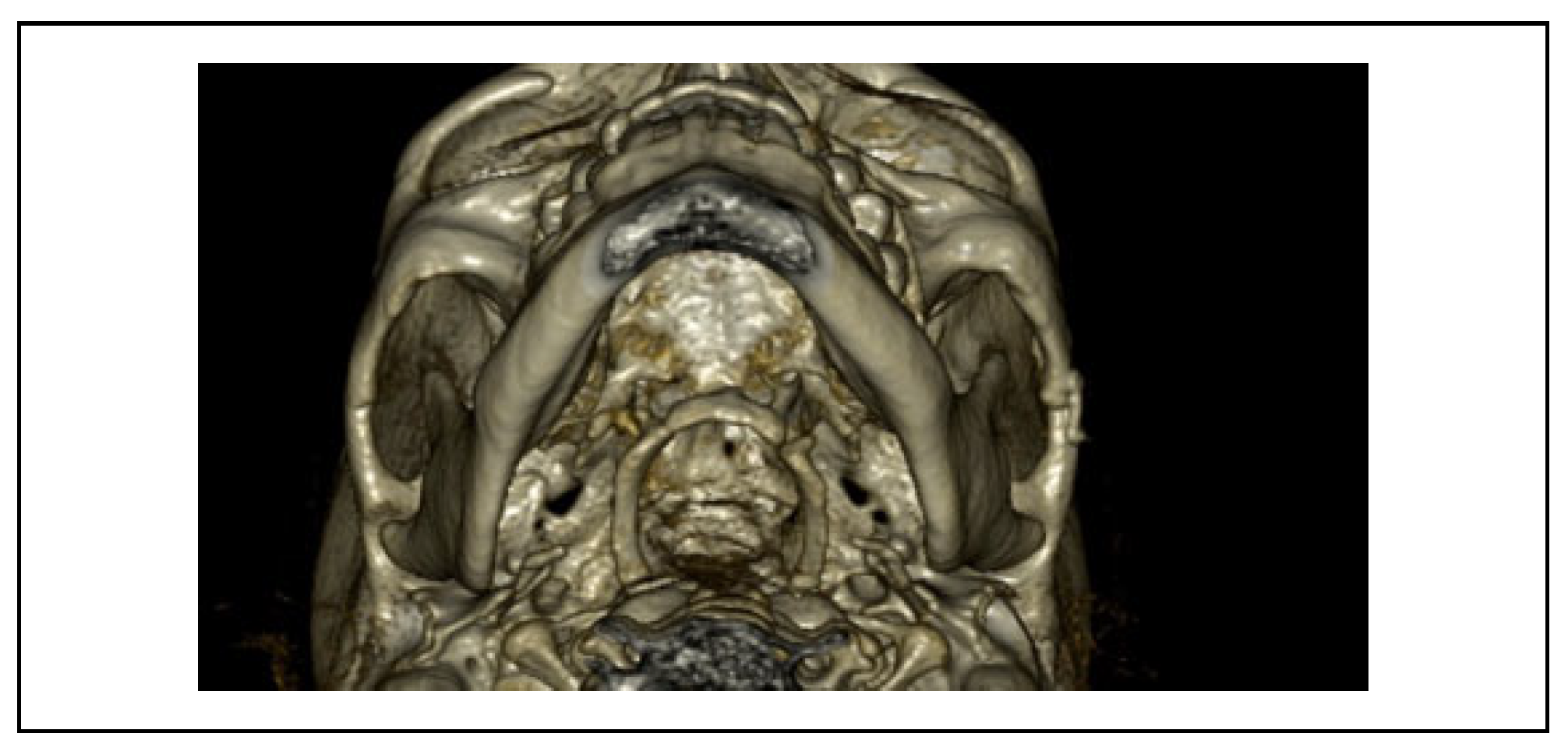
Closure of the intraoral wound was done with 3-0 Vicryl and transbuccal entry wound with a single, 5-0 Prolene suture. Postoperatively, the patient demonstrated good mouth opening, facial symmetry, and an imperceptible scar on the skin over the zygomatic arch. Postoperative com- puted tomography taken 4 weeks after the procedure demonstrated optimal reduction and fixation in axial view (Figure 4).

Discussion

Restoration of the zygomatic arch anatomy is essential for aesthetic purpose and functional stabilization of midface.[1] The approaches commonly followed to access the frac- tured arch for open reduction and fixation involve cuta- neous incisions,[1] namely (1) suprazygomatic Dingman’s incision (above the zygomatic arch and parallel to it), (2) suprazygomatic arch incision, along the skin crease, (3) preauricular, and (4) coronal. However, these approaches are associated with limitations; scarring is a major concern related to all skin incisions.[1,2] In addition, suprazygomatic incisions and preauricular incision pose potential risks to the facial nerve[1] and inability to access the arch fracture, which is located at the anterior third of the zygomatic arch (near the zygomaticotemporal suture).
Coronal approach is a relatively extensive procedure with increased surgical time and blood loss. It is generally indicated in the management of arch fractures with conco- mitant midface injuries. The associated risks of coronal approach are temporal hollowing, scalp numbness, and facial nerve injury. Further, the resultant scar may lead to alopecia and unesthetic scarring in bald patients.[1]
Endoscopic-assisted approach to reduce and fix zygo-matic arch fractures has been practiced for decades, with the aim of minimizing surgical morbidity and injury to the facial nerve. However, the endoscopic access to the arch also requires incisions which may be restricted to the tem- poral region or a preauricular incision with a scalp exten- sion and are associated with 7% to 13% of temporary frontal weakness.[3] Further, endoscopic fixation requires special armamentarium, training, and expertise. Xie et al[4] used a modified endoscopic-assisted approach with a pre- auricular incision but used a different plane of dissection that negated facial nerve injury. The use of “z instrument” along with endoscopy has been claimed to facilitate fixa- tion with better vision and less nerve damage, but the tech- nique requires 3 cutaneous incisions as well as special armamentarium.[5]

Author’s Approach Versus Conventional Approaches

The transbuccal approach is ideal to fix the fractured arch, which is displaced at the zygomaticotemporal suture or anterior one-third of the arch. As compared with the other techniques, the observed advantages of the author’s tech- nique are as follows: (1) avoiding scarring due to cutaneous approaches, (2) prevention of facial nerve injury, (3) mini- mizing blood loss, (4) ease and rapidity of technique, and (5) facilitates correct angulation of screw placement. How- ever, a prospective study involving a large sample of cases would validate the advantages of this technique.
The possibility of facial nerve damage (especially the temporal branch) during trocar placement may be an aspect of concern to surgeons. Nerve injury can be avoided using safe anatomical landmarks, as described by Dahlke and Murray.[6] The temporal branch of the facial nerve lies in the danger zone, which is bounded by 2 imaginary lines: inferior line extending from the lateral eyebrow to the ear- lobe and superior line connecting the tragus to the upper forehead crease. Introducing the trocar just below this zone would ensure safe transbuccal instrumentation and protec- tion of the temporal branch of the facial nerve. Limitation of the technique includes inadequate access for fixation of fracture involving the posterior third of the arch, which may necessitate a preauricular incision.

Conclusion

The management of zygomatic arch fractures by intraoral open reduction and transbuccal fixation is an effective method to address zygomatic arch fractures. The technique is simple and less invasive with reduced risk of facial nerve injury.

Funding

The author(s) received no financial support for the research, authorship, and/or publication of this article.

Patient Consent

Patient consent was obtained to publish the clinical intraoral photographs without any patient identification.

Conflicts of Interest

The author(s) declared no potential conflicts of interest with respect to the research, authorship, and/or publication of this article.

References

  1. Chen, C.T.; Lai, J.P.; Chen, Y.R.; Tung, T.C.; Chen, Z.C.; Rohrich, R.J. Application of endoscope in zygomatic fracture repair. Br J Plast Surg. 2000, 53, 100–105. [Google Scholar] [PubMed]
  2. Shikimori, M.; Motegi, K. Skin incision parallel with skin cleavage lines for access to the fractured zygomatic arch. J Maxillofac Surg. 1986, 14, 321–322. [Google Scholar]
  3. Lee, C.H.; Lee, C.; Trabulsy, P.P.; Alexander, J.T.; Lee, K. A cada- veric and clinical evaluation of endoscopically assisted zygo- matic fracture repair. Plast Reconstr Surg. 1998, 101, 333–345. [Google Scholar] [PubMed]
  4. Xie, L.; Shao, Y.; Hu, Y.; Li, H.; Gao, L.; Hu, H. Modification of surgical technique in isolated zygomatic arch fracture repair: seven case studies. Int J Oral Maxillofac Surg. 2009, 38, 1096–1100. [Google Scholar] [PubMed]
  5. Badillo, O.; Osben, R.; Vidal, C.; Duarte, V. Design and use of an instrument for video-assisted surgical treatment of unstable fractures of the zygomatic arch: the Z instrument. Br J Oral Maxillofac Surg. 2015, 53, 767–768. [Google Scholar] [CrossRef] [PubMed]
  6. Dahlke, E.; Murray, C.A. Facial nerve danger zone in dermato- logic surgery: temporal branch. J Cutan Med Surg. 2011, 15, 84–86. [Google Scholar] [PubMed]
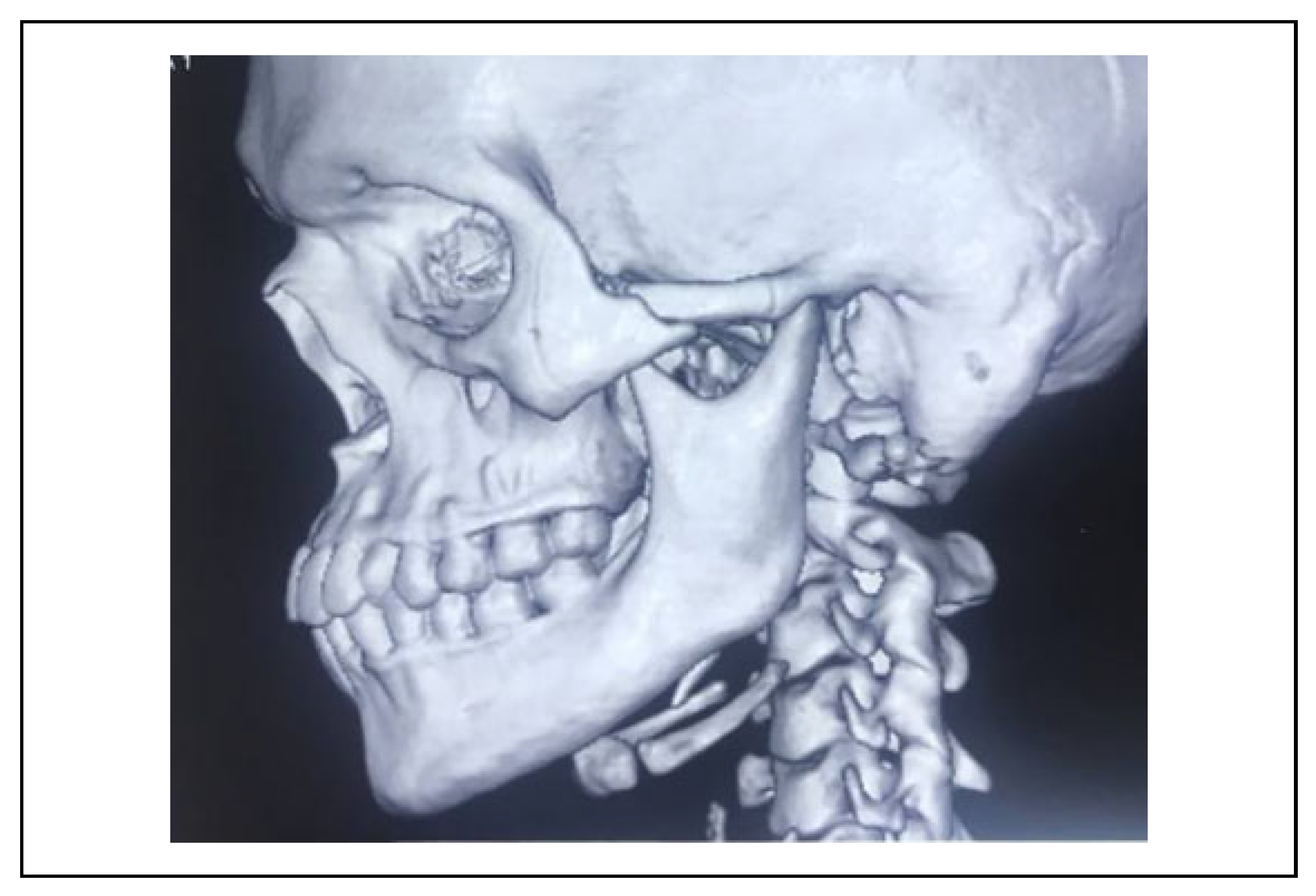
Figure 1. CT demonstrating arch fracture. CT indicates com- puted tomography.
Cmtr 13 00048 g001
Figure 2. Transbuccal instrumentation.
Cmtr 13 00048 g002
Figure 3. Mini plate fixation on the zygomatic arch.
Cmtr 13 00048 g003
Figure 4. Postoperative CT taken 4 weeks after fixation—axial view. CT indicates computed tomography.
Cmtr 13 00048 g004
Disclaimer/Publisher’s Note: The statements, opinions and data contained in all publications are solely those of the individual author(s) and contributor(s) and not of MDPI and/or the editor(s). MDPI and/or the editor(s) disclaim responsibility for any injury to people or property resulting from any ideas, methods, instructions or products referred to in the content.