Next Article in Journal
Total Temporomandibular Joint Replacement: Stick to Stock or Optimization by Customization?
Previous Article in Journal
Intact Periorbita Can Prevent Post-Traumatic Enophthalmos Following a Large Orbital Blow-Out Fracture
 
 
Craniomaxillofacial Trauma & Reconstruction is published by MDPI from Volume 18 Issue 1 (2025). Previous articles were published by another publisher in Open Access under a CC-BY (or CC-BY-NC-ND) licence, and they are hosted by MDPI on mdpi.com as a courtesy and upon agreement with Sage.
Article

A Clinical and Radiological Investigation of the Use of Dermal Fat Graft as an Interpositional Material in Temporomandibular Joint Ankylosis Surgery

by
Sajjad Abdur Rahman
,
Tabishur Rahman
*,
Ghulam Sarwar Hashmi
,
Syed Saeed Ahmed
,
Mohammad Kalim Ansari
and
Abdus Sami
Department of Oral and Maxillofacial Surgery, Faculty of Medicine, Aligarh Muslim University, Aligarh, Uttar Pradesh 202002, India
*
Author to whom correspondence should be addressed.
Craniomaxillofac. Trauma Reconstr. 2020, 13(1), 53-58; https://doi.org/10.1177/1943387520903876
Submission received: 1 December 2019 / Revised: 31 December 2019 / Accepted: 1 February 2020 / Published: 2 April 2020

Abstract

:
Management of temporomandibular joint (TMJ) ankylosis is mainly through surgical intervention. Interpositional materials are a necessity when it comes to prevention of TMJ re-ankylosis after arthroplasty. Early aggressive postoperative physiotherapy is essential for the prevention or treatment of TMJ hypomobility or ankyloses. Recently, it has been shown that abdominal dermis fat helps promote smooth, pain-free joint function and it is stable after interposition and less prone to fragmentation. The purpose of this study was to assess that whether dermal fat is a good choice of interpositional material when it comes to decreased pain perception during aggressive physiotherapy after release of ankyloses thus ensuring good compliance by the patient. We also assessed the fate of the graft material on computed tomography to evaluate any volume changes if occurred after interposition.

Introduction

Temporomandibular joint (TMJ) ankylosis is an intracapsular union of the disc condyle complex to the temporal articular surface that includes fibrous adhesion or bony fusion between condyle of mandible and glenoid fossa and articular eminence of squamous temporal bone.[1]
The causes include trauma, local or systemic infection, degenerative joint diseases, surgical intervention of the joint space, and neoplasms. TMJ ankylosis results in a vast array of problems including difficulty in mastication, digestion, and speech and hygiene maintenance. Dysmorphic development of the jaw alters the appearance of the patient and also has an impact on the psychologic development of patient.
Management of TMJ ankylosis is mainly through surgical intervention. Interpositional materials are a necessity when it comes to prevention of TMJ re-ankylosis after arthroplasty. Early aggressive postoperative physiotherapy is essential for the prevention or treatment of TMJ hypomobility or ankyloses.[2]
Recurrence of TMJ ankyloses could be attributed to the following: the high osteogenic potential of the condylar stump after removal of the ankylotic mass, the increased incidence of the heterotopic bone formation after surgery, and the surgery itself. Furthermore, the postoperative pain complicates the performance of physiotherapy as patients’ initial compliance is reduced. Also, the long-term compliance is reduced as aggressive physiotherapy by itself is a very painful procedure for patients to withstand.[3]
Recently, it has been shown that abdominal dermis fat helps promote smooth, pain-free joint function and it is stable after interposition and less prone to fragmentation.[4]
The purpose of this study was to assess that whether dermal fat is a good choice of interpositional material when it comes to decreased pain perception during aggressive physiotherapy after release of ankyloses thus ensuring good compliance by the patient. We also assessed the fate of the graft material using computed tomography (CT) to evaluate any volume changes if occurred after interposition.

Materials and Method
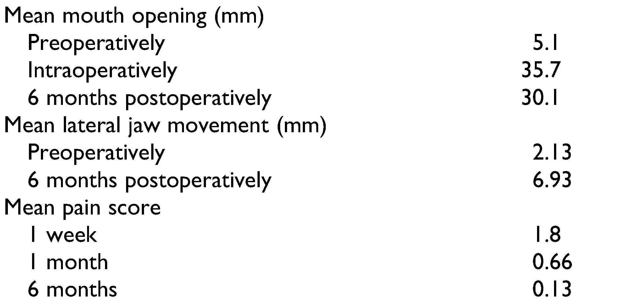
A prospective open-label interventional study involving patients who presented with true ankylosis of the TMJ confirmed on CT was undertaken. Only those with true osseous or fibro-osseous ankylosis directly involving the intracapsular structures of the TMJ were included in the study (Figure 1). Recurrent cases of TMJ ankyloses and patients with extra-articular fibrosis and pathological and mechanical obstruction were excluded from the study. A detailed history of the age, mode of onset, duration, and sex was recorded. Any facial asymmetry, scars, or deviation of chin was examined. The oral cavity was examined to see the deviation of midline; occlusion; any evidence of protrusive, retrusive, and lateral movements; gingival condition; caries; and any impacted or abnormally placed teeth. The extent of maximal incisal opening (MIO) was measured from the incisal edge of the mandibular central incisor to the incisal edge of maxillary central incisor (Figure 2). Both joints were assessed radiographically with orthopantomograms and computed tomograms. All patients had routine hematological examinations. This trial was approved by the institutional ethical committee and the patients gave informed and written consent for inclusion into the study. All patients were operated on under general anesthesia with nasoendotracheal intubation or elective tracheostomy. Through a standard preauricular incision as described by Risdon,[5] a gap arthroplasty was performed by excision of the ankylosed mass creating a gap of at least 1 centimeter between the proximal and distal segments. Contralateral coronoidectomy was done if intraoperative mouth opening was less than 35 centimeters. Dermis fat graft was harvested through an elliptical incision made just medial to iliac crest. Epidermis with a layer of dermis and subcutaneous fat was dissected. The epidermis was then dissected finely and discarded (Figure 3). The graft was then used to fill the gap created in the ankylosed area (Figure 4). The donor site was repaired by primary closure. The preauricular incision was closed in layers and pressure bandage was applied around the head with extra padding applied over the surgical site for 24 hours postoperatively. Physiotherapy to help mobilize the joint was commenced 7 days postoperatively. Assessment of pain was done by visual analogue scale (VAS) at 1-week, 1-month, and 6-month intervals. Pain was scored as 0 = no pain, 1 = mild pain (VAS 1-4), 2 = moderate pain (VAS 5-7), and 3 = severe pain (VAS 8-10). Maximal interincisal mouth opening was measured presurgically, intraoperatively, and 6 months post-operatively (Figure 5). Lateral movement of mandible was evaluated presurgically and at 6 months after the surgery. Assessment of graft material was done by CT scan of operated TMJ at least 6 months postsurgically.

Results

We studied 15 patients with ankylosis of the TMJ out of which 3 patients had bilaterally affected joints, making a total of 18 joints. The average duration of ankyloses was 6.28 years. The age of the patients ranged from 6 to 30 years with the mean age of 12.6 years. The male:female ratio was 1:2. Fall from height was the most common cause (93%) followed by infection of ear (7%). The mean duration of ankyloses was 6.28 years. The mean pain score was 1.8, 0.66, and 0.13 at 1-week, 1-month and 6-month postsurgical intervals. The mean mouth opening preoperatively, intraoperatively, and 6 months postsurgically was 5.1, 35.7, and 30.1 millimeters, respectively. The mean lateral jaw movement at 6-month post-intervention duration was 6.93 millimeters (Table 1, Figure 6, Figure 7 and Figure 8). Recurrence was observed in 1 patient involving 1 joint. Mild infection at donor site was seen in 1 case and at TMJ region in 2 cases which was effectively managed with antibiotics.

Discussion

The management of TMJ ankyloses is mainly through surgery coupled with aggressive physiotherapy postsurgically. Surgery removes the ankylotic mass thus creating a gap between the basal skull and the remaining condylar stump whereas physiotherapy improves range of motion and builds up muscle bulk. There are 3 main causes for development of re-ankylosis in these cases: high osteogenic potential of the condylar stump, increased incidence of heterotopic bone formation after surgery and pain (postsurgical as well as physiotherapy-induced) which limits physiotherapy. Therefore, it is necessary to use an interpositional material which may prevent TMJ re-ankylosis after arthroplasty, and facilitate easy performance of postsurgical physiotherapy. The search for an ideal interpositional material has led to the use of alloplastic/allogenic materials, xenografts, and autogenous tissue as the substitute of discal tissue.
The use of silastic and Teflon-proplast implants was very popular in the 1980s but unfortunately these materials were found to be associated with numerous notorious complications including a foreign body reaction with giant cell infiltrate surrounding the implant, gross destruction of surrounding structures including severe bone erosions around TMJ causing cerebrospinal fluid leakage, implant fragmentation and perforation, and increase in peripheral osseous proliferation leading to fibrous or fibro-osseous ankyloses.[6,7]
Auricular cartilage offers the advantage of being biologically inert and viability in addition to a shape favoring fit into the glenoid fossa. The bulk of the graft also partially compensates for partial loss of the condyle. Also, the graft is resistant to pressure and its surface is smooth, allowing sliding movement, thus facilitating physiotherapy.[8,9]
However, apart from a resulting donor site morbidity, recent studies have revealed that auricular grafts may fragment, proliferate, and result in fibrous ankylosis of the TMJ with progressive limitation of mouth opening.[10,11]
The dermis graft again offers the advantage of bulk. Also, it has been demonstrated by Stewart et al that it may provide a scaffold for subsequent soft tissue growth and healing between the head of the condyle and glenoid fossa if used as a replacement of discal tissue.[12]
The most obvious drawback is the donor site morbidity. The other drawbacks include difficulty in anchoring the graft to remaining tissues and rare incidences of dermoid cysts.[13] In a study by Dimitroulis involving magnetic resonance imaging (MRI) evaluation of dermis as an interpositional material, he has demonstrated that dermis is not a load-bearing tissue which was made evident by absence of dermis tissue in MRI of significant number of patients in his study.[14]
Similar to dermis graft, the use of full-thickness skin graft may lead to development of cyst. Popescu and Vasiliu and Chossegross et al used full-thickness skin grafts to treat TMJ ankyloses in humans and reported only a single case of cyst formation in the grafts.[15,16] But in a study by Dimitroulis involving full-thickness skin graft placed in the craniomandibular joint of rabbit, epidermoid cyst formation was seen within all the grafted joints.[14]
The temporalis muscle and fascia flap is the most popular interpositional material used for the reconstruction of the TMJ owing to the advantages such as autogenous nature, resilience, adequate blood supply, and proximity to the joint, allowing for a pedicled transfer of vascularized tissue into the joint area.[17,18,19]
However, the use of temporalis muscle and fascia results in scarring and trismus from the donor site with the fascia lacking sufficient bulk and the muscle itself undergoing degenerative fibrosis under compressive load.[20]
Studies have shown that autogenous free fat grafts prevent scar formation by acting as an effective hemostatic agent and a space filler that prevents the accumulation of blood and serum, which would otherwise turn into scar or bone.[21,22] But the disadvantages are that autogenous fat grafts are difficult to handle and long-term studies have shown that fat grafts often fragmented when placed into the joint cavity and on an average lost 45% of transplanted volume due to shrinkage.[23,24]
In 2004, Dimitroulis first reported the use of abdominal dermis fat grafts in TMJ surgery in 11 patients with TMJ ankyloses based on his observations that fat grafts alone are easily fragmented, but when attached to dermis the fat tends to be more stable and less likely to fragment during handling and manipulation. The other advantages offered by this graft include its contourability, survival (which is due to early establishment of vascular ingrowth facilitated by the removal of the surface epidermis), and maintenance of volume (the resorption rate of dermis fat is only 15%).[25]
As stated previously, 2 of the main reasons of recurrence of TMJ ankyloses is the increased incidence of the heterotopic bone formation after surgery and postoperative pain limiting the performance of physiotherapy. The first stage of the re-ankylosis is the formation of fibrous adhesions between the ramus stump and the base of the skull. Aggressive physiotherapy disrupts and prevents formation of more adhesions and contraction of the soft tissues. It also helps in stretching and lengthening of the formed maturing fibrous tissue until it achieves a length equal to the normal value of the mouth opening. It also allows normal muscle function, which results in regaining the intraoperative MIO and prevention of the bony re-ankylosis by preventing the ossification of the formed fibrous tissues. Apart from acting as an effective barrier to joint ankylosis by preventing new bone formation, the dermal fat helps smooth, pain-free joint function during the physiotherapy phase as made evident by the quality of life study by Dimitroulis et al.[26] This was corroborated by our study where we found that the patients experienced lesser degree of pain during the initial phase of physiotherapy and thus became more compliant to the physiotherapy schedule and hence were able to maintain good mouth opening even after an interval of 6 months. The postoperative CT scans obtained in our study have demonstrated the survival and stability of the graft confirming the physical presence of the graft (Figure 9) which reduces the chances of new bone formation by eliminating the dead space, thus preventing development and organization of a hematoma apart from providing a cushion for pain-free physiotherapy during the entire period of performance of physiotherapy.
The abdominal dermis fat graft, due to its excellent characteristics, appears to offer some promise as the material of choice and superiority over other grafts. The associated disadvantage of a visible abdominal scar, which is a major issue, particularly for young patients, is a concern when deciding to use this graft material for interposition.

Funding

The author(s) received no financial support for the research, authorship, and/or publication of this article.

Conflicts of Interest

The author(s) declared no potential conflicts of interest with respect to the research, authorship, and/or publication of this article.

References

  1. Long, X.; Li, X.; Cheng, Y.; et al. Preservation of disc for treatment of traumatic temporomandibular joint ankylosis. J Oral Maxillofac Surg. 2005, 63, 897–902. [Google Scholar] [CrossRef] [PubMed]
  2. Buchbinder, D.; Currivan, R.B.; Kaplan, A.J.; Urken, M.L. Mobilization regimens for the prevention of jaw hypomobility in the radiated patient: a comparison of three techniques. J Oral Maxillofac Surg. 1993, 51, 863–867. [Google Scholar] [CrossRef]
  3. Khalifa, G.A. Monitoring of incremental changes in maximum interincisal opening after gap arthroplasty omits the risk of reankylosis. J Craniomaxillofac Surg. 2018, 46, 75–81. [Google Scholar]
  4. Chuong, R.; Piper, M.A. Cerebrospinal fluid leak associated with proplast implant removal from the temporomandibular joint. Oral Surg Oral Med Oral Pathol. 1992, 74, 422–425. [Google Scholar] [PubMed]
  5. Risdon, F. Ankylosis of the temporo-mandibular joint. J Am Dent Assoc. 1934, 21, 1933. [Google Scholar]
  6. Dolwick, M.F.; Aufdemorte, T.B. Silicone induced foreign body reaction and lymphadenopathy after temporomandibular joint arthroplasty. Oral Surg Oral Med Oral Pathol. 1985, 59, 449–452. [Google Scholar] [CrossRef] [PubMed]
  7. Ioannides, C.; Freihofer, H.P.M. Replacement of the damaged inter-articular disc of the TMJ. J Craniomandib Pract. 1988, 16, 273–278. [Google Scholar]
  8. Matukas, V.J.; Lachner, J. The use of autologous auricular cartilage for temporomandibular joint disc replacement: a preliminary report. J Oral Maxillofac Surg. 1990, 48, 348–353. [Google Scholar]
  9. Sandler, N.A.; MacMillan, C.; Buckley, M.J.; Barnes, L. Histological and histochemical changes in failed auricular cartilage grafts used for a temporomandibular joint disc replacement. J Oral Maxillofac Surg. 1997, 55, 1014–1019. [Google Scholar] [CrossRef]
  10. Takatsuka, S.; Narinobou, M.; Nakagawa, K.; Yamamoto, E. Histological evaluation of auricular cartilage grafts after discectomy in the rabbit craniomandibular joint. J Oral Maxillofac Surg. 1996, 54, 1216–1225. [Google Scholar]
  11. Yih, W.-Y.; Zysset, M.; Merrill, R.G. Histopathological study of fate of autogenous auricular cartilage grafts in the human temporomandibular joint. J Oral Maxillofac Surg 1992, 50, 964–967. [Google Scholar] [PubMed]
  12. Stewart, H.M.; Hann, J.R.; DeTomasi, D.C.; Neville, B.W.; DeChamplain, R.W. Histological fate of dermal grafts following implantation for temporomandibular joint meniscal perforation: a preliminary study. Oral Surg Oral Med Oral Pathol. 1986, 62, 481–485. [Google Scholar] [PubMed]
  13. Muto, T.; Tomioka, K.; Michiya, H.; Kanazawa, M. Epidermoid cyst in the temporomandibular joint after dermal graft. J Craniomaxillofac Surg. 1992, 20, 270–272. [Google Scholar]
  14. Dimitroulis, G. The use of dermis grafts after discectomy for internal derangement of the temporomandibular joint. J Oral Maxillofac Surg. 2005, 63, 173–178. [Google Scholar] [PubMed]
  15. Popescu, V.; Vasiliu, D. Treatment of temporomandibular ankyloses with particular reference to the interposition of full thickness skin autotransplant. J Maxillofac Surg. 1977, 5, 3–14. [Google Scholar]
  16. Chossegros, C.; Guyot, L.; Cheynet, F.; Blanc, J.L.; Cannoni, P. Full-thickness skin graft interposition after temporomandibular joint ankylosis surgery: a study of 31 cases. Int J Oral Maxillofac Surg. 1999, 28, 330–334. [Google Scholar]
  17. Dimitroulis, G.; Slavin, J. Histological evaluation of full thickness skin as an interpositional graft in the rabbit craniomandibular joint. J Oral Maxillofac Surg. 2006, 4, 1075–1080. [Google Scholar] [CrossRef]
  18. Omura, S.; Fujita, K. Modification of the temporalis muscle and fascia flap for the management of ankylosis of the temporomandibular joint. J Oral Maxillofac Surg. 1996, 54, 794–795. [Google Scholar] [CrossRef]
  19. Pogrel, M.A.; Kaban, L.B. The role of a temporalis fascia and muscle flap in temporomandibular joint surgery. J Oral Maxillofac Surg. 1990, 48, 14–19. [Google Scholar]
  20. Feinberg, S.E.; Larsen, P.E. The use of a pedicled temporalis muscle-pericranial flap for replacement of the TMJ disc: preliminary report. J Oral Maxillofac Surg. 1989, 47, 142–146. [Google Scholar]
  21. Van Akkerveeken, P.F.; Van Der Kraan, W.; Muller, J.W. The fate of the free fat graft: a prospective clinical study using CT scanning. Spine 1986, 11, 501–504. [Google Scholar] [CrossRef] [PubMed]
  22. Aagenskiold, A.; Kiviluoto, O. Prevention of epidural scar formation after operations on the lumbar spine by means of free fat transplants: a preliminary report. Clin Orthop Relat Res. 1976, 115, 92–95. [Google Scholar]
  23. Billings, E.; May, J.W. Historical review and present status of free fat graft autotransplantation in plastic and reconstructive surgery. Plast Reconstr Surg. 1989, 83, 368–381. [Google Scholar]
  24. Lexer, E. Zwanzig jahre transplantations furschung in der chirurgie. Langenbecks Arch Klin Chir. 1925, 138, 297–302. [Google Scholar]
  25. Dimitroulis, G. The interpositional dermis-fat graft in the management of temporomandibular joint ankylosis. Int J Oral Maxillofac Surg. 2004, 33, 755–760. [Google Scholar]
  26. Dimitroulis, G.; McCullough, M.; Morrison, W. Quality of life survey of patients prior to and following temporomandibular joint discectomy. J Oral Maxillofac Surg. 2010, 68, 101–106. [Google Scholar]
Figure 1. 3-D CT image of a patient demonstrating TMJ ankylosis of right side. CT indicates computed tomography; TMJ, temporomandibular joint.
Cmtr 13 00010 g001
Figure 2. Maximum interincisal opening of a patient preoperatively.
Cmtr 13 00010 g002
Figure 3. Harvested dermal fat graft folded and sutured to conform to the superior and inferior surfaces of the gap created after removal of ankylotic mass.
Cmtr 13 00010 g003
Figure 4. Dermal fat graft in situ.
Cmtr 13 00010 g004
Figure 5. Maximum interincisal opening of the patient 6 months after the surgery.
Cmtr 13 00010 g005
Figure 6. Graph demonstrating number of patients experiencing pain at different time intervals with corresponding VAS scores. VAS indicates visual analogue scale.
Cmtr 13 00010 g006
Figure 7. Graph demonstrating 95% confidence interval in relation to mouth opening measurements.
Cmtr 13 00010 g007
Figure 8. Graph demonstrating 95% confidence interval in relation to lateral jaw movement measurements.
Cmtr 13 00010 g008
Figure 9. Coronal section of CT image obtained 6 months after surgery. CT indicates computed tomography.
Cmtr 13 00010 g009
Table 1. Comparison of the Preoperative and Postoperative Results.
Cmtr 13 00010 i001

