Next Article in Journal
Beta-Catenin Mutation with Complex Chromosomal Changes in Desmoid Tumor of the Scalp: A Case Report
Previous Article in Journal
Craniomaxillofacial Trauma Experience in Otolaryngology Residency: A National Survey of Program Directors
 
 
Craniomaxillofacial Trauma & Reconstruction is published by MDPI from Volume 18 Issue 1 (2025). Previous articles were published by another publisher in Open Access under a CC-BY (or CC-BY-NC-ND) licence, and they are hosted by MDPI on mdpi.com as a courtesy and upon agreement with Sage.
Case Report

Extranasopharyngeal Angiofibroma Arising from the Anterior Nasal Septum in a 35-Year-Old Woman

by
Tarik Kujundžić
1,2,
Aleksandar Perić
1,* and
Biserka Vukomanović Đurđević
3
1
Department of Otorhinolaryngology, Military Medical Academy School of Medicine, Crnotravska 17, 11000 Belgrade, Serbia
2
ENT Unit, General Hospital Pljevlja, Pljevlja, Montenegro
3
Institute for Pathology, Military Medical Academy School of Medicine, Belgrade, Serbia
*
Author to whom correspondence should be addressed.
Craniomaxillofac. Trauma Reconstr. 2019, 12(2), 141-145; https://doi.org/10.1055/s-0038-1675809
Submission received: 23 August 2018 / Revised: 1 September 2018 / Accepted: 27 September 2018 / Published: 12 November 2018

Abstract

:
Angiofibroma is a relatively rare vascular lesion originating predominantly in the nasopharynx and occurs typically in male adolescents. Extranasopharyngeal sites of origin, such as nasal cavity, paranasal sinuses, pharynx, and larynx, are extremely rarely seen in clinical practice. In the nasal cavity, extranasopharyngeal angiofibromas (ENAs) have been reported to originate from the nasal septum, as well as from the middle and inferior turbinate. In this study, we report a case of a 35-year-old female patient with angiofibroma originating from the anterior part of the nasal septum with left-sided nasal obstruction and mild-to-moderate epistaxis as main complaints. After the radiological diagnostics and embolization of sphenopalatine artery, we performed the excision of the lesion with removal of small part of septal mucosa and perichondrium around the attachment of tumor pedicle. Histopathological examination suggested the presence of angiofibroma and diagnosis was confirmed by immunohistochemical analysis. This is the fifth reported case of septal ENA in female population in the English-language literature.

Angiofibroma is a histologically benign but locally aggressive, nonencapsulated, highly vascular tumor, usually originating in nasopharynx and affecting mostly male adolescents.[1] The most common form of angiofibroma with primary nasopharyngeal localization is termed “juvenile nasopharyngeal angiofibroma (JNA).” According to recent investigations, JNA does not originate in the nasopharynx, as previously assumed, but rather from a fibrovascular nidus in the posterior part of the nasal cavity, near by the sphenopalatine foramen, representing approximately 0.05% of all tumors of the head and neck.[2] The incidence in other age group and in women is extremely low. Concerning the origin and pathogenesis of JNA, developmental factors, hormonal imbalance, and genetic causes are still being discussed.[1,2]
Angiofibromas with extranasopharyngeal primary localization are extremely rare lesions and different sites of origin of these tumors can be found in the international literature: maxillary and ethmoid sinus, nasal septum, middle and inferior turbinate, oral cavity, tonsil, larynx, hypopharynx, lacrimal sac, middle cranial fossa, etc. [1,2] About 170 cases of extranasopharyngeal angiofibroma (ENA) are described in the literature and this tumor is histologically very similar, but clinically and epidemiologically very different from, to JNA.[1] According to the literature, the oldest patient with ENA is 87 years, and the youngest one is 8 years. As well as in JNA, ENA is more common in male population. However, the sex ratio is more balanced (male:female = 2.13:1) in ENA, compared with JNA. [1]
Nasal septal angiofibroma as a subclass of ENA is rare and a PubMed and Medline research revealed only 20 cases of ENA originating from the nasal septum in the English-language medical literature. [3,4,5,6,7,8,9,10,11,12,13,14,15,16,17,18] Of those reported patients, only four were female with one case of a female child and one case of a postmenopausal woman. [15,16,17,18] This study reported a case of a woman with ENA arising from the anterior part of the nasal septum. We also discuss the etiology, pathogenesis, clinical characteristics, diagnosis, and treatment of ENA originating from the nasal septum.

Case Report

A 35-year-old woman consulted the Department of Otorhinolaryngology, complaining of a 1-year history of progressive leftsided nasal obstruction, facial pain, and mild-to-moderate intermittent epistaxis. Several days before ENT examination, she had few episodes of moderate left-sided nasal bleeding, but the patient did not require hospitalization for any of these episodes. At the time of examination, the left-sided nasal obstruction was complete and constant for 5 months. The patient had no other health problems. She did not have any history of bleeding disorders and the coagulation status was normal. The patient had two normal pregnancies. On anterior rhinoscopy, a big purple, smooth, nonfriable, nonulcerated lobular mass was seen through the left nostril, adhering to the anterior portion of the nasal septum, occupying completely theleft side of the nasal cavity. Therewasnotumor extending in the nasopharynxonposterior rhinoscopy. Duetothe dimension of the mass, the nasal endoscopy was not possible. Computed tomographic (CT) scan of the paranasal sinuses showed a mild contrast-enhancing mass with soft-tissue density that completely fills the anterior parts of the left-sided nasal cavity, middle and inferior nasal meatus. The dimension of the mass was 40.2 × 39.5 × 25.6 mm. There was no radiological evidence of extension to the paranasal sinuses (Figure 1a–c). A homogenous and intense contrast enhancement of the lesion was noted on T1/T2-weighted magnetic resonance imaging (MRI). A presumptive diagnosis of a vascular lesion was made. Due to the potential bleeding, we did not attempt to undertake biopsy of the lesion. We performed a preoperative digital subtraction angiography (DSA). The tumor vascularization by branch of left sphenopalatine artery was identified. After the selective embolization of this artery, the patient underwent surgical excision of the tumor under general anesthesia. We found a big, dense fibrous mass that completely fills the anterior part of the left nasal cavity, pedunculated to the mucosa of the anterior part of the nasal septum. The pedicle was cauterized after removal of the mass. About 5 mm of septal mucosa around the attachment site was elevated off of the septal cartilage and resected. The patient received oral antibiotic cefuroxime (Zinnat film tablets; Glaxo Operations UK LTD, Durham, United Kingdom) twice a day for 5 days after surgery. On the fifth day, the anterior nasal packing was removed and the patient was discharged with suggestion to start of nasal irrigation. The rhinoscopic finding seemed good.
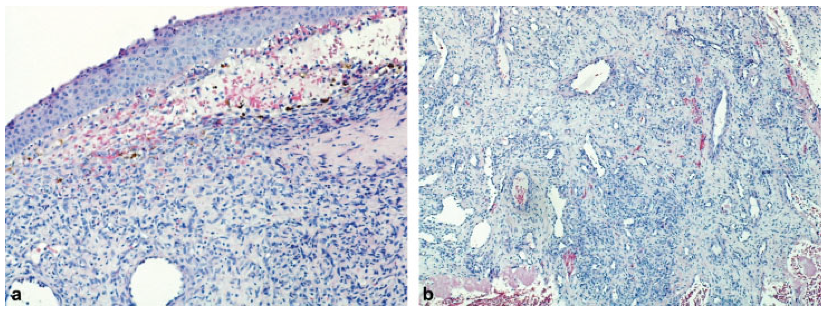
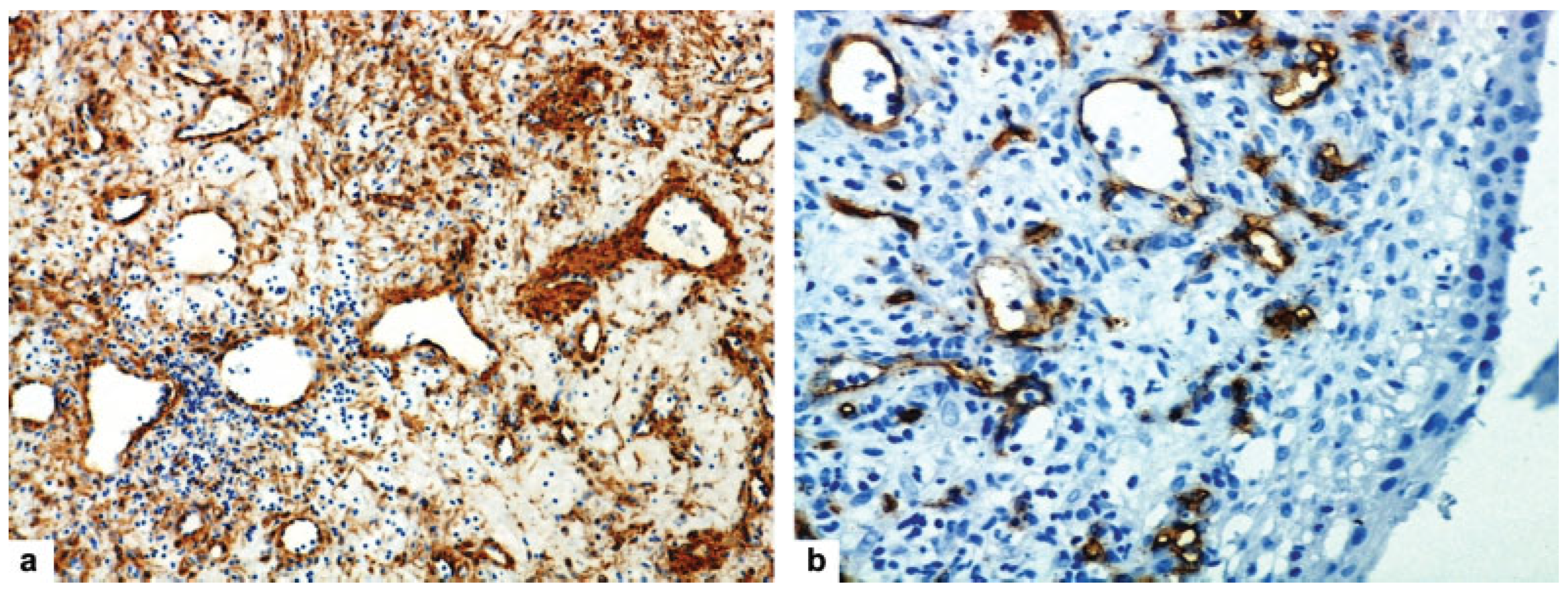
Histopathological examination of the excised mass (hematoxylin–eosin stain) showed a metaplastic squamous epithelium with the respiratory epithelium remnants on the tumor surface (Figure 2a). Under the epithelium, we found that many irregular blood vessels range from capillaries and sinusoids to large vessels, lined with one layer of tat endothelial cells lying in a fibrous stroma (Figure 2b). Several fields of proliferate fibrosis were seen. This suggested the presence of angiofibroma. However, the diagnosis of angiofibroma was confirmed by immunohistochemical analysis. The avidin–biotin peroxidase technique was performed. The finding was positive for αactin. In thick smooth muscle layers, the orientation of muscle cells is frequently disturbed. Individual muscle cells frequently differ in size and shape. The musclelayers disperse peripherally into individual cells and create the impression of vessel-independent smooth muscle cells within the stroma (Figure 3a). All vascular cells were positive for “cluster of differentiation 34” (CD34; Figure 3b).
Four years later, the patient was free of symptoms and there was no rhinoscopic, endoscopic, and CT evidence of recurrence.

Discussion

Many hypotheses have been proposed to explain the etiology of angiofibroma, but none of them found general acceptance. Some studies with findings of chromosome 17 deletions, including regions of thesuppressor gene p53 and the oncogene Her-2/Neu, suggest that genetic factors may be involved in the origin of angiofibroma.[19,20] Developmental theories suggest angiofibroma to be a vascular malformation deriving from an incomplete regression of a primitive artery of the first branchial arch.[21] This hypothesis is supported by histological finding of high irregularity of the smooth muscle layers, focal lack of pericytes, and discontinuous vascular basal lamina.[21] Some authors speculated angiofibroma to be a testosteronedependent tumor originating from a fibrovascular nidus in the nasopharynx inactive until the onset of puberty.[22] This explanation could be accepted in cases of angiofibroma in male patients and in postmenopausal women, because hormonal hypothesis suggests that high estrogen levels protect females. However, this hypothesis does not explain the presence of angiofibroma in female in reproductive period. Another hypothesis suggests that angiofibroma originates from the periosteum of the anterior margin of the atlas at the lower surface of the sphenoid bone, which is called fascia basalis.[2] During its development, this tissue extends up to the posterior part of the vomer and the ethmoid bone, including the lamina perpendicularis and middle turbinate. Akbas and Anadolu [15] suggested that extension of fascia basalis anteriorly, as a developmental anomaly, results in formation of small islands of ectopic tissue which could be the sites of origin of angiofibroma of the nasal septum.
Clinical characteristics of nasal septal ENA are different from that of JNA. Nasal bleeding in patients with septal ENA, preoperatively and during the surgical excision of the tumor, is less intense.[1,3,4,5,6,7,8,9,10,11,12,13,14,15,16,17,18] The other symptoms of JNA, including headache, facial pain, Eustachian tube dysfunction with secondary otitis media, hyposmia, swelling of the cheek, etc, are rarely seen in patients with septal ENA.[1,2] In contrast to other tumors, diagnosis of angiofibroma is not based on biopsy. The most important diagnostic procedures are CT and MRI with intravenously administered contrast. While JNA is associated with a strong and homogenous enhancement on CT and MRI, contrast enhancement is less and inhomogeneous in most patients with ENA. Accordingly, Tasca and Compadretti suggested that DSA and preoperative embolization in patients with septal ENA may be needed only if high vascularity is suspected by CT and MRI.[16]
Differential diagnosis in relation to ENA of the nasal septum is not so simple, because unilateral nasal obstruction with or without nasal bleeding, associated with the presence of pinkish or reddish nasal mass, may also be caused by many different nasal entities, including angiomatous choanal polyp (ACP), capillary hemangioma, and hemangiopericytoma. ACP, a rare subclass ofchoanalpolyps, originates usuallyfromantrochoanal polyp after the occlusion of stromal vessels, infarction, and neovascularization.[23] A contrast-enhanced CT scan of the paranasal sinuses with angiography should be used to differentiate angiofibroma from ACP. On angiography, ACP has only a few demonstrable feeding vessels compared with the rich irregular vascular supply of an angiofibroma. So, preoperative embolization is not necessary for management of ACPs.[23] The histopathological examination of ACP demonstrates a collagenous matrix with many intammatory cells, without the dilated vascular channels.[23] On the other hand, a dense fibrous stroma with spindled or stellate cells and variably sized, thin-walled vascular spaces are main histological characteristics of angiofibroma. In contrast to angiofibroma, capillary hemangioma shows the lobulation and not excessive stromal fibrosis. Also, anastomosing vessel architecture, which is very specific for capillary hemangioma, is not found in angiofibroma.[24] Immunohistochemical analysis may bevery helpful for differentiation between angiofibroma and sinonasal-type hemangiopericytoma.[25] Sinonasal-type hemangiopericytoma is a rare vascular tumor, arising from pericytes surrounding capillaries, situated usually in the nasal cavity and paranasal sinuses region, originating in the most cases from nasal septal mucosa. An immunohistochemical analysis of this lesion shows positive staining for vimentin, and negative staining for CD34 and smooth muscle α-actin.[26] Angiofibromas have positive staining for vimentin, CD34, and α-actin. Then, sinonasal-type hemangiopericytoma can be considered to be distinct from angiofibroma of the nasal cavity.
The main treatment modality for septal ENA is simple surgical excision of the lesion with cauterization of the pedicle. In our case, due to the experience that dimension of lesion frequently is not in correlation with intensity of bleeding, we decided to perform preoperative DSA and embolization of sphenopalatine artery. In our previous case, we surgically treated a postmenopausal woman with ENA originating from the left middle turbinate.[27] Despite small dimension of the tumor (length: 15 mm, diameter: 9 mm), excision of the lesion was followed by profuse bleeding and the blood loss was estimated at 300 mL.[27] After the removal of septal angiofibroma, several millimeters of mucosa around the pedicle attachment should be elevated to the septal cartilage or bone and removed, due to the evidence that lesion involves the perichondrium or periosteum. In contrast to JNA with recurrence rate of approximately 25%, the recurrence rate for ENA is only 2.3%.[1] However, in review of the literature, we found no patients with septal ENA with recurrence after the surgical treatment.

Conclusion

Angiofibromas originating from the nasal septum are clinically distinct from nasopharyngeal angiofibromas and can therefore be misdiagnosed. Nasal septal angiofibromas should be included in the differential diagnosis of vascular lesions of the nasal/paranasal sinuses area, especially from ACPs, capillary hemangiomas, and sinonasal-type hemangiopericytoma. Immunohistochemical analysis is important in all doubtful cases, especially in those with atypical location and may be very helpful in distinction from nasal hemangiopericytomas. Surgical excision of the mass with cauterization of the tumor pedicle and removal of the mucosa and perichondrium/periosteum several millimeters around the pedicle attachment is the treatment of choice for ENA of the nasal septum.

Previous Presentation

This article was presented at the Third Symposium of Rhinology and Allergy Association of Serbia, Belgrade, Serbia, March 16, 2018.

Funding

None declared.

Conflicts of Interest

No contict of interest was declared by the authors.

References

  1. Windfuhr, J.P.; Vent, J. Extranasopharyngeal angiofibroma revisited. Clin. Otolaryngol. 2018, 43, 199–222. [Google Scholar] [CrossRef] [PubMed]
  2. Windfuhr, J.P.; Remmert, S. Extranasopharyngeal angiofibroma: Etiology, incidence and management. Acta Otolaryngol. 2004, 124, 880–889. [Google Scholar] [CrossRef]
  3. Hiraide, F.; Matsubara, H. Juvenile nasal angiofibroma: A case report. Arch. Otorhinolaryngol. 1984, 239, 235–241. [Google Scholar] [CrossRef]
  4. Sarpa, J.R.; Novelly, N.J. Extranasopharyngeal angiofibroma. Otolaryngol. Head Neck Surg. 1989, 101, 693–697. [Google Scholar] [CrossRef]
  5. Handa, K.K.; Kumar, A.; Singh, M.K.; Chhabra, A.H. Extranasopharyngeal angiofibroma arising from the nasal septum. Int. J. Pediatr. Otorhinolaryngol. 2001, 58, 163–166. [Google Scholar] [CrossRef] [PubMed]
  6. Somdas, M.A.; Ketenci, I.; Unlu, Y.; Canoz, O.; Guney, E. Extranasopharyngeal angiofibroma originating from the nasal septum. Otolaryngol. Head. Neck Surg. 2005, 133, 647. [Google Scholar] [CrossRef] [PubMed]
  7. Castillo, M.P.; Timmons, C.F.; McClay, J.E. Autoamputation of an extranasopharyngeal angiofibroma arising from the nasal septum. Int. J. Pediatr. Otorhinolaryngol. Extra 2006, 1, 267–270. [Google Scholar] [CrossRef]
  8. Mohindra, S.; Grover, G.; Bal, A.K. Extranasopharyngeal angiofibroma of the nasal septum: A case report. Ear Nose Throat J. 2009, 88, E17–E19. [Google Scholar]
  9. Uyar, M.; Turanli, M.; Pak, I.; Bakir, S.; Osma, U. Extranasopharyngeal angiofibroma originating from the nasal septum: A case report. Kulak Burun Bogaz Ihtis. Derg. 2009, 19, 41–44. [Google Scholar]
  10. Hamdan, A.L.; Moukarbel, R.V.; Kattan, M.; Natout, M. Angiofibroma of the nasal septum. Middle East. J. Anaesthesiol. 2012, 21, 653–655. [Google Scholar]
  11. Garcia-Rodriguez, L.; Rudman, K.; Cogbill, C.H.; Loehrl, T.; Poetker, D.M. Nasal septal angiofibroma, a subclass of extranasopharyngeal angiofibroma. Am. J. Otolaryngol. 2012, 33, 473–476. [Google Scholar] [CrossRef] [PubMed]
  12. Correia, F.G.; Simões, J.C.; Mendes-Neto, J.A.; Seixas-Alves, M.T.; Gregório, L.C.; Kosugi, E.M. Extranasopharyngeal angiofibroma of the nasal septum–uncommon presentation of a rare disease. Rev. Bras. Otorrinolaringol. (Engl. Ed.) 2013, 79, 646. [Google Scholar] [CrossRef] [PubMed]
  13. Doğan, S.; Yazici, H.; Baygit, Y.; Metin, M.; Soy, F.K. Extranasopharyngeal angiofibroma of the nasal septum: A rare clinical entity. J. Craniofac, Surg. 2013, 24, e390–e393. [Google Scholar] [CrossRef]
  14. Atmaca, S.; Bayraktar, C.; Yıldız, L. Extranasopharyngeal angiofibroma of the posterior nasal septum: A rare clinical entity. Kulak Burun Bogaz Ihtis. Derg. 2013, 23, 295–298. [Google Scholar] [CrossRef] [PubMed]
  15. Akbas, Y.; Anadolu, Y. Extranasopharyngeal angiofibroma of the head and neck in women. Am. J. Otolaryngol. 2003, 24, 413–416. [Google Scholar] [CrossRef]
  16. Tasca, I.; Compadretti, G.C. Extranasopharyngeal angiofibroma of nasal septum. A controversial entity. Acta Otorhinolaryngol. Ital. 2008, 28, 312–314. [Google Scholar]
  17. Ewe, S.; Dayana, F.; Fadzilah, F.M.; Gendeh, B.S. Nasal septal angiofibroma in a post-menopausal woman: A rare entity. J. Clin. Diagn. Res. 2015, 9, MD03–MD05. [Google Scholar] [CrossRef]
  18. Singh, G.B.; Shukla, S.; Kumari, P.; Shukla, I. A rare case of extranasopharyngeal angiofibroma of the septum in a female child. J. Laryngol. Otol. 2018, 132, 184–187. [Google Scholar] [CrossRef]
  19. Barnes, L.; Brandwein, M.; Som, P.M. Diseases of the nasal cavity, paranasal sinuses and nasopharynx. In Surgical Pathology of the Head and Neck; Barnes, L., Ed.; Marcel Dekker: New York, NY, USA, 2001; pp. 440–516. [Google Scholar]
  20. Schick, B.; Veldung, B.; Wemmert, S.; et al. p53 and Her-2/neu in juvenile angiofibromas. Oncol. Rep. 2005, 13, 453–457. [Google Scholar] [CrossRef]
  21. Beham, A.; Beham-Schmid, C.; Regauer, S.; Auböck, L.; Stammberger, H. Nasopharyngeal angiofibroma: True neoplasm or vascular malformation? Adv. Anat. Pathol. 2000, 7, 36–46. [Google Scholar] [CrossRef]
  22. Schiff, M. Juvenile nasopharyngeal angiofibroma: A theory of pathogenesis. Laryngoscope 1959, 69, 981–1016. [Google Scholar] [CrossRef]
  23. Perić, A.; Jovanovski, A.; Vukomanović Đurđević, B. Giant angiomatous choanal polyp originating from the middle turbinate: A case report. ENT Updates 2017, 7, 53–56. [Google Scholar] [CrossRef]
  24. Pilch, B.Z. Soft tissue pathology of the head and neck. In Head and Neck Surgical Pathology; Keel, S.B., Rosenberg, A.E., Eds.; Lippincott Williams & Wilkins: Philadelphia, PA, USA, 2001; pp. 389–430. [Google Scholar]
  25. Perić, A.; Sotirović, J.; Cerović, S.; Zivić, L. Immunohistochemistry in diagnosis of extranasopharyngeal angiofibroma originating from nasal cavity: Case presentation and review of the literature. Acta Med. (Hradec Kralove) 2013, 56, 133–141. [Google Scholar] [CrossRef] [PubMed]
  26. Yokoi, H.; Arakawa, A.; Kuribayashi, K.; Inoshita, A.; Haruyama, T.; Ikeda, K. An immunohistochemical study of sinonasal hemangiopericytoma. Auris Nasus Larynx 2011, 38, 743–746. [Google Scholar] [CrossRef]
  27. Perić, A.; Baletić, N.; Cerović, S.; Vukomanović-Durdević, B. Middle turbinate angiofibroma in an elderly woman. Vojnosanit. Pregl. 2009, 66, 583–586. [Google Scholar] [CrossRef] [PubMed]
Figure 1. Coronal (a), axial (b), and sagittal (c) plane of computed tomography scan of the paranasal sinuses showing a mild contrast-enhancing mass with soft-tissue density that completely fills the anterior part of the left-sided nasal cavity, as well as the anterior parts of the left middle and inferior nasal meatus. The dimension of the mass was 40.2 × 39.5 × 25.6 mm. There was no radiological evidence of bony erosion or extension to the paranasal sinuses and we also found no opacities of the paranasal sinuses mucosa.
Cmtr 12 00020 g001
Figure 2. (a) Histopathological examination of the excised mass showed a metaplastic squamous epithelium with the respiratory epithelium remnants on the tumor surface (hematoxylin and eosin staining, magnification ×100). (b) Under the epithelium, we found many irregular blood vessels range from capillaries and sinusoids to large vessels, lined with one layer of flat endothelial cells lying in a fibrous stroma. Several fields of proliferate fibrosis were seen (hematoxylin and eosin staining, magnification ×50).
Cmtr 12 00020 g002
Figure 3. (a) Immunohistochemical finding of excised mass was positive for α-actin. In thick smooth muscle layers, the orientation of muscle cells is frequently disturbed. Frequently, individual cells differ in size and shape. The muscle layers disperse peripherally into individual cells and create the impression of vessel-independent smooth muscle cells within the stroma (avidin–biotin peroxidase technique, magnification ×100). (b) By immunohistochemistry, we found in the excised tumor that all vascular cells were positive for CD34 (avidin–biotin peroxidase technique, magnification ×200).
Cmtr 12 00020 g003
Disclaimer/Publisher’s Note: The statements, opinions and data contained in all publications are solely those of the individual author(s) and contributor(s) and not of MDPI and/or the editor(s). MDPI and/or the editor(s) disclaim responsibility for any injury to people or property resulting from any ideas, methods, instructions or products referred to in the content.