Next Article in Journal
Comparison of Imaging Softwares for Upper Airway Evaluation: Preliminary Study
Previous Article in Journal
Two- Versus Three-Point Internal Fixation of Displaced Zygomaticomaxillary Complex Fractures
 
 
Craniomaxillofacial Trauma & Reconstruction is published by MDPI from Volume 18 Issue 1 (2025). Previous articles were published by another publisher in Open Access under a CC-BY (or CC-BY-NC-ND) licence, and they are hosted by MDPI on mdpi.com as a courtesy and upon agreement with Sage.
Article

Analysis of the Changing Patterns of Midface Fractures Using 3D Computed Tomography: An Observational Study

by
Preeti Satish
*,
Kavitha Prasad
,
R. M. Lalitha
,
Krishnappa Ranganath
and
Parimala Sagar
Department of Oral and Maxillofacial Surgery, Faculty of Dental Sciences, MS Ramaiah University of Applied Sciences, Room No. 10, MSRIT Nagar, New Bel Road, Bangalore, Karnataka 560054, India
*
Author to whom correspondence should be addressed.
Craniomaxillofac. Trauma Reconstr. 2018, 11(4), 265-272; https://doi.org/10.1055/s-0037-1606250
Submission received: 14 March 2017 / Revised: 1 April 2017 / Accepted: 27 May 2017 / Published: 25 August 2017

Abstract

:
This article aims to analyze the changing Le Fort fracture patterns using computed tomography (CT) scans with three-dimensional (3D) reconstruction. A prospective observational study was conducted on 60 patients with midface trauma, who had reported to MS Ramaiah Group of Hospitals, Bangalore, between January 2015 and October 2016. CT scans using 1.6 mm axial, sagittal, coronal sections were taken and their 3D reconstruction was made. The images were studied and compared with the standard Le Fort lines. The deviations from the classical Le Fort lines were analyzed and recorded. A note was also made of any additional fixation that was required for these deviations. Descriptive analysis was done and the results expressed in numbers and percentages. Study revealed that the most common cause for the midface fractures was found to be road traffic accidents (81.7%) with a male preponderance (88.3%) and peak incidence in 21 to 30 years of age (40%). Among the 60 patients, 18 (30%) patients had fracture patterns similar to the ideal Le Fort lines, 4 (6.6%) had a combination of Le Fort patterns, and 38 (66.3%) patients had deviations seen from the ideal Le Fort lines. Four types of deviations were recorded, namely, D1(60%), D2(5.4%), D3(10.9%), and D4(23.6%). It was observed that D1 and D3 required additional fixation. Majority of the cases presented as a deviation from ideal Le Fort fractures. CT was a valuable tool in the assessment of these fracture patterns. Deviations, if any, could be better analyzed using the 3D reconstruction images. Proper diagnosis and detection of these deviations make the planning for fixation easier. Repetition of these deviations could propose a newer or modified classification system for Le Fort fractures.

In 1901, Rene Le Fort conducted experimental studies on 35 cadavers and proposed the three great lines of weaknesses, popularly known as Le Fort fracture lines.[1] Although the etiology for midface trauma remains the same as proposed by Rene Le Fort, what has changed over the years is the mass and velocity of the wounding object.[2,3] This change has brought about a change in the fracture patterns that are being encountered. The severity and pattern of the fracture depends on the magnitude of the causative force, impact duration, acceleration imparted by it to the part of the body struck, and the rate of acceleration change.[4]
Recently, radiologists and surgeons often have observed varied patterns of facial fractures, which do not coincide with the ideal Le Fort patterns. Comprehensive and extended classifications and biomechanics have also been proposed in literature[5,6,7] to involve these patterns of facial injuries; however, variations in fracture patterns are being encountered in clinical practice.
Therefore, a systematic descriptive study of the Le Fort fracture patterns using computed tomography (CT) scans with three-dimensional (3D) reconstruction was conducted.

Subjects and Method

After obtaining ethics clearance from the institutional ethics committee, a prospective observational study was conducted on patients who reported to Accident and Emergency/Casualty, MS Ramaiah Group of Hospitals, Bangalore, during the period January 2015 to October 2016. CT scans with 3D reconstruction of the facial bones was done as a preliminary investigation. A thorough head to toe examination was done in these patients to identify all the associated injuries. Informed consent was taken from the patients. Patients diagnosed with midface fractures clinically and radiographically with or without associated injuries were included in the study. Patients with isolated maxillary dentoalveolar or nasal bone fracture, mandibular fractures, malunited fractures, and old fractures were excluded from the study.
The CT scans were done using Toshiba Asteion Single Slice CT machine and Siemens Somatom Perspective (128-slice CT scanner). The images obtained were studied using the software Syno-Via DICOM Viewer and RadiAnt DICOM Viewer. The CT scans with 3D reconstruction were analyzed by a radiologist and two oral and maxillofacial surgeons. The fracture patterns were then compared with the classical Le Fort fracture lines to determine deviation, if any, from the classical Le Fort fracture pattern. The patients were categorized based on the fracture patterns:
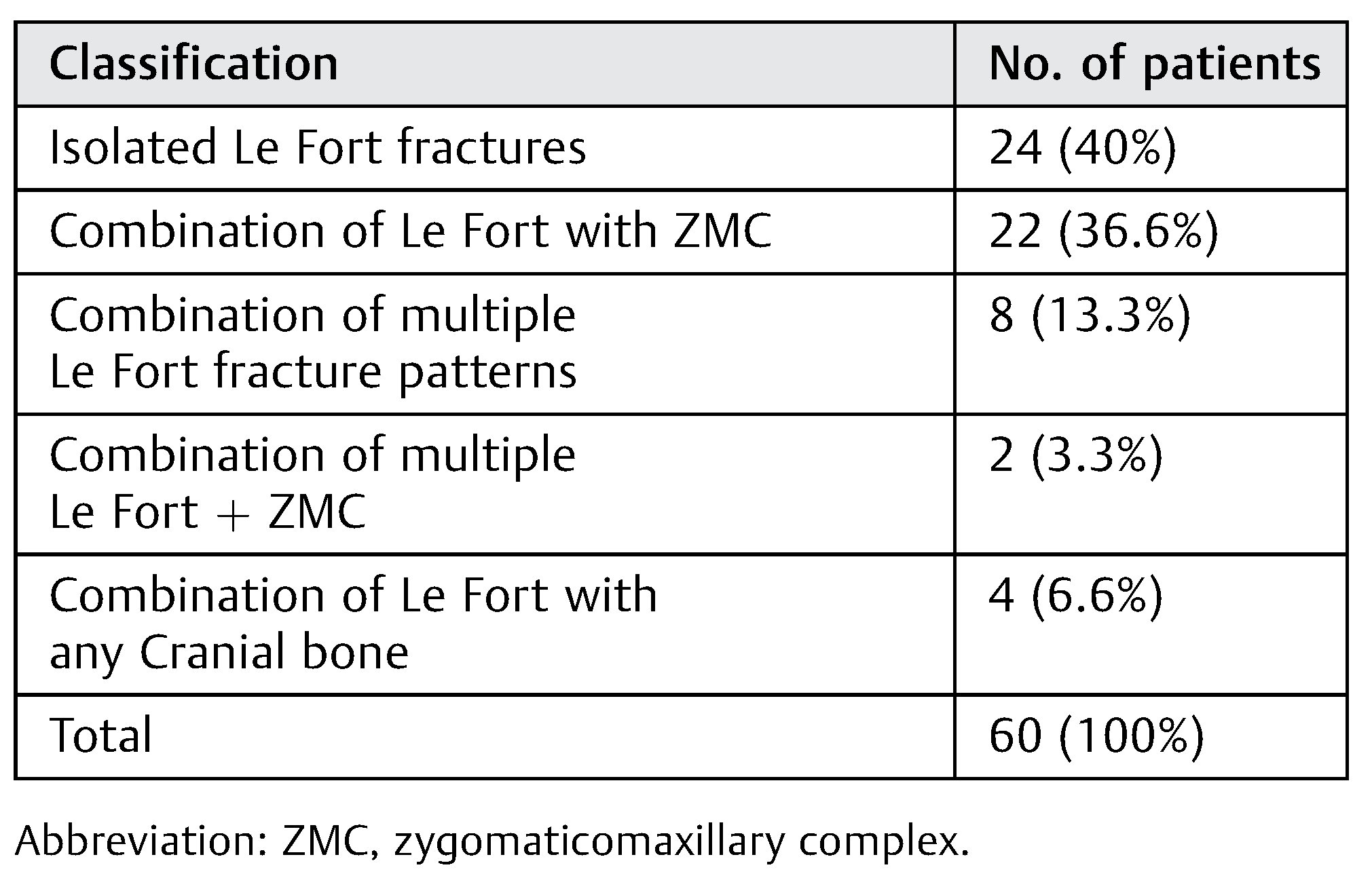
  • Similar to Le Fort fracture patterns.
  • Combination of Le Fort fracture patterns.
  • Deviation from Le Fort fracture patterns.
The deviations were analyzed and categorized accordingly. The deviations from Le Fort fracture patterns requiring additional fixation were also recorded. The statistical software SPSS version 20.0 was used to calculate descriptive data. The results were expressed in percentages and graphs.

Results

Demographical data were recorded in terms of age, gender, etiology, type of automobile, type of helmet, alcohol history, and treatment given. The distribution of patients according to age and gender is shown in Figure 1.
The most common etiology was road traffic accidents (81.7%) followed by assault (6.7%) and falls (5%). In the road traffic accidents, two wheelers (60%) were most commonly involved with non-helmeted drivers under the influence of alcohol (46.7%).
A total of 60 patients with midface fractures were analyzed by subjecting them to CT and obtaining sectional and 3D-reconstructed images. Among the 60 patients, a total of 129 fractures of facial bones were seen. The isolated and combinations of the facial bone fractures have been mentioned in Table 1. Distribution of fractures according to classification of facial fractures is given in Table 2. The CT scans and 3D images were used to analyze the fracture pattern and compare them to the classical Le Fort fracture lines and were classified accordingly (Table 3).
Majority of the cases (63.3%) were deviations with the Le Fort patterns. Among them, five types of deviations were the most predominant. These deviations were categorized as D1, D2, D3, D4, and D5:
D1:
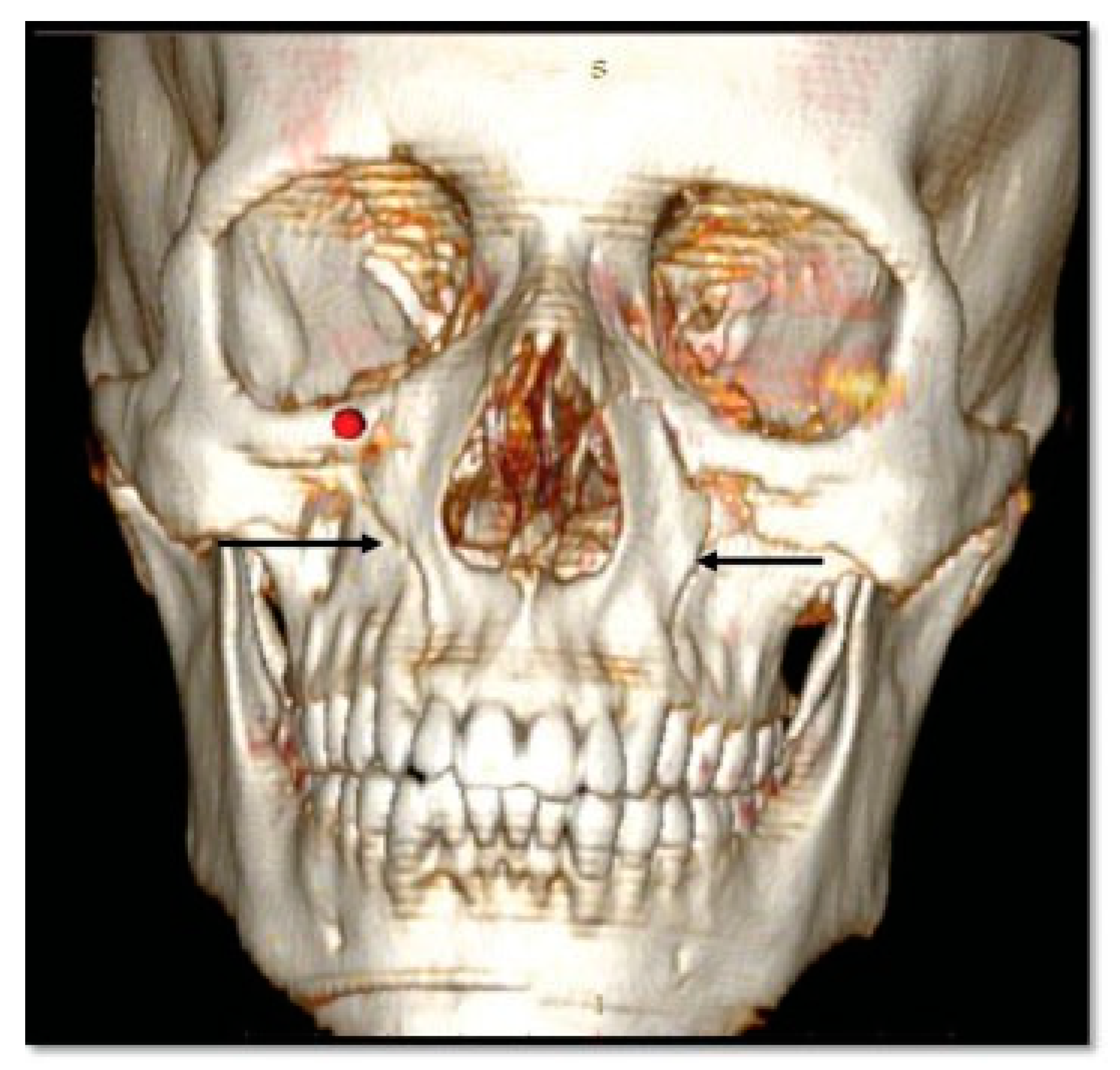
Additional line extending from the pyriform aperture to the infraorbital rim of the same side (60%).
D2:
Additional line extending from the infraorbital rim to the Le Fort fracture line.
D3:
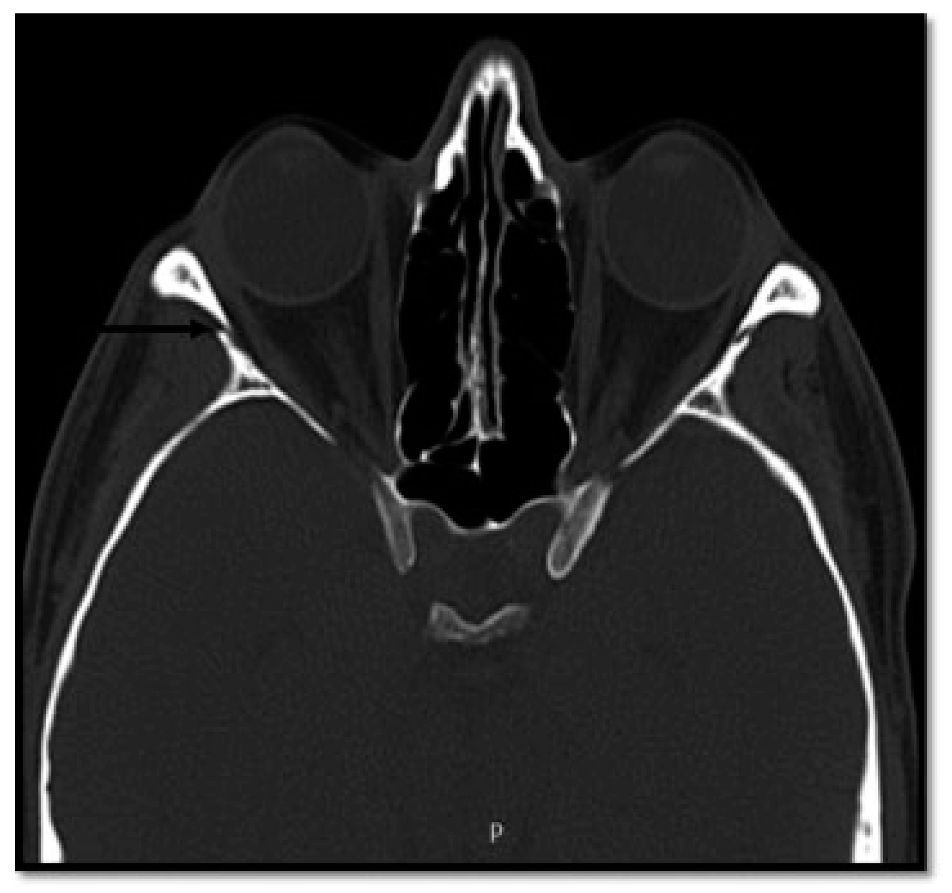
Additional line extending from the pyriform aperture to the lateral wall of orbit, without breaking the infraorbital rim of the same side (5.4%).
D4:
Additional line extending from the infraorbital rim through the canine fossa to terminate as a fractured dentoalveolar segment of the same side, involving the fractured segment (10.9%).
D5:
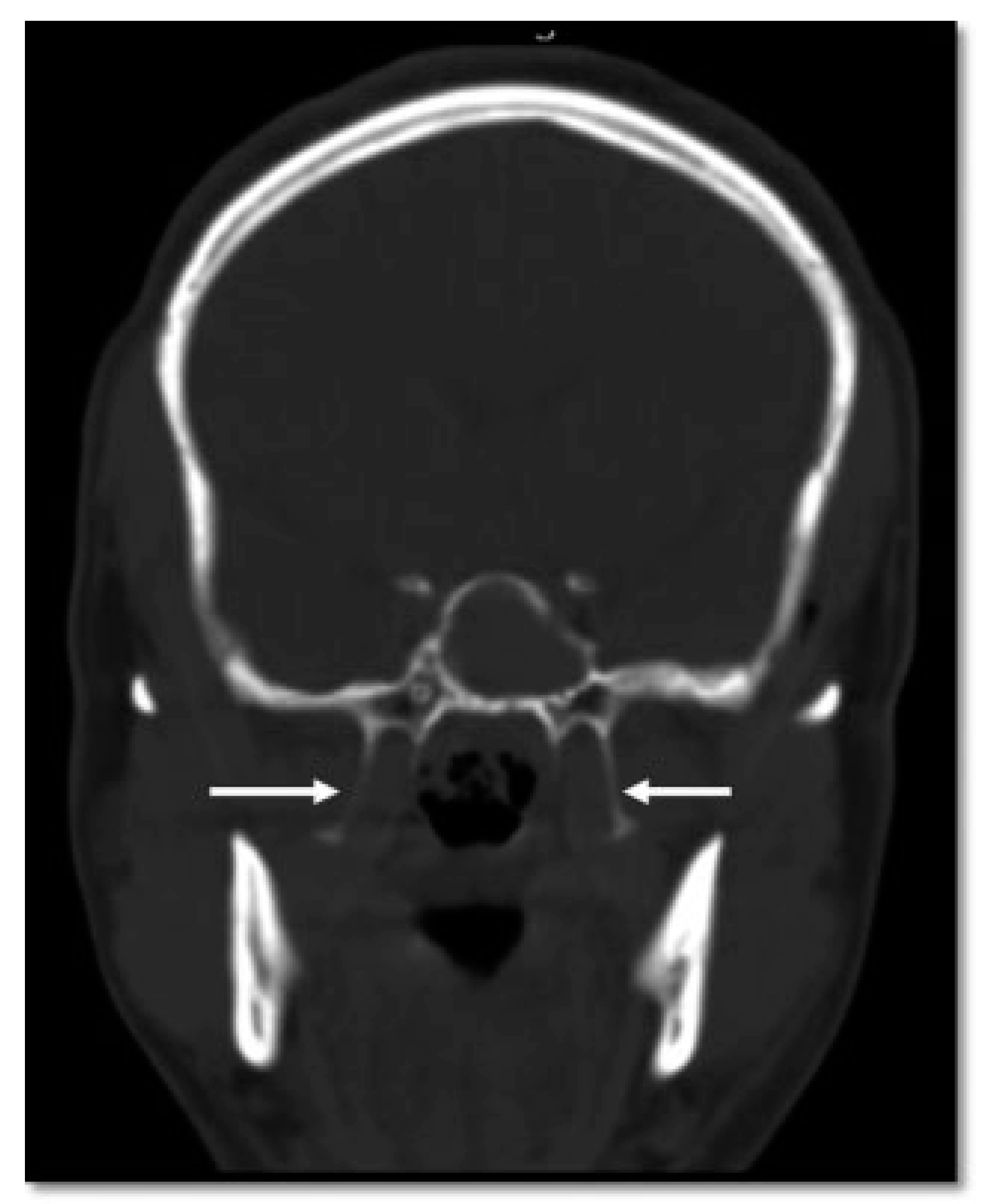
Pterygoid plates are not fractured (23.6%).
The distribution of these deviations according to site is exhibited in Table 4.
A total of 55 deviations were observed in the 33 cases which showed deviation from classical Le Fort lines. Some cases exhibited a combination of these deviations, either on the same side or on the opposite side. Fourteen cases had a combination of multiple deviations, among which bilateral D1 deviation predominated in 42.8% (n = 6) of cases; followed by bilateral D5 in 2 cases; D1 + D5, D1 + D3 + D5 in 2 (14.2%) cases each; and D3 + D4, D2 + D4 in 1 case (7.1%) each, respectively.
It was seen that D1, D2, and D4 deviations required additional fixation for better stability. The details of the fixation have been dealt in “Discussion” section (Figure 2, Figure 3, Figure 4, Figure 5, Figure 6 and Figure 7).

Discussion

Today, the majority of the fractures that occur rarely correlate to the classical Le Fort fracture lines. This could be attributed to the change in the mass and the velocity of the wounding object. In motor vehicle accidents, three types of collision can occur. First, when the victim is static and the wounding object is moving, second when the victim is moving and the wounding agent is static, and third when both are moving at varying velocities.[8] The third type of collision leads to comminuted fractures.
Our study showed a male:female ratio of 8:1 which was found to be slightly lower than the findings that were observed by Kadkhodaie[9] (10:1), Al Ahmed et al[10] (10.5:1), Patil et al[11] (11.5:1), and Hächl et al[12] (11.8:1). Majority (40%) of the patients were in the 21- to 30-year-old age group; the studies done by Maladière et al,[13] Ajike et al,[14] and Subhashraj et al[15] resonated with similar findings.
The most common etiology was road traffic accidents in 81.7% (n = 50), followed by assault in 6.7% (n = 5), and falls in 5.0% (n = 3) of the cases. Among the road traffic accidents, two-wheeler accidents were the maximum (60%), followed by four-wheeler accidents (20%) and pedestrian–vehicle accidents (20%). These findings were similar to studies done by Al Ahmed et al10 and Toivari et al[16] that were done on demographics of facial fractures. On the contrary, a study conducted by Olasoji et al[17] on the changing picture of facial fractures in Nigerian population found interpersonal violence to be the major etiological factor.
Of the 36 patients with history of two-wheeler accidents, 5% (n = 3) of the patients were wearing full-face helmets, 46.7% (n = 28) were without helmets, and 10% (n = 6) with half helmets. Our study showed that the non-helmeted motorcyclists were more than nine times likely and half helmeted motorcyclists almost two times more likely to sustain facial injuries when compared with the full-helmeted motorcyclists. This was in accordance with studies by Cook et al,[18] Yu et al,[19] and Tsai et al.[20]
Among the 60 patients, 45% (n = 27) of the patients were under the influence of alcohol and 55% (n = 33) were not under the influence of alcohol. Hence, “a male patient in his third decade of life, riding a twowheeler without helmet under the influence of alcohol” is most likely to sustain midface fractures.
All the 60 patients underwent radiographic imaging. CT scans were mandatorily done as a primary investigation for all the patients. The CT was done in all the three planes, that is, axial, coronal, and sagittal with 1.6-mm slice thickness. These slices were then used for 3D image reconstruction. This choice of slice thickness was decided after thorough literature search[11,21,22,23] according to which better visualization of the facial fractures would be with each slice thickness ranging from 1 to 2.4 mm and slice distance of less than 1.5 mm. However, according to Bernhardt et al,[24] slice thickness of 0.4 to 0.5 mm would be ideal for the radiographic diagnosis of facial fractures. The images obtained were evaluated and compared with the standard Le Fort fracture lines.
We observed that even though 3D images were very helpful in providing an exact picture of the course of the fracture lines, it alone was not sufficient for the diagnosis of the fracture pattern. 2D images, however, provided excellent information of every minute fracture line. Soft-tissue injuries such as fat or muscle entrapment and hematoma could also be appreciated in 2D views as opposed to the 3D view. Our experience with the imaging modalities resonated with other studies[11,21,22,25] which used CT for the assessment of facial fractures. Le Fort I fractures were best studied in the coronal cuts, Le Fort II in the axial and coronal cuts, and Le Fort III in the axial and sagittal cuts.[22] But, according to Daffner[23] Le Fort I fractures were to be assessed using the coronal cuts, Le Fort II using coronal and axial, and Le Fort III using axial cuts. The results of our assessment of the fracture lines resonated with findings of Daffner et al.
Although the Le Fort classification was given 100 years ago, till date it is considered as the gold standard in classifying midface fractures. However, in recent times, classical Le Fort fractures are rarely being encountered, with most of the fractures being a permutation and combination of the Le Fort lines. Also, the facial skeletal components articulate and interdigitate in a complex fashion, and it is rare to find an isolated facial bone fracture without the disruption of its neighbor. This finding was in accordance with the large retrospective observational studies done by Shankar et al[26] and Salentijn.[27]
In our study, it was observed that in only 22 cases the ideal Le Fort fracture lines were followed. Surprisingly, majority of the cases (n = 38) showed deviation from Le Fort pattern. These deviations were additional fracture lines which occurred either in isolation or in association with Le Fort fractures. The presence of this additional line altered the treatment plan, as in many of the cases it required additional fixation. Hence, diagnosis of these additional deviations should be done carefully, by studying the sectional as well as 3D-reconstructed images so as to classify and treat them accordingly. The deviations encountered were classified as D1 (n = 19), D2 (n = 14), D3 (n = 3), D4 (n = 6), and D5 (n = 13).
This finding resonated with other studies on different fracture patterns.6,11 However, the deviations that were encountered in our study have not been mentioned in any literature till now. Doi et al[6] did find two deviated patterns which were quite similar to the deviations that we found. Patil et al[11] discussed a set of cases, which showed deviated patterns, but no comment on the appearance or prevalence of any pattern was made.
The deviations were then compared and confirmed intraoperatively. We assessed the need for additional fixations when these deviations were present. The point of fixation of the fractures depends on the accurate assessment and the degree of instability of the fractures.

Case Discussions

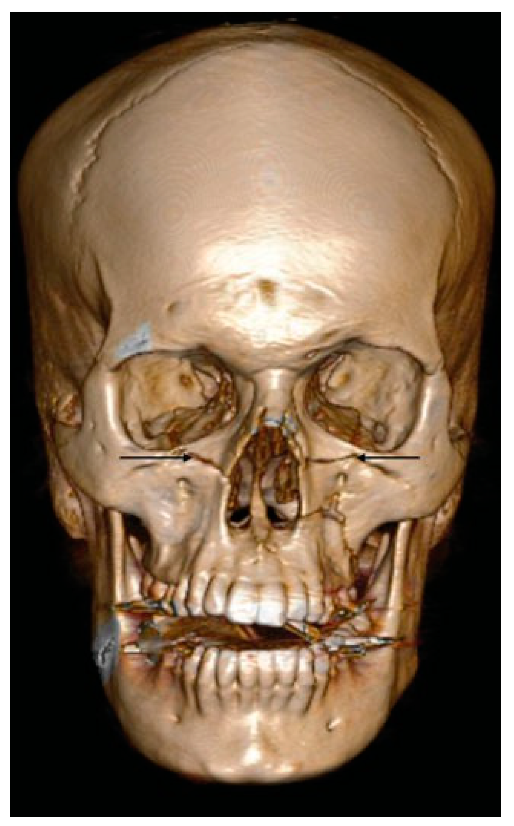
  • D1: Additional Line Extending from the Pyriform Aperture to the Infraorbital Rim of the Same Side (34.5%)
The D1 type of deviation was seen to occur either in isolation or in association with Le Fort I or comminuted Le Fort II fractures (Figure 2, Figure 3 and Figure 4).
When this type of deviation occurred in association with Le Fort I fracture, then an additional fixation had to be done at the infraorbital rim. However, when this deviation was associated with Le Fort II fracture, an additional fixation was done at the pyriform rim. In all our cases of D1 deviation, fixation of the additional line was done (100%).
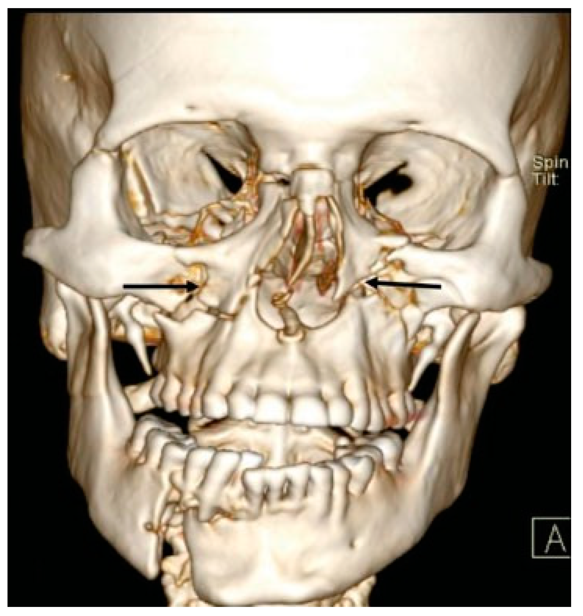
  • D2: Additional Line Extending from Infraorbital Rim to the Le Fort Fracture Line (25.4%)
D2 deviation was most frequently seen to be associated with Le Fort I and high level Le Fort I fractures (Figure 5 and Figure 6).
In these cases, an additional fixation had to be done at the infraorbital rim. In all our cases of D2 deviation, fixation of the additional line was done (100%).
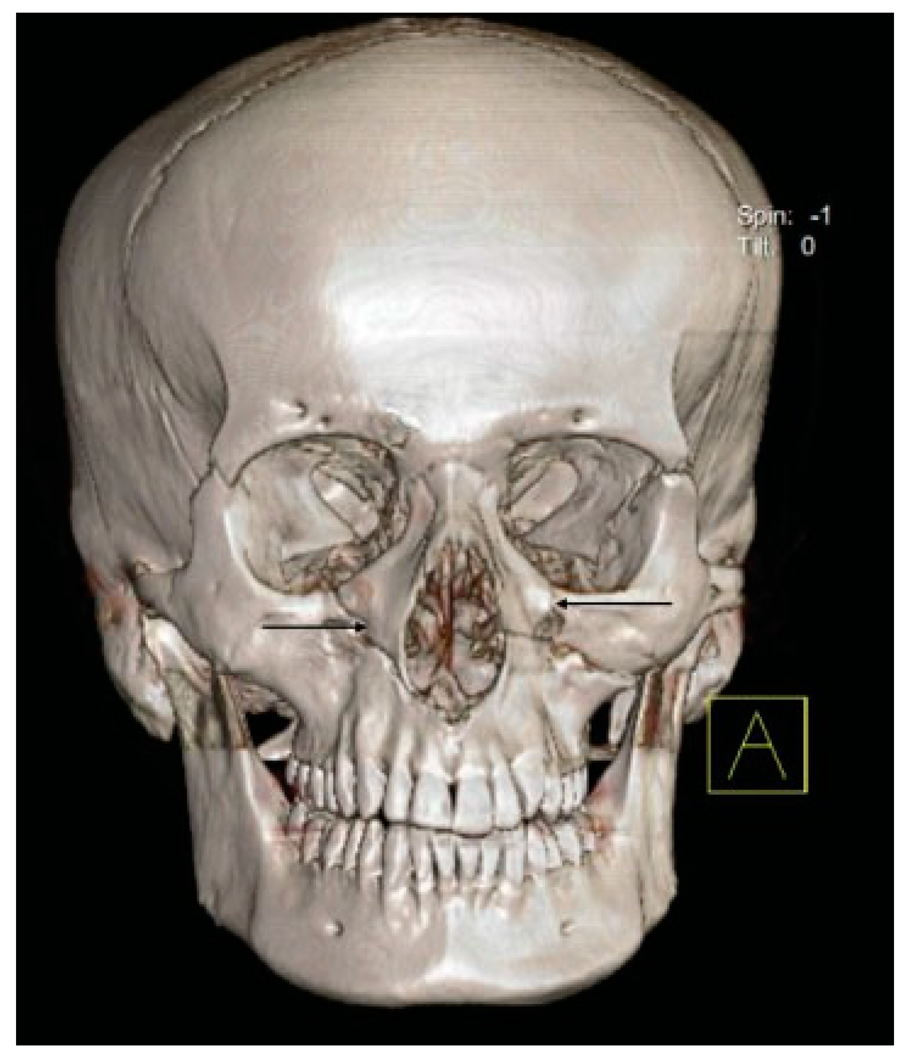
  • D3: Additional Line Extending from Pyriform Fossa to Lateral Wall of Orbit, without the Fracture of the Infraorbital Rim of the Same Side (5.4%)
This additional line was usually seen to be associated with Le Fort I fractures. It was not seen to occur in isolation (Figure 7, Figure 8 and Figure 9).
In these cases, it was observed that after reduction and fixation at the pyriform rim, good stability was achieved. In our study, two cases did not require fixation at the frontozygomatic (FZ) region/lateral border of the eye. Only in one case, fixation was done at the FZ, as the fracture at the FZ was comminuted.
  • D4: Additional Line Running from the Infraorbital Rim through the Canine Fossa to Terminate as a Fractured Dentoalveolar Segment of the Same Side (10.9%)
D4 deviation is seen to be associated with Le Fort I, II, and III fractures (Figure 10 and Figure 11).
Dentoalveolar fractures do occur in isolation, but many a times they are seen to be associated with the Le Fort fracture line and thus being a deviation of Le Fort pattern. The dentoalveolar segment fracture is in continuity with the Le Fort fracture line and hence required additional fixation along with the Le Fort fracture.
In this type of deviation, an additional fixation of the fracture line was done just above the apices of the teeth, stabilizing the dentoalveolar split. This was then followed by splinting of the teeth in the fractured dentoalveolar segment to the stable segment of the maxilla. In all the six cases, additional fixation was done using either a four- or two-holed 2.0-mm miniplate. In all the cases, splinting of the teeth was done, except in one case in which the teeth in the segment were avulsed.
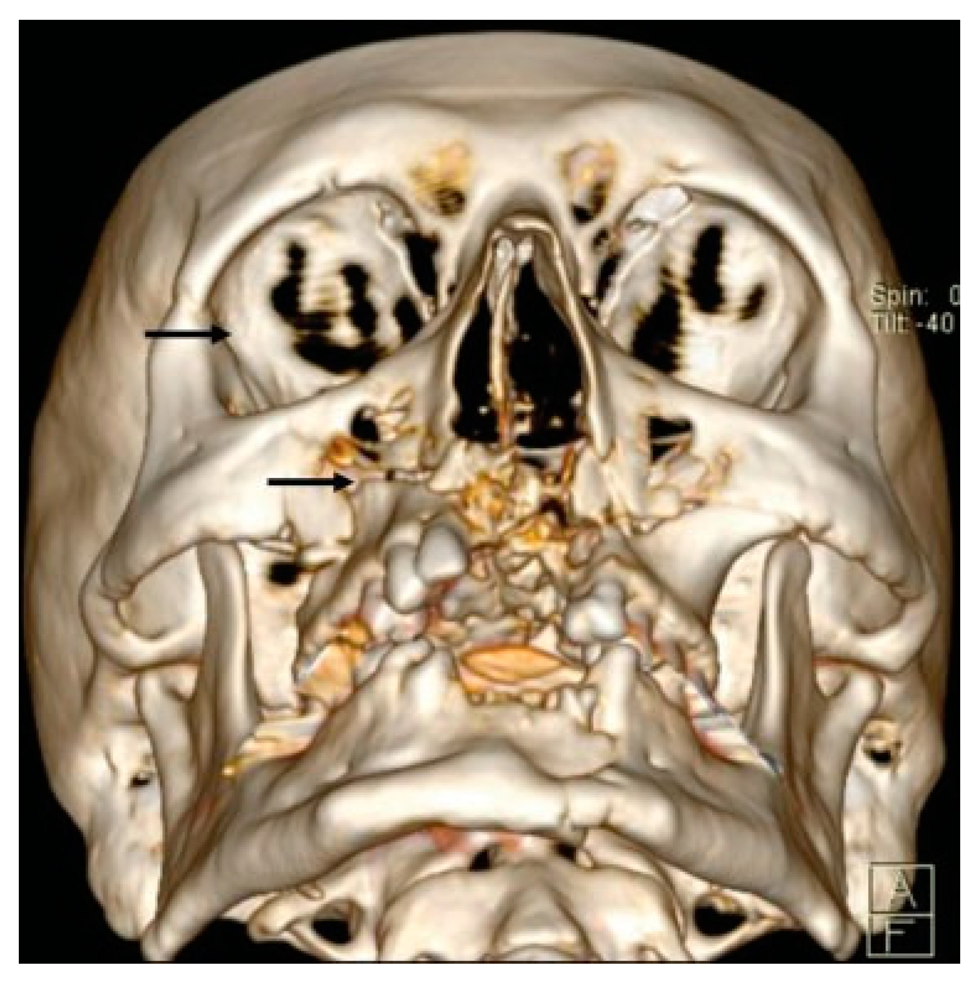
  • D5: Pterygoid Plates Are Not Fractured (23.6%)
Fracture of the pterygoid plates causing pterygomaxillary dysjunction is the main characteristic feature of a Le Fort fracture. However, in our study, we observed Le Fort fractures mimicking the ideal Le Fort lines, but the pterygoid plates were intact. Most of these cases were accompanied with a palatal split, which caused mobility of the maxilla giving the impression of a dysjunction, thus a deviation. No additional treatment was required for this deviation (Figure 12, Figure 13 and Figure 14).

Conclusion

A thorough clinical and radiological evaluation should be mandatory for all patients with facial fractures. CT serves as a valuable tool in the diagnosis of midface fractures; studying the sectional images of the scans along with 3D-reconstructed images is required for exact determination of fracture patterns. Deviations from classical Le Fort patterns are being encountered quite frequently mandating additional points of fixation, warranting for more studies and research in this field. A repetitive finding of similar deviations can prompt a modification in the classification of Le Fort frac tures. We acknowledge that the limitation of our study is the small sample size. Hence, more studies should be conducted to record and detect the changing patterns of Le Fort lines with a larger sample size and spanning over a longer duration of time.

Funding

The authors received no funding from any organization.

References

  1. Gartshore, L. A brief account of the life of René Le Fort. Br J Oral Maxillofac Surg 2010, 48, 173–175. [Google Scholar] [CrossRef] [PubMed]
  2. Noffze, M.J.; Tubbs, R.S. René Le Fort 1869-1951. Clin Anat 2011, 24, 278–281. [Google Scholar] [CrossRef] [PubMed]
  3. Rowe, N.L.; Williams, J.L.; Hobbs, J.A. Williams, J.L., Ed.; Etiology of injury. In Rowe and Williams Maxillofacial Injuries, 2nd ed.; Churchill Livingstone: New York, NY, USA, 1994; Volume 1, pp. 34–36. [Google Scholar]
  4. Simpson, D.A.; McLean, A.J. Mechanisms of injury. In Craniomaxillofacial Trauma; David, D.J., Simpson, D.A., Eds.; Churchill Livingstone: New York, NY, USA, 1995; Volume 101. [Google Scholar]
  5. Marciani, R.D.; Gonty, A.A. Principles of management of complex craniofacial trauma. J Oral Maxillofac Surg 1993, 51, 535–542. [Google Scholar] [CrossRef]
  6. Doi, Y.; Tomitsuka, S.; Asano, K.; Sakai, E.; Sasaki, T.; Imai, Y. A study on the classification of the maxillary fractures—new proposal for revised Le Fort classification. Int J Oral Maxillofac Surg 2005, 34, 159–160. [Google Scholar] [CrossRef]
  7. Zhang, Y.; Gu, X.M.; Cai, Y.J.; Duan, D.H. [A revised scheme for LeFort classification of upper jaw fractures]. Zhonghua Kou Qiang Yi Xue Za Zhi 2005, 40, 134–136. [Google Scholar]
  8. Roccia, F.; Servadio, F.; Gerbino, G. Maxillofacial fractures following airbag deployment. J Craniomaxillofac Surg 1999, 27, 335–338. [Google Scholar] [CrossRef]
  9. Kadkhodaie, M.H. Three-year review of facial fractures at a teaching hospital in northern Iran. Br J Oral Maxillofac Surg 2006, 44, 229–231. [Google Scholar] [CrossRef]
  10. Al Ahmed, H.E.; Jaber, M.A.; Abu Fanas, S.H.; Karas, M. The pattern of maxillofacial fractures in Sharjah, United Arab Emirates: a review of 230 cases. Oral Surg Oral Med Oral Pathol Oral Radiol Endod 2004, 98, 166–170. [Google Scholar] [CrossRef]
  11. Patil, R.S.; Kale, T.P.; Kotrashetti, S.M.; Baliga, S.D.; Prabhu, N.; Issrani, R. Assessment of changing patterns of Le fort fracture lines using computed tomography scan: an observational study. Acta Odontol Scand 2014, 72, 984–988. [Google Scholar] [CrossRef]
  12. Hächl, O.; Tuli, T.; Schwabegger, A.; Gassner, R. Maxillofacial trauma due to work-related accidents. Int J Oral Maxillofac Surg 2002, 31, 90–93. [Google Scholar] [CrossRef]
  13. Maladière, E.; Bado, F.; Meningaud, J.P.; Guilbert, F.; Bertrand, J.C. Aetiology and incidence of facial fractures sustained during sports: a prospective study of 140 patients. Int J Oral Maxillofac Surg 2001, 30, 291–295. [Google Scholar] [CrossRef] [PubMed]
  14. Ajike, S.O.; Adebayo, E.T.; Amanyiewe, E.U.; Ononiwu, C.N. An epidemiologic survey maxillofacial fractures and concomitant injuries in Kaduna, Nigeria. Niger J Surg Res 2005, 7, 251–255. [Google Scholar] [CrossRef]
  15. Subhashraj, K.; Nandakumar, N.; Ravindran, C. Review of maxillofacial injuries in Chennai, India: a study of 2748 cases. Br J Oral Maxillofac Surg 2007, 45, 637–639. [Google Scholar] [CrossRef] [PubMed]
  16. Toivari, M.; Suominen, A.L.; Lindqvist, C.; Thorén, H. Among patients with facial fractures, geriatric patients have an increased risk for associated injuries. J Oral Maxillofac Surg 2016, 74, 1403–1409. [Google Scholar] [CrossRef]
  17. Olasoji, H.O.; Tahir, A.; Arotiba, G.T. Changing picture of facial fractures in northern Nigeria. Br J Oral Maxillofac Surg 2002, 40, 140–143. [Google Scholar] [CrossRef]
  18. Cook, L.J.; Kerns, T.; Burch, C.; et al. Motorcycle Helmet Use and Head and Facial Injuries: Crash Outcomes in CODES-Linked Data; National Highway Traffic Safety Administration: Washington, DC, USA, 2009. [Google Scholar]
  19. Yu, W.Y.; Chen, C.Y.; Chiu, W.T.; Lin, M.R. Effectiveness of different types of motorcycle helmets and effects of their improper use on head injuries. Int J Epidemiol 2011, 40, 794–803. [Google Scholar] [CrossRef]
  20. Tsai, Y.J.; Wang, J.D.; Huang, W.F. Case-control study of the effectiveness of different types of helmets for the prevention of head injuries among motorcycle riders in Taipei, Taiwan. Am J Epidemiol 1995, 142, 974–981. [Google Scholar] [CrossRef]
  21. Hoeffner, E.G.; Quint, D.J.; Peterson, B.; Rosenthal, E.; Goodsitt, M. Development of a protocol for coronal reconstruction of the maxillofacial region from axial helical CT data. Br J Radiol 2001, 74, 323–327. [Google Scholar] [CrossRef]
  22. Hopper, R.A.; Salemy, S.; Sze, R.W. Diagnosis of midface fractures with CT: what the surgeon needs to know. Radiographics 2006, 26, 783–793. [Google Scholar] [CrossRef]
  23. Daffner, R.H. Imaging of facial trauma. Semin Musculoskelet Radiol 1998, 2, 65–82. [Google Scholar] [CrossRef]
  24. Bernhardt, T.M.; Rapp-Bernhardt, U.; Fessel, A.; Ludwig, K.; Reichel, G. Grote RCTscanning of the paranasal sinuses: axial helical CTwith reconstruction in the coronal direction versus coronal helical, CT. Br J Radiol 1998, 71, 846–851. [Google Scholar] [CrossRef] [PubMed]
  25. Reuben, A.D.; Watt-Smith, S.R.; Dobson, D.; Golding, S.J. A comparative study of evaluation of radiographs, CT and 3D reformatted CT in facial trauma: what is the role of 3D? Br J Radiol 2005, 78, 198–201. [Google Scholar] [CrossRef] [PubMed]
  26. Shankar, A.N.; Shankar, V.N.; Hegde, N.; Prasad, R. The pattern of the maxillofacial fractures: a multicentre retrospective study. J Craniomaxillofac Surg 2012, 40, 1–5. [Google Scholar]
  27. Salentijn, E.G.; van den Bergh, B.; Forouzanfar, T. A ten-year analysis of midfacial fractures. J Craniomaxillofac Surg 2012, 40, 51–54. [Google Scholar] [CrossRef]
Figure 1. Distribution of patients according to age and gender.
Cmtr 11 00044 g001
Figure 2. Bilateral isolated D1 deviation (both the arrows indicate isolated D1 deviation).
Cmtr 11 00044 g002
Figure 3. Left D1 deviation associated with Le Fort I and right combination of Le Forts. Right arrow indicates D2 deviation associated with LeFort I. Left arrow indicates D1 deviation associated with LeFort I.
Cmtr 11 00044 g003
Figure 4. Left D1 associated with comminuted Le Fort II. Left arrow indicates deviation D1 associated with comminuted LeFort III fracture.
Cmtr 11 00044 g004
Figure 5. Right isolated D1 deviation and left deviation D2 associated with Le Fort I. Right arrow indicates isolated D1 deviation. Left arrow indicates deviation D2 associated with LeFort I.
Cmtr 11 00044 g005
Figure 6. D2 deviation associated with high-level Le Fort I. Right arrow indicates D2 deviation fracture line associated with high level LeFort I.
Cmtr 11 00044 g006
Figure 7. D3 deviation associated with Le Fort I. Right lower arrow indicates D3 deviation line starting from the pyriform aperture associated with LeFort I. Right upper arrow indicates break in the Lateral orbital wall without fracture of infraorbital rim.
Cmtr 11 00044 g007
Figure 8. Arrows indicate fracture at the FZ and the lateral wall of the orbit breaking the frontal process of the zygomatic bone at two places.
Cmtr 11 00044 g008
Figure 9. Arrow indicates fracture of the lateral wall of orbit.
Cmtr 11 00044 g009
Figure 10. Right high dentoalveolar fracture and left D4 deviation with Le Fort I fracture. Right arrow indicates right high dentoalveolar fracture. Left arrow indicates deviation D4 associated with LeFort I fracture.
Cmtr 11 00044 g010
Figure 11. Bilateral arrows indicate the involvement of the dentoalveolar fragment extending through the canine fossa but not running completely through the palate.
Cmtr 11 00044 g011
Figure 12. Right Le Fort I and left deviation D4 associated with Le Fort I. Right arrow indicates right LeFort I. Left arrow indicates D4 Deviation associated with LeFort I.
Cmtr 11 00044 g012
Figure 13. Arrows indicate intact (not fractured) pterygoid plates: deviation D5.
Cmtr 11 00044 g013
Figure 14. Arrows indicate intact pterygoid plates: deviation D5.
Cmtr 11 00044 g014
Table 1. Distribution of patients according to the category of facial fractures.
Cmtr 11 00044 i001
Table 2. Distribution of facial fractures according to classification.
Cmtr 11 00044 i002
Table 3. Classification of patients based on fracture patterns.
Cmtr 11 00044 i003
Table 4. Distribution of these deviations according to site.
Cmtr 11 00044 i004

