Next Article in Journal
Pseudoaneurysm following Endoscopic-Assisted Repair of Subcondylar Fracture
Previous Article in Journal
New Algorithm for the Management of Orbital Blowout Fracture Based on Prospective Study
 
 
Craniomaxillofacial Trauma & Reconstruction is published by MDPI from Volume 18 Issue 1 (2025). Previous articles were published by another publisher in Open Access under a CC-BY (or CC-BY-NC-ND) licence, and they are hosted by MDPI on mdpi.com as a courtesy and upon agreement with Sage.
Case Report

Traumatic Anterosuperior Dislocation of the Intact Mandibular Condyle into the Temporal Fossa

by
Roger Lanes Silveira
1,2,
Ivan Ranuzia
3,*,
Marcelo Fernandes S. Melo
4,
Rogerio Araujo de Oliveira
5,
Antonio Alburquerque de Brito
6 and
Victor Laviola Vidigal
7
1
Department of Oral and Maxillofacial Surgery, FHEMIG, Belo Horizonte, Brazil
2
Department of Otorhinolaryngology—Head and Neck Surgery, Santa Casa BH, Belo Horizonte, Brazil
3
Odontology School of Centro Universitário Newton Paiva, Av. Augusto de Lima, N.1135/104, Barro Preto, Belo Horizonte 30190-002, Brazil
4
Department of Oral and Maxillofacial Surgery, Pontificial Catholic University of Porto Alegre (PUC–RS), Porto Alegre, Brazil
5
Otorhinolaryngology—Head and Neck Surgery, Private Service, Belo Horizonte, Brazil
6
Oral and Maxillofacial Surgery/Head and Neck Surgery, Private Service, Belo Horizonte, Brazil
7
Odontology School of Pontificial Catholic University of Minas Gerais (PUC–MG), Belo Horizonte, Brazil
*
Author to whom correspondence should be addressed.
Craniomaxillofac. Trauma Reconstr. 2018, 11(4), 296-301; https://doi.org/10.1055/s-0037-1607067
Submission received: 15 April 2017 / Revised: 1 June 2017 / Accepted: 17 June 2017 / Published: 27 October 2017

Abstract

:
Temporomandibular joint (TMJ) dislocation, or luxation, occurs when the condyle crosses the articular eminence in such a way that it does not return to its correct anatomical position, unless aided by a reduction in external forces for TMJ. The diagnosis of condylar luxation is clinical; however, image exams are important in classifying the types of condylar luxation and associated fractures. Displacement of the TMJ can occur due to either an exaggerated mouth opening or a forced opening and occasionally is associated with a high-impact trauma to the jaw, the latter being an extremely rare condition. Few cases of anterosuperior dislocation of the intact mandibular condyles into the temporal fossa (ADIMC) have been documented in medical literature, many of which are associated with craniofacial trauma. This study describes the case of an ADIMC of the left side combined with facial fractures, as well as the treatment performed. A review of cases found in the literature from 1969 to 2017 was conducted through a detailed bibliographical study.

The way the condyle is displaced is closely linked to its physical shape and how the mouth was positioned upon impact, where the condyle strikes the glenoid fossa with intense force, being the condylar fractures more common than this displacement.[1] Although uncommon, the superolateral and medial cranial fossa dislocation is better described in the literature than the ADIMC, which is rare and poorly documented.[1,2,3,4,5] The first reported cases by Allen and Young[6] were divided into two types: type I (lateral subluxation), in which the condyle is laterally dislocated out of the fossa, and type II (complete dislocation), in which the condyle passes laterally and above zygomatic arch, before actually entering the temporal fossa. Satoh et al[7] reported one case, and further specified it from type II dislocation to type IIA, in which the condyle is not hooked above the zygomatic arch; type IIB, in which the condyle is hooked above the zygomatic arch; and type IIC, in which the condyle is lodged inside the zygomatic arch, which is fractured. In addition, Prabhakar and Singla[2] classified the type III dislocation, which describes the ADIMC with no zygomatic arch fracture and no mandible fracture.
This study aims to describe a case of ADIMC of the left side associated with maxillofacial fractures. This study, through a retrospective analysis, also presents a review of cases found in English-language literature from 1969 to 2017. In addition, this article describes a reduction technique, as well as the treatment sequence used in the clinical case, and suggests a subclassification of “displacement type III.”

Case Report

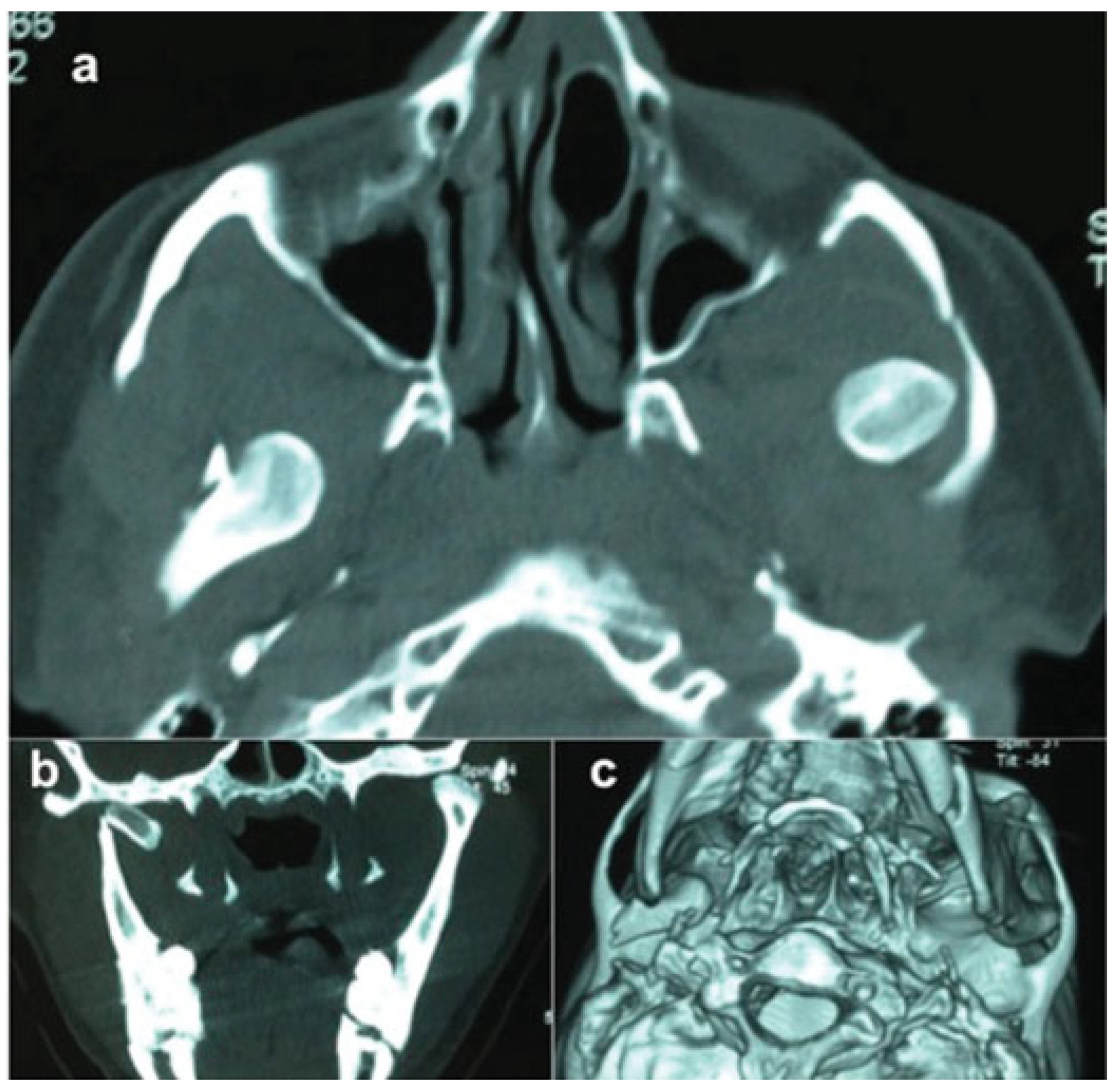
A 27-year-old male patient suffered a car accident on April 29, 2012, while sitting in the back seat without a seatbelt. He was admitted to the hospital, lucid and aware, with no change in his vital signs, hemodynamically stable. Upon maxillofacial examination, he presented facial trauma and mandibular deviation to the right when fully opening his mouth, and no spontaneous reduction. The patient presented with mild bleeding in the gums of the left posterior mandibular teeth, pain upon palpation of the right preauricular region, and a visible exacerbated edema in the area; the left condyle was not palpable and also presented crackling in left mandibular body. Pain medication was prescribed, and a computed tomography (CT) of the face was requested. The results were as follows: left zygomatic arch fracture without dislocation; fracture of the right process condylar; fracture of the left mandibular body; and anterior and superior dislocation of the left condyle, now located in the temporal fossa (Figure 1 and Figure 2). Both the patient and his family were advised about the surgical approach in the right TMJ for the second surgery, because an exacerbated edema had been identified. The patient was taken to the operating room, and after attempts to reduce the left TMJ, the surgeon then applied digital pressure on the coronoid process (using the thumb and forefinger of his left hand, intraorally), while using a bone hook in a minimally invasive reduction (was performed by means of a 3-mm skin incision slightly posterior to the coronoid process, below the zygomatic arch, and a zygomatic bone hook was inserted onto the mandibular notch), described by Melo et al (2015).[8] Digital pressure was applied because there was a fracture of the left mandibular body. After having reduced the left TMJ dislocation, arch bars were utilized, and fixation of fracture in the mandibular body was performed with the use of 02 plates and 8 screws of the 2.0 fixation system.
Stable occlusion was maintained on the postoperative follow-up of the maxillomandibular fixation, using elastics for 2 weeks. The patient and his family excluded the proposed surgery in the right condyle; after 15 days, the patient was sent to functional myofascial treatment with speech therapy.
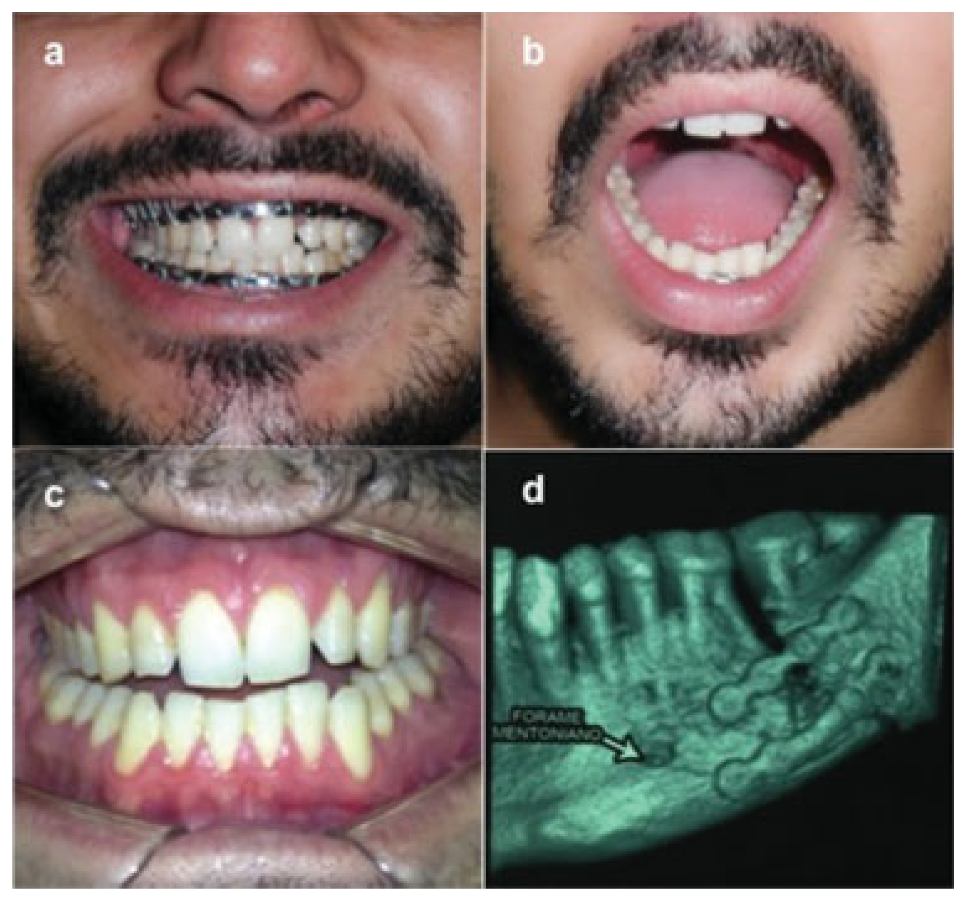
The arch bars were removed 45 days after the first surgery, with stable occlusion, mouth opening of 40 mm, and deviation to the right. After 70 days, during postoperative control, an anterior open bite, of approximately 2 mm, with premature touch molars was observed. A CT exam of the patient’s face was requested, which revealed a resorption of the bone crest between teeth 36 and 37, as well as vertical osteolysis (Figure 3). To resolve this condition, a modeling prototype and a 2.4 locking plate and screws were proposed to the patient. Intraoral access surgery was performed 115 days after the accident, the inferior alveolar and the mental nerves were preserved, the fibrous connective tissue in the osteolysis area was debrided, and a fixation system (2.4 and 2.0) was performed. A particulate graft from the iliac crest was used, without applying a surgical approach in the fractured right condyle, as requested by the patient and his family (Figure 4), despite the information of potential changes related to the nonsurgical approach of the right TMJ. In 5 years of postoperative follow-ups, the patient showed no functional disturbances or ankylosis, and he had occlusal stability with no mouth opening restrictions. A discrete deviation from the median line to the right during the buccal opening that may be related to the presence of condylar fracture not surgically approached was observed. The left condyle remains well positioned and the right condyle is stable, presenting bone remodeling (Figure 5).
A retrospective analysis was performed through a systematic search based on two different methods: (1) main search: made in PubMed and Embase databases; (2) search handling: all references of the included studies were read to find articles not found on PubMed or Embase. Including the present case report, nine cases were deemed acceptable for evaluation and analysis, showing the ADIMC as a rare condition in the literature. The diagnosis, management, and correct classification of this type of dislocation has also been discussed in the literature, being that the open reductions [2,9,10,11,12] were more applied than the closed reductions,[8,13,14] being directly related to the time of approach in relation to the trauma, as shown in Table 1.[2,8,9,10,11,12,13,14]
The anterior mandibular dislocation is the most common dislocation of the TMJ and is connected to common extension movements, such as yawning or hypermotility of the joint. [15,16,17,18] Occasionally, it is associated with a high-impact trauma to the jaw, the latter being an extremely rare condition. [12,19] The diagnosis of condylar luxation is clinical; however, image exams are important in classifying the types of condylar luxation and associated fractures.[20] Superior dislocation of the condyle into medial cranial fossa and lateral and posterior displacements are rare and usually associated with facial trauma.[21] Shen et al[22] collected 23 cases of superolateral dislocation (SD) of the mandibular condyle from the literature (1969–2011). Recently, Sharma et al (2016) reported one case of SD.[23] The present study reported only eight cases of anterosuperior dislocation of mandibular condyles into the temporal fossa.
Worthington[9] suggested that traumatic dislocations occur due to a high-energy impact, or if the patient’s mouth is open at the moment of trauma. Mandible fractures are also commonly associated with these types of dislocations.[6,7,24] Prabhakar and Singla[2] suggested an additional subtype classification proposed by Allen and Young[6] and Satoh et al.[7] The authors suggested a type III dislocation, which they described as “the clinical condition when the condyle has passed anteriorly and then superiorly to enter the temporal fossa and is lodged between the unfractured zygomatic arch on the lateral side and the temporal fossa and its contents on the medial side, with an intact mandible.” In the present study, regarding Table 1, the authors agreed that the nomenclature of anterosuperior dislocation is more appropriate than SD, as reported by other authors.13,22,24–26 However, mandible fracture was not considered in the Prabhakar and Singla classification. Thus, the present study proposes a new type III classification by dividing it into two categories: type IIIA, as described by Prabhakar and Singla, and type IIIB, which could be described as a “clinical condition when the condyle has passed anteriorly and then superiorly to enter the temporal fossa and is between the zygomatic arch fracture but without displacement to the lateral side and the temporal fossa and its contents on the medial side are associated with a mandible fracture.”
Radiographs and CT exams can help confirm and classify the type of condylar dislocation, as well as identify maxillofacial fractures.[25] In the present study, the CT proved to be useful in the classification of the condylar dislocation (type III). Closed reduction is the first choice of treatment for anterosuperior dislocation. It is less traumatic and safer than all other techniques.[25,26] However, condylar luxation may cause muscle spasms and, depending on the time span between the injury and treatment, fibrous adhesion in the TMJ, which in turn disturbs condyle reduction.[27] In the present case, the approach was performed under general anesthesia, and manual and minimally invasive reduction were used together to reduce the left condyle and then stabilize the left mandibular body fracture.
Different dislocations require different approaches, as the closed techniques to open approaches can be observed in the approaches of different authors. Choosing the right approach depends on the displacement’s evolution time. Shorter times appear to be linked to better results.8,13 The present literature review (Table 1) showed that the time between injury and reduction ranged from 1 to 45 days, and most cases were treated with open reduction presenting a delayed approach, which was different from the cases treated with closed reduction, using less development time.[2,8,9,13]
The reduction of late time seems to be related to increased difficulty reduction by manual techniques.[28]

Conclusion

Different approaches can be used for different types of mandibular condyle displacements, whether fractured or not. Based on the case presented herein, as well as on literature review, it can be concluded that the anterosuperior specific offset for temporal fossa is a rare occurrence, and the treatment can range from a noninvasive approach to even more complex surgical approaches.
We observed in this clinical case (mandibular fracture that needs to be stabilized with a 2.0 fixation system—load sharing) that it was possible to perform a closed reduction under general anesthesia and the minimally invasive reduction, simultaneously, before load sharing (to avoid plate deformation). Such results were obtained in a scenario where treatment started in a small time span after the trauma. Some factors may make it difficult, or even impossible, to achieve a noninvasive/minimally invasive reduction: time elapsed, fractures in the condyle, and other mandibular fractures, among other relevant factors.[28]

References

  1. He, Y.; Zhang, Y.; Li, Z.L.; An, J.G.; Yi, Z.Q.; Bao, S.D. Treatment of traumatic dislocation of the mandibular condyle into the cranial fossa: development of a probable treatment algorithm. Int J Oral Maxillofac Surg 2015, 44, 864–870. [Google Scholar] [CrossRef]
  2. Prabhakar, V.; Singla, S. Bilateral anterosuperior dislocation of intact mandibular condyles in the temporal fossa. Int J Oral Maxillofac Surg 2011, 40, 640–643. [Google Scholar] [CrossRef] [PubMed]
  3. Vaezi, T.; Rajaei, S.A.; Hosseini Abrishami, M.; Erfanian Taghvaei, M. Dislocation of the mandibular condyle into the middle cranial fossa: a case report. Oral Maxillofac Surg 2014, 18, 69–73. [Google Scholar] [CrossRef]
  4. Oberman, B.; Setabutr, D.; Goldenberg, D. Traumatic dislocation of intact mandibular condyle into middle cranial fossa. Am J Otolaryngol 2014, 35, 251–253. [Google Scholar] [CrossRef] [PubMed]
  5. Rahman, T. Unusual superolateral dislocation of mandibular condyle. Craniomaxillofac Trauma Reconstr 2017. [CrossRef] [PubMed]
  6. Allen, F.J.; Young, A.H. Lateral displacement of the intact mandibular condyle. A report of five cases. Br J Oral Surg 1969, 7, 24–30. [Google Scholar] [CrossRef]
  7. Satoh, K.; Suzuki, H.; Matsuzaki, S. A type II lateral dislocation of bilateral intact mandibular condyles with a proposed new classification. Plast Reconstr Surg 1994, 93, 598–602. [Google Scholar] [CrossRef]
  8. Melo, M.F.S.; Oliveira, R.B.; Silveira, R.L.; Amaral, M.B.F. Anterosuperior dislocation of the intact condyles into the temporal fossa. J Craniofac Surg 2015, 26, 990–991. [Google Scholar] [CrossRef]
  9. Worthington, P. Dislocation of the mandibular condyle into the temporal fossa. J Maxillofac Surg 1982, 10, 24–27. [Google Scholar] [CrossRef]
  10. DeVita, C.L.; Friedman, J.M.; Meyer, S.; Breiman, A. An unusual case of condylar dislocation. Ann Emerg Med 1988, 17, 534–536. [Google Scholar] [CrossRef]
  11. Yoshii, T.; Hamamoto, Y.; Muraoka, S.; Teranobu, O.; Shigeta, Y.; Komori, T. Traumatic dislocation of the mandibular condyle into the temporal fossa in a child. J Trauma 2000, 49, 764–766. [Google Scholar] [CrossRef] [PubMed]
  12. Özalp, B.; Elbey, H.; Durgun, M.; Selçuk, C.T. An alternative surgical procedure for anterosuperior dislocation of intact mandibular condyle. J Craniofac Surg 2014, 25, e382–e384. [Google Scholar] [CrossRef] [PubMed]
  13. Bu, S.S.; Jin, S.L.; Yin, L. Superolateral dislocation of the intact mandibular condyle into the temporal fossa: review of the literature and report of a case. Oral Surg Oral Med Oral Pathol Oral Radiol Endod 2007, 103, 185–189. [Google Scholar] [CrossRef]
  14. Alemán Navas, R.M.; Martínez Mendoza, M.G. ‘Inverse’ temporo-mandibular joint dislocation. Int J Oral Maxillofac Surg 2011, 40, 877–879. [Google Scholar] [CrossRef]
  15. Chan, T.C.; Harrigan, R.A.; Ufberg, J.; Vilke, G.M. Mandibular reduction. J Emerg Med 2008, 34, 435–440. [Google Scholar] [CrossRef] [PubMed]
  16. Myrhaug, H. A new method of operation for habitual dislocation of the mandible; review of former methods of treatment. Acta Odontol Scand 1951, 9, 247–260. [Google Scholar] [CrossRef]
  17. Young, A.L.; Khan, J.; Thomas, D.C.; Quek, S.Y. Use of masseteric and deep temporal nerve blocks for reduction of mandibular dislocation. Anesth Prog 2009, 56, 9–13. [Google Scholar] [CrossRef]
  18. Li, Z.; Li, Z.B.; Shang, Z.J.; Wu, Z.X. An unusual type of superolateral dislocation of mandibular condyle: discussion of the causative mechanisms and clinical characteristics. J Oral Maxillofac Surg 2009, 67, 431–435. [Google Scholar] [CrossRef]
  19. Ogawa, M.; Kanbe, T.; Kano, A.; et al. Conservative reduction by lever action of chronic bilateral mandibular condyle dislocation. Cranio 2015, 33, 142–147. [Google Scholar] [CrossRef]
  20. Shorey, C.W.; Campbell, J.H. Dislocation of the temporomandibular joint. Oral Surg Oral Med Oral Pathol Oral Radiol Endod 2000, 89, 662–668. [Google Scholar] [CrossRef]
  21. Tutela, J.P.; Verbist, D.E.; Kelishadi, S.; Little, J.A. Traumatic dislocation of the mandibular condyle into the middle cranial fossa in an elderly patient. J Craniofac Surg 2013, 24, 1703–1705. [Google Scholar] [CrossRef]
  22. Shen, L.; Li, P.; Li, J.; Long, J.; Tian, W.; Tang, W. Management of superolateral dislocation of the mandibular condyle: a retrospective study of 10 cases. J Craniomaxillofac Surg 2014, 42, 53–58. [Google Scholar] [CrossRef] [PubMed]
  23. Sharma, D.; Khasgiwala, A.; Maheshwari, B.; Singh, C.; Shakya, N. Superolateral dislocation of an intact mandibular condyle into the temporal fossa: case report and literature review. Dent Traumatol 2017, 33, 64–70. [Google Scholar] [CrossRef] [PubMed]
  24. Tauro, D.; Lakshmi, S.; Mishra, M. Superolateral dislocation of the mandibular condyle: report of a case with review of literature and a proposed modification in the classification. Craniomaxillofac Trauma Reconstr 2010, 3, 119–123. [Google Scholar] [CrossRef] [PubMed]
  25. Kapila, B.K.; Lata, J. Superolateral dislocation of an intact mandibular condyle into the temporal fossa: a case report. J Trauma 1996, 41, 351–352. [Google Scholar] [CrossRef]
  26. Amaral, M.B.; Bueno, S.C.; Silva, A.A.; Mesquita, R.A. Superolateral dislocation of the intact mandibular condyle associated with panfacial fracture: a case report and literature review. Dent Traumatol 2011, 27, 235–240. [Google Scholar] [CrossRef]
  27. Huang, I.Y.; Chen, C.M.; Kao, Y.H.; Chen, C.M.; Wu, C.W. Management of long-standing mandibular dislocation. Int J Oral Maxillofac Surg 2011, 40, 810–814. [Google Scholar] [CrossRef]
  28. Lello, G.E. Treatment of long-standing mandibular dislocation. J Oral Maxillofac Surg 1987, 45, 893–896. [Google Scholar] [CrossRef]
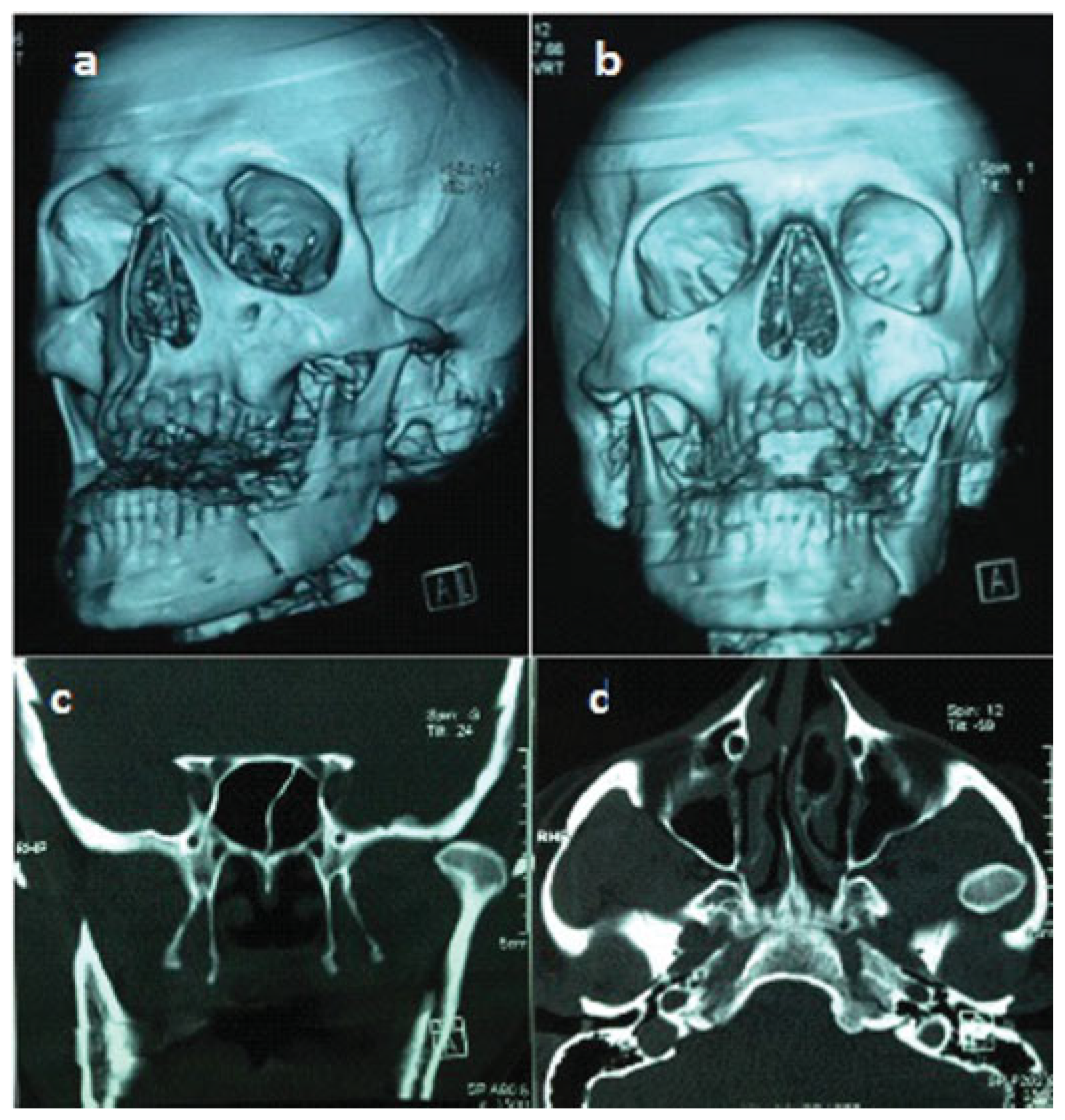
Figure 1. (a, b) Three-dimensional face CT exam, showing mouth opening, with right deviation, right condylar fracture and left mandibular body, left zygomatic arch (no displacement), and dislocation of the left mandibular condyle; (c, d) coronal and axial section, showing displacement of the left mandibular condyle, in contact with the temporal fossa and left zygomatic arch fracture without dislocation.
Cmtr 11 11i301 g001
Figure 2. Details of both condyle positions—right condylar fracture and left condyle dislocation: (a) axial; (b) coronal; (c) 3D face CT exam.
Cmtr 11 11i301 g002
Figure 3. (a, b) Patient ~40 days after the surgery in the initial approach, with Erich maxillary and mandibular bars, stable occlusion, and maximum mouth opening, with a slight mandibular deviation to the right. (c) Occlusion at 70 days postoperatively, with anterior open bite and premature contacts on posterior molars. (d) CT image showing osteolysis between molars on the left.
Cmtr 11 11i301 g003
Figure 4. (a, b) Prototyping model with simulation surgery of osteolysis area; this served as the basis for the locking of the 2.4 plate modeling on approach for intraoral access; (c, d) stable dental occlusion and panoramic postoperative radiograph (right condyle partially visible, with displacement and left condyle well positioned).
Cmtr 11 11i301 g004
Figure 5. (ac) Current picture of maximum mouth opening (45 mm, with a slight mandibular deviation to the right—condylar fracture not surgically approached) and stable occlusion. (d) Current panoramic radiograph: details of the left condyle remains well positioned and the right remodeling condylar.
Cmtr 11 11i301 g005
Table 1. Reported cases of anterosuperior dislocation of intact mandibular condyles into the temporal fossa (from 1969 to 2017).
Cmtr 11 11i301 i001
Abbreviations: F, female; M, male.