Next Article in Journal
Removal of a Frontal Sinus Osteoma and Reconstruction by a Custom-Made Implant with Neuronavigation Assistance
Previous Article in Journal
Traumatic Anterosuperior Dislocation of the Intact Mandibular Condyle into the Temporal Fossa
 
 
Craniomaxillofacial Trauma & Reconstruction is published by MDPI from Volume 18 Issue 1 (2025). Previous articles were published by another publisher in Open Access under a CC-BY (or CC-BY-NC-ND) licence, and they are hosted by MDPI on mdpi.com as a courtesy and upon agreement with Sage.
Case Report

Pseudoaneurysm following Endoscopic-Assisted Repair of Subcondylar Fracture

by
Emily Ambrose
1,*,
Mofiyinfolu Sokoya
1 and
Marcia Eustaquio
2
1
Department of Otolaryngology, University of Colorado, 12631 E. 17th Avenue B-205, Aurora, CO 80045, USA
2
Department of Otolaryngology, Denver Health and Hospital Authority, Denver, CO, USA
*
Author to whom correspondence should be addressed.
Craniomaxillofac. Trauma Reconstr. 2018, 11(4), 302-304; https://doi.org/10.1055/s-0037-1607064
Submission received: 20 January 2017 / Revised: 1 April 2017 / Accepted: 17 June 2017 / Published: 27 October 2017

Abstract

:
Pseudoaneurysms are a known complication following facial trauma and orthognathic surgery. Few reports exist of this clinical entity following traditional open techniques of repair, and none have been associated with endoscopic-assisted open reduction and internal fixation (ORIF) of a subcondylar fracture. We present a case of a 33-year-old man who developed pseudoaneurysm after endoscopic-assisted ORIF as well as a review of the literature on this topic. While uncommon, this is a potential complication that should be recognized with the use of endoscopic-assisted repair of mandibular fractures.

Pseudoaneurysms are partial disruptions in the wall of a blood vessel leading to collection of blood in the adventitia.[1] They have been reported in the literature following penetrating trauma, as well as following orthognathic surgery, particularly sagittal split osteotomy. A small number of reports describe pseudoaneurysm formation as a complication of open reduction and internal fixation (ORIF) techniques for mandibular fractures, though description of the external approach used and proposed mechanism of injury are limited. There are currently no published reports that describe this clinical entity in association with endoscopicassisted ORIF. Here, we present a case of pseudoaneurysm following endoscopic-assisted ORIF of a subcondylar fracture as well as a review of the literature.

Presentation of Case

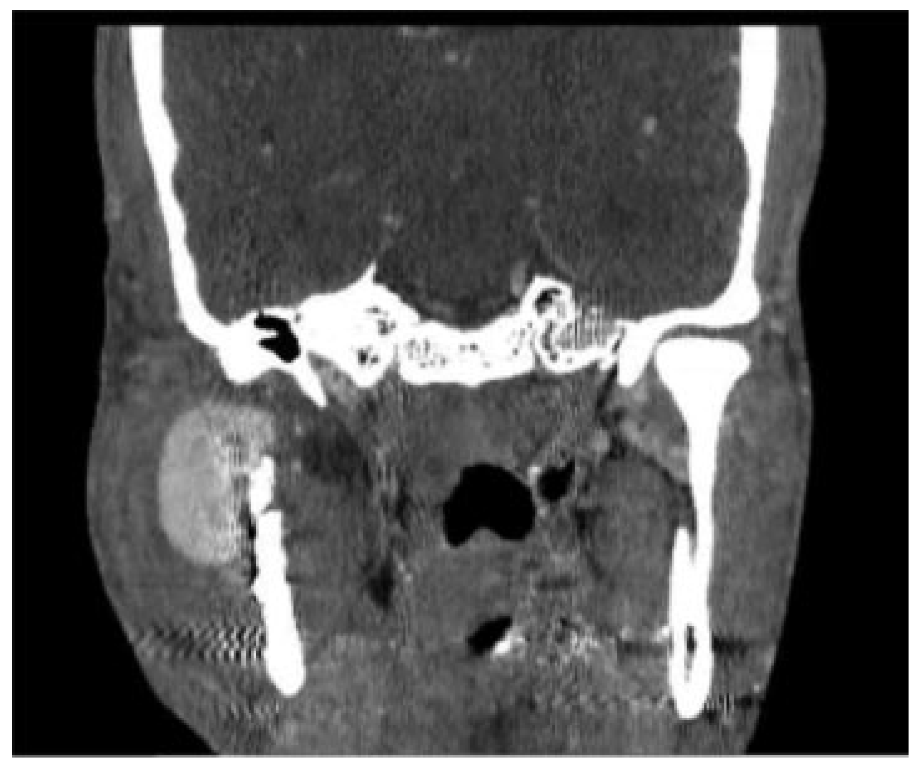
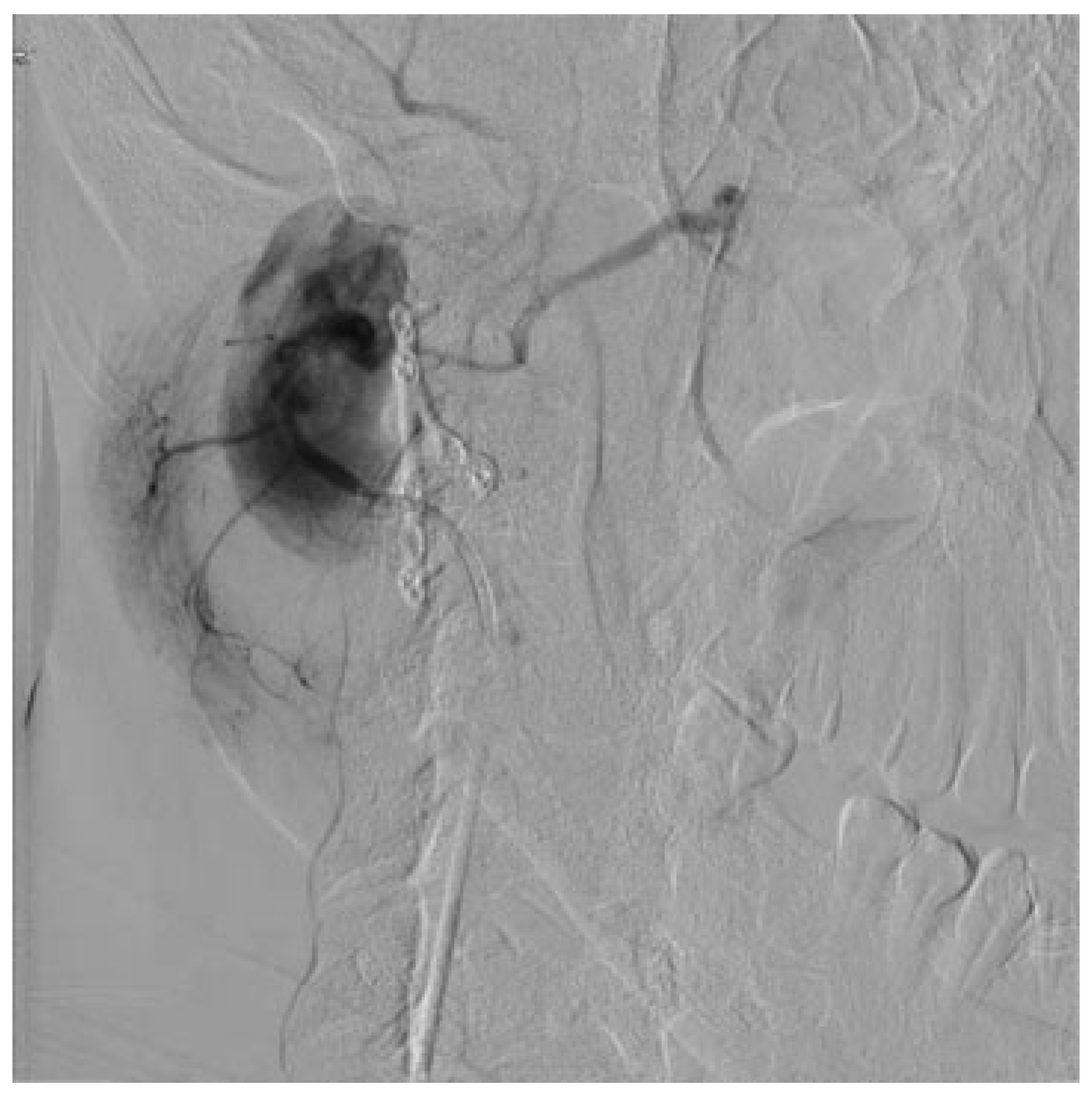
A previously healthy 33-year-old man in the department of corrections (DOC) suffered a right subcondylar fracture following assault with a fist. The patient presented for evaluation 2.5 weeks following the injury and underwent endoscopicassisted ORIF of the isolated right subcondylar fracture. Following the procedure, hewas found to have good occlusion and was returned to the prison infirmary for recovery. While in the infirmary, he was treated with oral antibiotics given for cheek swelling and possible infection with improvement in symptoms. He then presented for his first postoperative visit at approximately 2 weeks with moderate cheek edema, concerning for a possible hematoma. A panoramic X-ray was obtained, showing mild posterior displacement of the condyle, but good occlusion (Figure 1). He was instructed to continue a no-chew diet and follow up in 1 month. Two weeks later, the patient developed worsening right cheek swelling and pain. Considering his worsening cheek swelling, incision and drainage were attempted at the DOC infirmary. The swelling did not improve and 3 days later, he was transferred to the hospital for further management. A CT of the neck was obtained which was concerning for right cheek pseudoaneurysm (see Figure 2). Ultrasound Doppler was performed which confirmed the diagnosis ofa right external carotid artery branch pseudoaneurysm. The patient underwent angiography which demonstrated the pseudoaneurysm at the level of the bifurcation of the internal maxillary and superficial temporal arteries (Figure 3). This was embolized by interventional radiology. He had no additional swelling and decreased trismus over the next 2 days and was returned to the prison infirmary. On further follow-up, he was found to have malocclusion due to condylar head rotation, possible loss of bony height, and scarring of the masseter.

Discussion/Review of Literature

Pseudoaneurysms following trauma or orthognathic surgery have been described extensively in literature. Reports of pseudoaneurysm following ORIF of mandible fractures are more limited, and there are no prior reports of a pseudoaneurysm as a result of endoscopic-assisted ORIF as described here. Shetty et al[2] presented a case of large postoperative pseudoaneurysms of the facial artery requiring treatment with sclerotherapy. The patient discussed in their review suffered from a facial artery pseudoaneurysm after traditional ORIF of a condylar fracture. Alonso et al[3] described a patient with delayed internal maxillary artery pseudoaneurysm following a gunshot wound to the face with multiple facial fractures and ORIF of ramus fracture. Katakol and Govindaraj[4] presented a case of delayed pseudoaneurysm formation of the internal maxillary artery after ORIF of parasymphyseal fracture and maxillomandibular fixation of condylar fractures. Lastly, El et al[5] described a patient who sustained a pseudoaneurysm of the external carotid artery after a retromandibular approach to a subcondylar fracture. In all the cases reported in the literature, extent of trauma and open approach were noted as likely reasons of the pseudoaneurysms. A unique characteristic of the case we present in this article is the use of an endoscope and a percutaneous trocar in the intraoperative management of the subcondylar fracture.
When reviewing the endoscopic-assisted ORIF literature, studies have assessed the safety and efficacy of this technique, but none have reported this complication.[6,7,8,9] In a series reviewing endoscopic-assisted ORIF of subcondylar fractures, Kang et al[9] found that nearly 70% of patients had temporary complications (<3 months) with 23% having long-term (>6 months) complications including infection, open bite, facial paresthesia, and weakness, but there were no reports of vascular injury. In this case, we believe that given the location and timing of the aneurysm, the likely etiology was the percutaneous trocar used to assist with endoscopic screw placement. While rare, this is a new complication to consider and discuss prior to endoscopic-assisted ORIF of subcondylar fractures.

Conclusion

Pseudoaneurysm of a branch of the external carotid artery is a potential complication following endoscopic-assisted ORIF of subcondylar fracture, likely due to percutaneous trocar placement and manipulation during surgery.

References

  1. Cox, M.W.; Whittaker, D.R.; Martinez, C.; Fox, C.J.; Feuerstein, I.M.; Gillespie, D.L. Traumatic pseudoaneurysms of the head and neck: early endovascular intervention. J Vasc Surg 2007, 46, 1227–1233. [Google Scholar] [CrossRef] [PubMed]
  2. Shetty, N.K.; Shandilya, R.; Pawar, S.; Gadre, P.K.; Gadre, K.; Singh, D. Management of late post-traumatic facial artery pseudoaneurysmal cyst: review of literature. J Maxillofac Oral Surg 2015, 14, 201–205. [Google Scholar] [CrossRef] [PubMed]
  3. Alonso, N.; de Oliveira Bastos, E.; Massenburg, B.B. Pseudoaneurysm of the internal maxillary artery: a case report of facial trauma and recurrent bleeding. Int J Surg Case Rep 2016, 21, 63–66. [Google Scholar] [CrossRef] [PubMed]
  4. Katakol, B.; Govindaraj, E. Pseudoaneurysm of the internal maxillary artery following mandibular condylar fracture. Ann Maxillofac Surg 2014, 4, 201–204. [Google Scholar] [PubMed]
  5. El, A.S.; Guo, W.; Loveless, T.; et al. Pseudoaneurysm of the external carotid artery secondary to subcondylar fracture. Int J Oral Maxillofac Surg 2011, 40, 644–646. [Google Scholar] [CrossRef] [PubMed]
  6. Arcuri, F.; Brucoli, M.; Baragiotta, N.; Benech, R.; Ferrero, S.; Benech, A. Analysis of complications following endoscopically assisted treatment of mandibular condylar fractures. J Craniofac Surg 2012, 23, e196–e198. [Google Scholar] [CrossRef] [PubMed]
  7. Troulis, M.J. Endoscopic open reduction and internal rigid fixation of subcondylar fractures. J Oral Maxillofac Surg 2004, 62, 1269–1271. [Google Scholar] [PubMed]
  8. Haug, R.H.; Brandt, M.T. Traditional versus endoscope-assisted open reduction with rigid internal fixation (ORIF) of adult mandibular condyle fractures: a review of the literature regarding current thoughts on management. J Oral Maxillofac Surg 2004, 62, 1272–1279. [Google Scholar] [PubMed]
  9. Kang, S.H.; Choi, E.J.; Kim, H.W.; Kim, H.J.; Cha, I.H.; Nam, W. Complications in endoscopic-assisted open reduction and internal fixation of mandibular condyle fractures. Oral Surg Oral Med Oral Pathol Oral Radiol 2012, 113, 201–206. [Google Scholar] [PubMed]
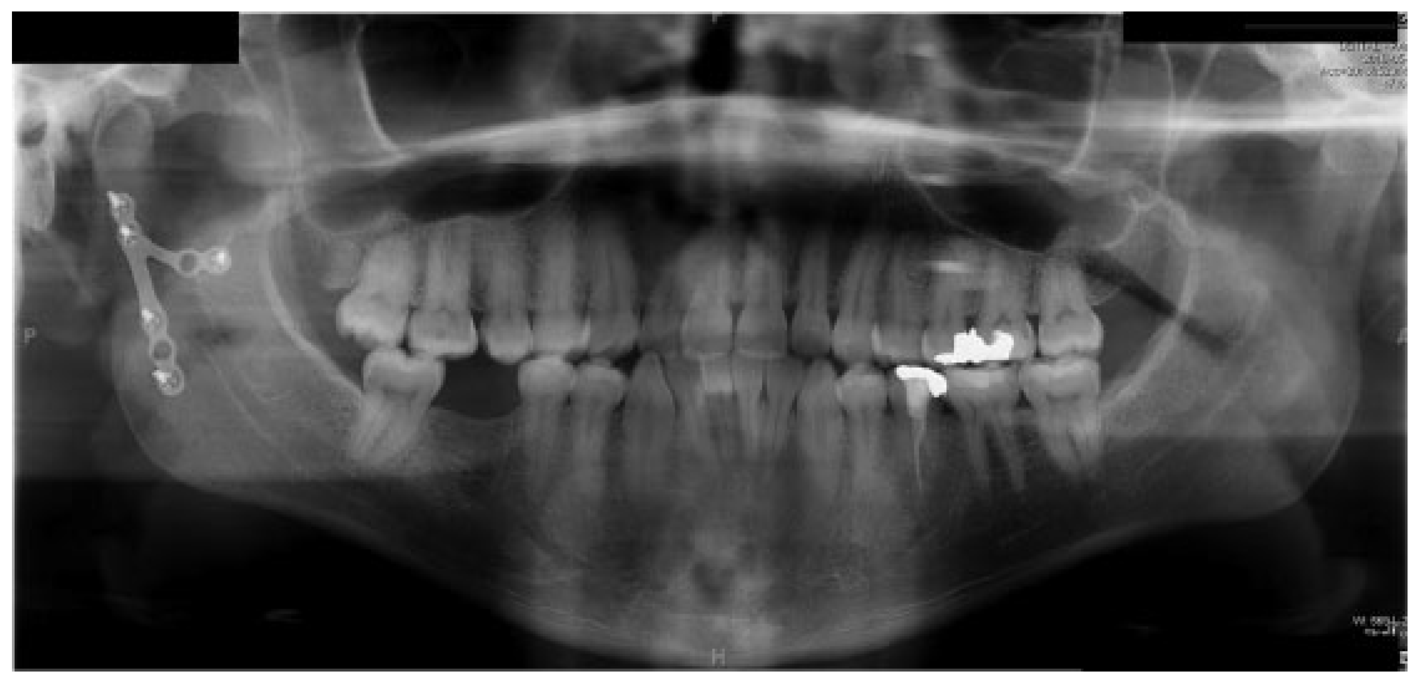
Figure 1. Postoperative panorex.
Cmtr 11 11i296 g001
Figure 2. CT of the neck with hyperintense homogenous collection in cheek concerning for pseudoaneurysm.
Cmtr 11 11i296 g002
Figure 3. Angiography demonstrating pseudoaneurysm at the level of the bifurcation of the internal maxillary and superficial temporal arteries.
Cmtr 11 11i296 g003