Next Article in Journal
Posttraumatic Parotid Sialocele Complicating a Mandibular Fracture: A Case Report
Previous Article in Journal
Submental Perforator Flap for Soft-Tissue Reconstruction in Bisphosphonate-Related Osteonecrosis of the Jaws
 
 
Craniomaxillofacial Trauma & Reconstruction is published by MDPI from Volume 18 Issue 1 (2025). Previous articles were published by another publisher in Open Access under a CC-BY (or CC-BY-NC-ND) licence, and they are hosted by MDPI on mdpi.com as a courtesy and upon agreement with Sage.
Case Report

Bone Allograft: An Option for Total Mandibular Reconstruction

by
Masoud Fallahi Motlagh
1,*,
Mohamad Bayat
2 and
Siamak Naji
3
1
Department of Oral and Maxillofacial Surgery, Urmia Medical University, Urmia, Iran
2
Department of Oral and Maxillofacial, Tehran University of Medical Science, Tehran, Iran
3
Department of Pathology, Urmia University of Medical Sciences Ringgold Standard Institution, Urmia, Iran
*
Author to whom correspondence should be addressed.
Craniomaxillofac. Trauma Reconstr. 2017, 10(4), 306-313; https://doi.org/10.1055/s-0036-1593474
Submission received: 20 May 2016 / Revised: 30 June 2016 / Accepted: 4 July 2016 / Published: 4 November 2016

Abstract

:
Total mandibular reconstruction is one of the most challenging modalities in maxillofacial surgery. In this article, we try to introduce a method of total mandible reconstruction. We report a 25-year-old male patient with complete involvement of the mandible by Langerhans cell tumor. The patient underwent total mandibulectomy and reconstruction with bone allograft harvested from a donor who had died the day before. The patient has good functional and aesthetic results in a long time. It seems that bone allograft can be a good alternative to other methods in total mandibular reconstruction.

Total mandibular reconstruction is one of the most challenging modalities in oral and maxillofacial surgery. When performing mandibular reconstruction, the restoration of bony continuity should not be considered as the only measure of success. The functions of mastication, swallowing, speech, articulation, and oral competence must also be addressed. However, it is not always possible to achieve such conditions.
Various methods are available for mandibular reconstruction, including autotransplantation, allotransplantation, and biomaterials. Autogenous bone grafting is a gold standard for mandibular reconstruction,[1] and is a reliable treatment modality for the reconstruction of mandibular bone defects with predictable aesthetic and functional results.[2] But when total reconstruction, especially condyle, is needed, no favorable outcomes can be achieved using this method. Autogenous bone grafts may also increase the risk of morbidity by increasing the risk of infection, pain, and length of hospitalization.[3,4,5] As a result, recently interest has been shown for the development of new grafting materials using allogeneic, xenogeneic, and synthetic biomaterials for reconstructive bony procedures.[6]
Some studies have compared the effectiveness of these alternatives as potential replacements for autogenous bone grafts.[7,8] Allogeneic bone, such as demineralized bone matrix (DBM), harvested from one subject to transfer to another subject of the same species, was first used to reconstruct the skull defects in dogs more than 100 years ago.[9,10] This method was not so popular until Dr. Marshall Urist reported the results of his research in 1965. He showed that demineralized segments of bone induced new bone formation when implanted in muscle in the rabbit. Then, scientists seriously considered demineralized allogeneic bone as a potential bioimplant for osseous repair.[11,12,13,14] Herein we report a patient with Langerhans cell tumor, which had involved the mandible completely. The mandible was reconstructed using the allograft. The case described in this study is a new method that we cannot find in anywhere in the literature review.

Case Presentation

A 25-year-old man presented with a history of progressive, painless mandibular swelling with 6-month duration. He reported no paresthesia and the mandible was not tender on physical examination. There was neither trismus nor enlarged lymph node over the submandibular and neck region. The patient had poor oral hygiene and all of the mandibular teeth were mobile. Panoramic radiography revealed a multilocular lesion that affected the entire mandible except the right condyle (►Figure 1a). Three-dimensional computed tomography (3D CT scan) showed massive destruction of the mandibular bone (►Figure 1b).
Incisional biopsy of the oral cavity was done under local anesthesia and eosinophilic granuloma was reported. The patient was then scheduled for total mandibulectomy via submandibular incision accompanied by reconstruction with a transplant bone. We have taken a surgical consent from the patient that was approved by the Urmia University Ethical Committee.
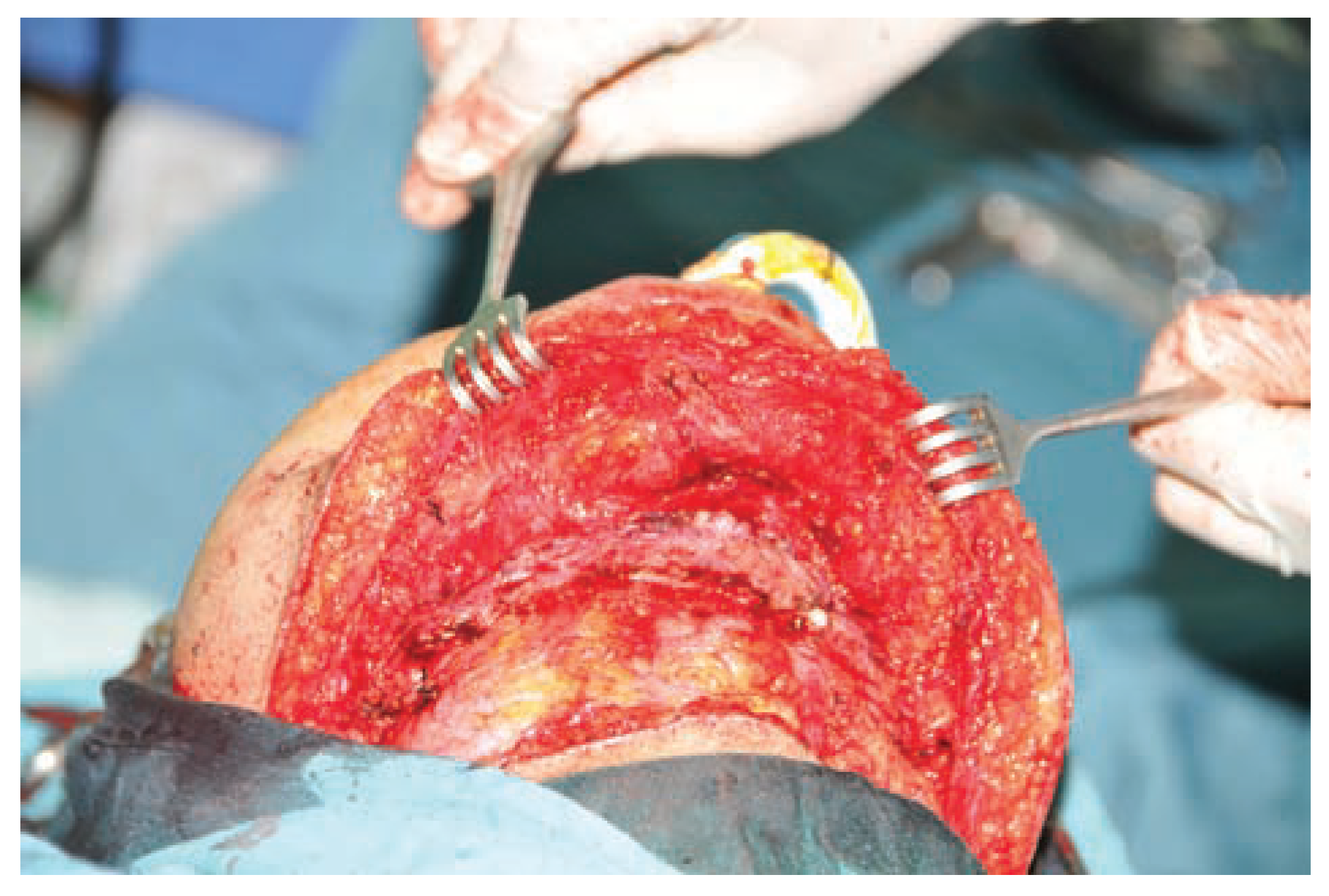
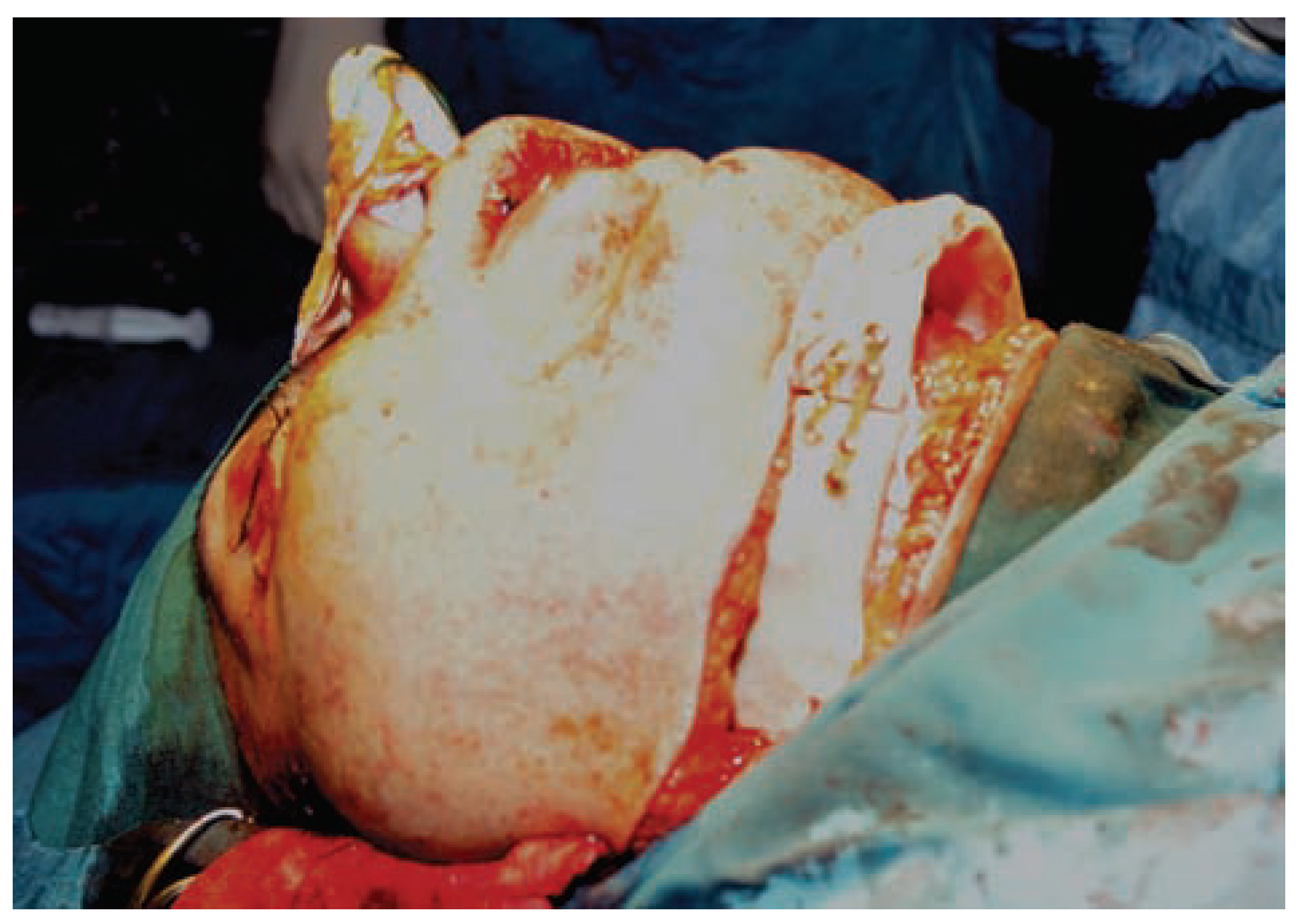
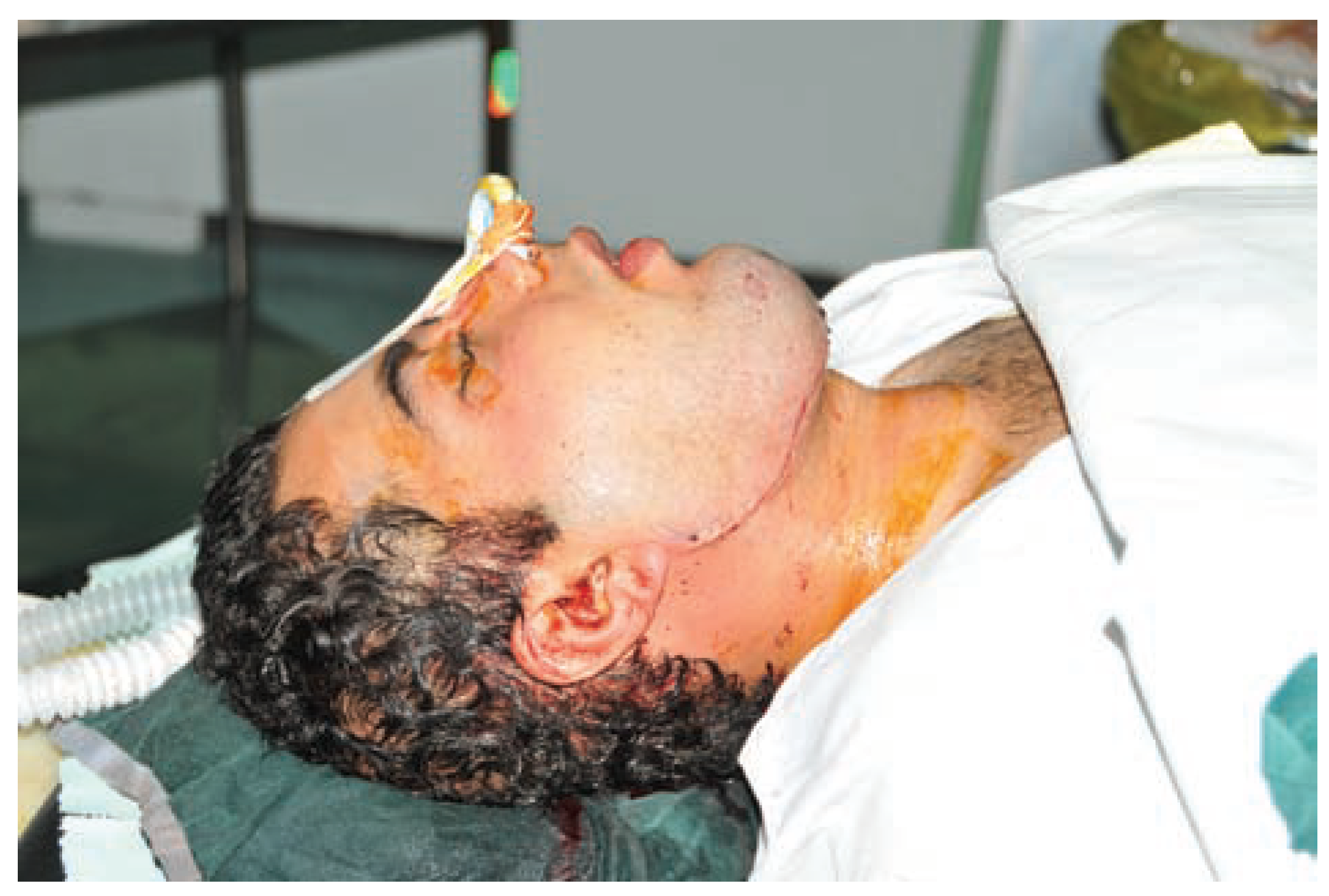
During the operation, the skin flap was raised and retracted superiorly exposing the whole mandible (►Figure 2). Then total mandibulectomy was performed and the transplanted mandible was located in the site of the mandible. Inferior alveolar dental nerve was not preserved, but the discs of temporomandibular joints were preserved bilaterally. As the inter-condylar distance of the new mandible was greater than the previous one, osteotomy was done in the right mandibular body region and the mandibular condyles were adjusted in glenoid fossa bilaterally (►Figure 3). Coronoidectomy was done on both sides, and at the end of the surgery, all masticatory muscles were intact and replaced to their positions. All muscles were fixed to the bone by 4–0 PDS sutures. The skin flap was sutured with 5–0 nylon (►Figure 4).
The transplanted bone was freeze-dried bone prepared according to the latest version of the guidelines of the American Association of Tissue Banks (AATB).[15]
The donor was a 38-year-old man who had died in a car accident and his mandible was harvested on the next day. Before harvesting his mandible, physical examination and required laboratory examinations were performed. The donor did not have any high-risk behavior and his laboratory tests for anti-HIV1, anti-HIV2, anti-hepatitis C virus (anti-HCV), hepatitis B surface antigen (HBsAg), cytomegalovirus, and fungal culture were negative.
Figure 2. Intraoperative image shows involvement and destruction of the mandible.
Cmtr 10 00048 g002
Figure 3. Osteotomy was done in the right site and fixed with miniplate.
Cmtr 10 00048 g003
The preparation, including physical and chemical treatment, was done by the Iranian Tissue Bank and Research Center. It was sterilized by 25 kGy gamma irradiation. The postoperative period was uneventful and feeding was started via nasogastric tube for 3 days and then the tube was removed and a soft diet was started. He was discharged from the hospital on day 5 after operation.
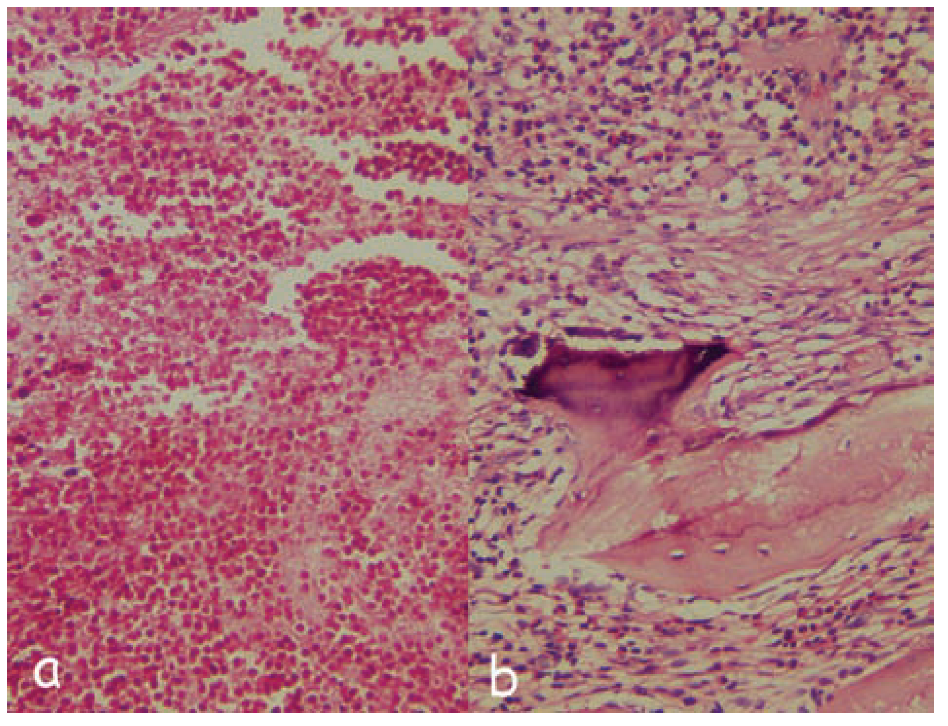
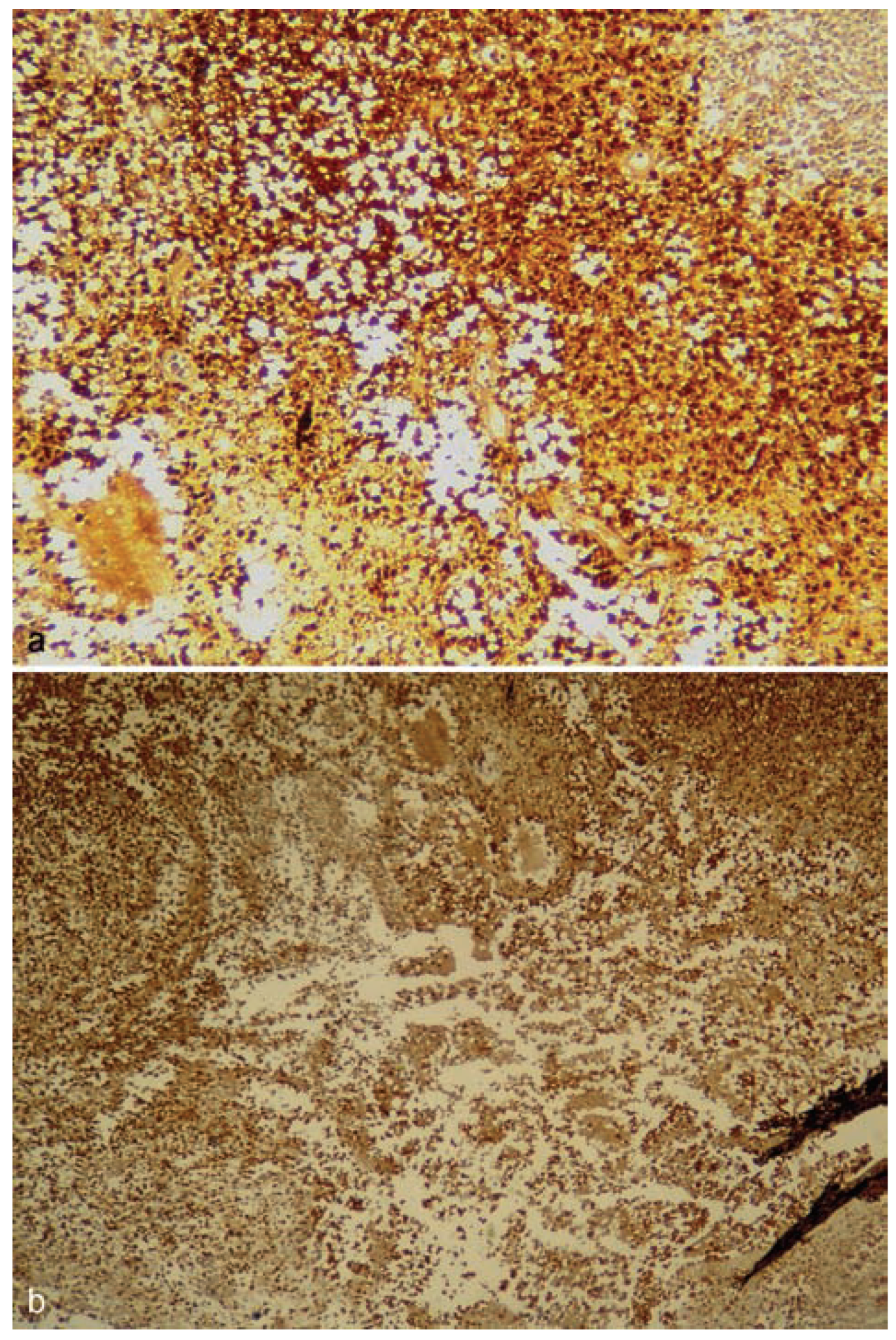
Histopathologic evaluation showed clusters and sheets of uniform, ovoid histocytes with abundant acidophilic cytoplasm and lobulated nuclei admixed with chronic inflammatory cells especially eosinophils. Osteoclast-like giant cells, necrosis, and fibrosis were present as well (►Figure 5a, b). These cells were positive for CD1a (►Figure 6a) and S100 (►Figure 6b). Based on the immunohistochemical finding, the diagnosis of Langerhans cell histiocytosis was made.
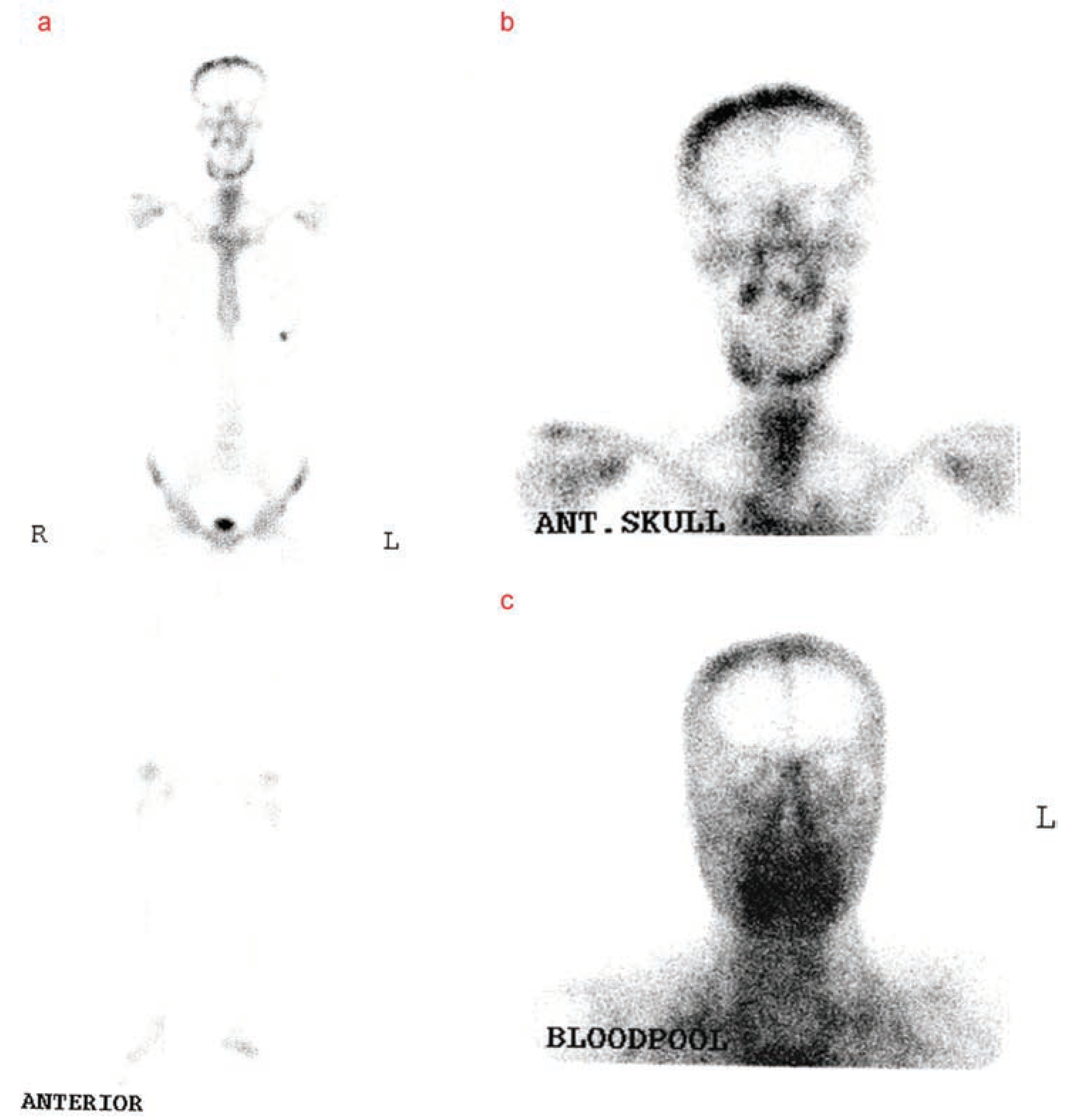
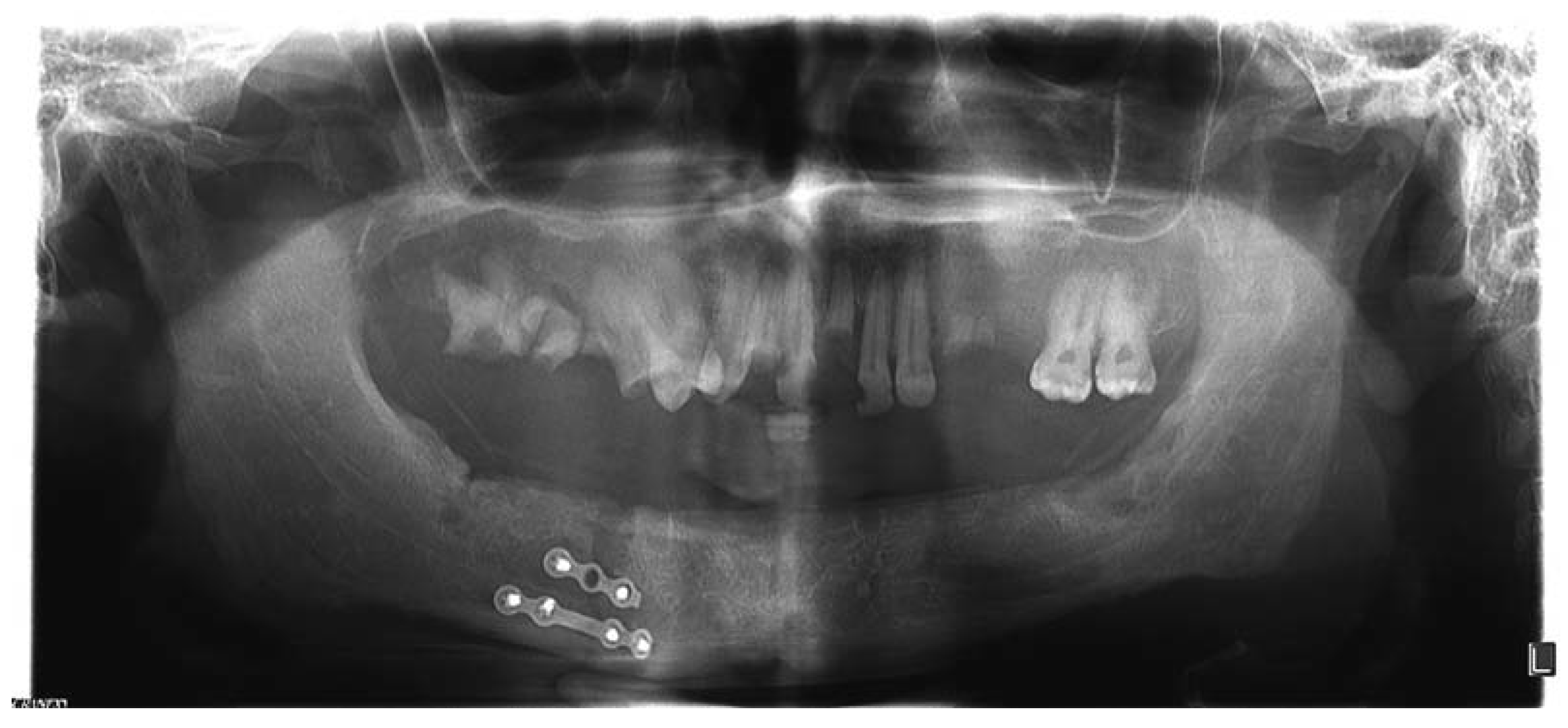
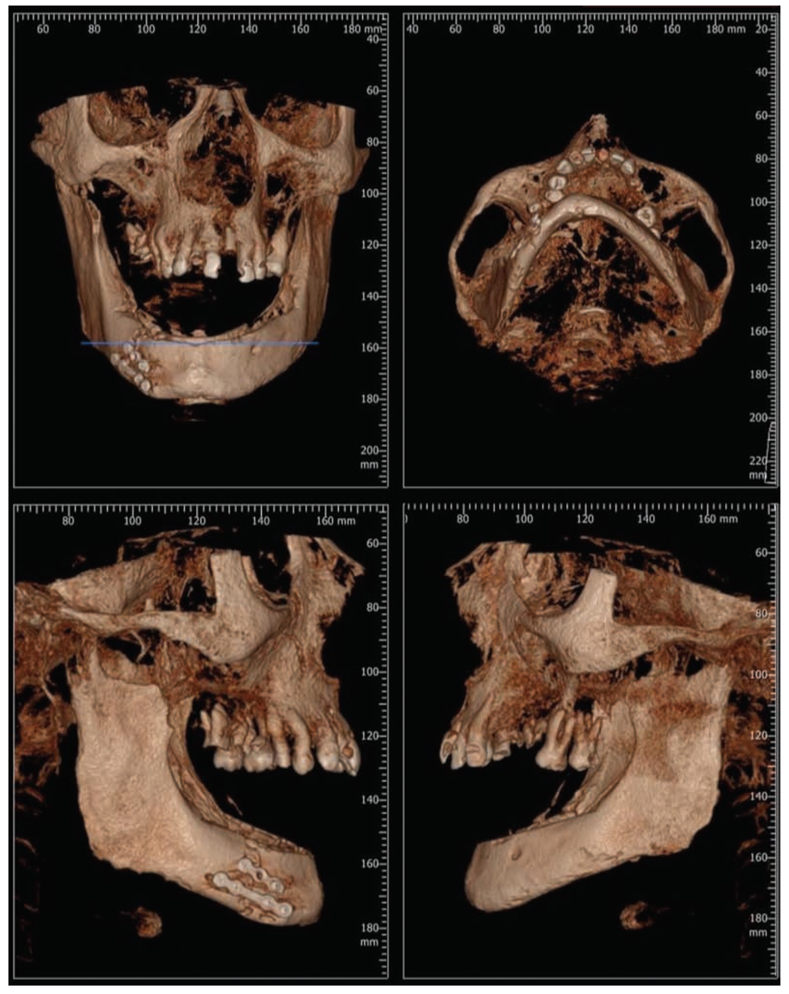
At the first follow-up (2 weeks later), the patient had no significant complaint, except a mild, clear discharge from the incision site on the right side. The diagnosis was parotid fistula which was treated by compression bandage for 10 days. Three months after the surgery, radiographic examination showed no bone resorption (►Figure 7) and in the follow-up 6-month postsugery, an adequate and stable restoration of facial counter was evident. The maximum inter-incisional opening was 40 mm. Tc-99m MPD bone scintigraphy was performed 9 months postoperatively (►Figure 8). There was a mild bone resorption in the panoramic radiography and 3D CT scan 2 years after operation (►Figure 9 and Figure 10). The mouth movements including opening and closing were normal 2 years after surgery. Our future plan is construction of the occlusion by dental implant.
Figure 4. Photography of the lateral view after surgery.
Cmtr 10 00048 g004
Figure 5. (a) Histopathologic evaluation showed a Langerhans cell histiocytosis, large histiocytes surrounded by an inflammatory infiltrate of lymphocytes, eosinophils, and osteoclast-like giant cells. (b)A mixture of Langerhans cells, eosinophil, mandibular bone, and fibrosis.
Cmtr 10 00048 g005
Figure 6. (a, b) Langerhans cells are positive for CD1a and S100 by immunohistochemical staining.
Cmtr 10 00048 g006
Figure 7. Three months postoperative panoramic view.
Cmtr 10 00048 g007

Discussion

Mandibular reconstruction after total resection can be achieved by various methods. There are various types of material for bone reconstruction, including autograft, allograft, xenograft, and synthetic materials. The successful use of these modalities is dependent on various biological, physiological, and biomechanical factors, each of which affects the result of the transplant.[16,17] The most common method for reconstruction of the mandible is use of reconstruction plate with or without bone graft.[18] It has some disadvantages, including plate exposure, fistula, and interface with radiation therapy.[19,20] Vascularized free fibula flap has become the preferred method of mandible reconstruction after oncologic surgical ablation. The fibula flap was first described by Taylor et al[21] and was used for the first time in mandibular reconstruction by Hidalgo.[22] It has some disadvantages, such as increased operating time and complications of the harvested site. Despite the high success rate of total mandibular reconstruction with vascularized fibula flap,[23,24] it is not suitable for condylar reconstruction.
There are three mechanisms in which a bone graft may possess: osteoconduction, osteoinduction, and osteogenesis.[25] Osteoconduction is the process by which the graft provides a 3D structure that facilitates the ingrowth of capillaries and mesenchymal stem cells to support new bone formation on the graft. Osteoblasts from the margin of the defect that is being grafted utilized the bone graft material as a scaffold upon which to spread and generate new bone. Osteoinduction is the process by which mesenchymal stem cells from the graft or the host tissue are induced to differentiate into chondroblasts or osteoblasts that then begin new bone formation. Growth factors such as bone morphogenic proteins (BMPs), interleukins, and platelet-derived growth factor can influence the recruitment and differentiation of mesenchymal stem cells.[26] A bone graft material that is osteoconductive and osteoinductive will not only serve as a scaffold for currently existing osteoblasts but will also trigger the formation of new osteoblasts, theoretically promoting faster integration of the graft. Osteogenesis is the process of new bone formation by cells from the host or the graft. Both autologous cancellous and cortical grafts can provide cells capable of producing bone, but cancellous autografts with their trabecular structure lined with osteoblasts and large surface area provide for much more potent osteogenesis.
There are three forms of bone allograft available: fresh or fresh-frozen bone, freeze-dried bone allograft (FDBA), and demineralized freeze-dried bone allograft (DFDBA).
FDBA provides an osteoconductive scaffold and elicits resorption when implanted in mesenchymal tissues.[27] DFDBA also provides an osteoconductive surface. In addition, it provides a source of osteoinductive factors. Therefore, it elicits mesenchymal cell migration, attachment, and osteogenesis when implanted in well-vascularized bone. It also induces endochondral bone formation when implanted in tissues that would otherwise not form bone.[11] The decision about which form of allograft to use should be based on the clinical condition of the site to be grafted. Because it is still mineralized, FDBA may have better physical characteristics. The effect of drying on the mechanical properties of cortical bone is very limited and consider as insignificant method on the mechanical properties of trabecular bone.[28] Although no significant differences have been found clinically between FDBA and DFDBA in primarily intraosseous defects, in sites where regeneration may be more problematic, DFDBA may be a more appropriate choice.[29,30]
Figure 8. Scintigraphy 9 months postoperative: (a) whole body scan; (b) flow phase; (c) pool phase.
Cmtr 10 00048 g008
Figure 9. There was a mild bone resorption in the panoramic radiography 2 years after operation.
Cmtr 10 00048 g009
One of the most important problems in bone allotransplant is the potential for transmission of infections as HIV; therefore, various techniques are used for this purpose, including irradiation, ethylene oxide, and chemical sterilization. The effect of radiation is more controversial. Although a 15- to 25-kGy dose is commonly used, many authors stated that 15 kGy would not reliably inactivate HIV in bone.[31,32,33] Bacterial sterilization is achieved at the usual dose of 25 kGy.[33] Irradiation of dried bone will substantially reduce its mechanical capacity.[34] Gamma radiation adversely affects the mechanical and biological properties of bone allografts by degrading the collagen in bone matrix. Specifically, gamma rays split polypeptide chains.[35] In the frozen state, damage to collagen is reduced because of the smaller amounts of free radicals generated by ionization of frozen water, in comparison to the room temperature.[36] The American Association of Tissue Bank (AATB) sets guidelines for tissue procurement, processing, and sterilization of bone grafts (available online at: www.aatb.org/American-Association-of-Tissue-Banks). AATB’s guidelines apply to quality control and compliance, ensuring safety. The U.S. Food and Drug Administration does have guidelines for implants manufactured from biomaterials, indicating upper limits on residuals and contaminants introduced during processing, including ethylene oxide (ETO) sterilization, At least with respect to ETO, these guidelines must be applied to banked bone.[15] The AATB29 advocates excluding collection of bone from donors with the following circumstances:
Figure 10. There was a mild bone resorption in the 3D CT scan 2 years after operation.
Cmtr 10 00048 g010
  • Donors from high-risk groups, as determined by medical testing and/or behavioral risk assessments
  • Donors with positive test for HIV antibody confirmed by ELISA
  • Donors whose autopsy reveals occult disease
  • Donors with positive bone tests for bacterial contamination
  • Donors with positive bone test for HBsAg or HCV
  • Donor with positive tests for syphilis
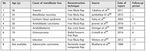
Table 1. Summary of the reported cases of total mandibular reconstruction
Cmtr 10 00048 i001
There have been no reports of virus contamination or acquired pathology from FDBA, although this material is in wide use clinically.[32,37,38] Although nowadays partial reconstruction of the mandible is a common treatment, there are a few literature about total mandibular reconstruction (►Table 1). The case described in this study is a new method of total mandibular reconstruction that we cannot find in anywhere in the literature review. It seems that it can be the first choice when total mandibular with condylar reconstruction is needed.

Funding

None.

Conflicts of Interest

None.
Ethical Approval: Not required. I have received patient consent to re-use the image.

Acknowledgments

The authors thank Amir Hossein Tavakoli, assistant professor of TUMS and medical director of Iranian Tissue Bank and Research Center for excellent assistance to this study. The authors also thank the Iranian Tissue Bank and Research Center.

References

  1. Genden, E.; Haughey, B.H. Mandibular reconstruction by vascularized free tissue transfer. Am J Otolaryngol 1996, 17, 219–227. [Google Scholar] [CrossRef]
  2. Rana, M.; Warraich, R.; Kokemüller, H.; et al. Reconstruction of mandibular defects - clinical retrospective research over a 10- year period. Head Neck Oncol 2011, 3, 23. [Google Scholar] [CrossRef]
  3. Younger, E.M.; Chapman, M.W. Morbidity at bone graft donor sites. J Orthop Trauma 1989, 3, 192–195. [Google Scholar] [CrossRef]
  4. Banwart, J.C.; Asher, M.A.; Hassanein, R.S. Iliac crest bone graft harvest donor site morbidity. A statistical evaluation. Spine 1995, 20, 1055–1060. [Google Scholar] [CrossRef] [PubMed]
  5. Sàndor, G.K.; Rittenberg, B.N.; Clokie, C.M.; Caminiti, M.F. Clinical success in harvesting autogenous bone using a minimally invasive trephine. J Oral Maxillofac Surg 2003, 61, 164–168. [Google Scholar] [CrossRef] [PubMed]
  6. Kinoshita, Y.; Maeda, H. Recent developments of functional scaffolds for craniomaxillofacial bone tissue engineering applications. Sci- entificWorldJournal 2013, 2013, 863157. [Google Scholar] [CrossRef]
  7. Burwell, R.G. Studies in the transplantation of bone. VII. The fresh composite homograft-autograft of cancellous bone; an analysis of factors leading to osteogenesis in marrow transplants and in marrow-containing bone grafts. J Bone Joint Surg Br 1964, 46, 110–140. [Google Scholar] [CrossRef] [PubMed]
  8. Bolander, M.E.; Balian, G. The use of demineralized bone matrix in the repair of segmental defects. Augmentation with extracted matrix proteins and a comparison with autologous grafts. J Bone Joint Surg Am 1986, 68, 1264–1274. [Google Scholar] [CrossRef]
  9. Huggins, C. The formation of bone under the influence of epithelium of the urinary tract. Arch Surg 1931, 22, 377–408. [Google Scholar] [CrossRef]
  10. Clokie, C.M.; Sándor, G.K. Reconstruction of 10 major mandibular defects using bioimplants containing BMP-7. J Can Dent Assoc 2008, 74, 67–72. [Google Scholar]
  11. Urist, M.R. Bone: formation by autoinduction. Science 1965, 150, 893–899. [Google Scholar]
  12. Huggins, C.; Wiseman, S.; Reddi, A.H. Transformation of fibroblasts by allogeneic and xenogeneic transplants of demineralized tooth and bone. J Exp Med 1970, 132, 1250–1258. [Google Scholar] [PubMed]
  13. Neigel, J.M.; Ruzicka, P.O. Use of demineralized bone implants in orbital and craniofacial reconstruction and a review of the literature. Ophthal Plast Reconstr Surg 1996, 12, 108–120. [Google Scholar] [PubMed]
  14. Mulliken, J.B.; Glowacki, J.; Kaban, L.B.; Folkman, J.; Murray, J.E. Use of demineralized allogeneic bone implants for the correction of maxillocraniofacial deformities. Ann Surg 1981, 194, 366–372. [Google Scholar]
  15. American Association of Tissue Banks. In 1998 Edition Standards for Tissue Banking; American Association of Tissue Banks: McLean, VA, USA, 1998.
  16. Horowitz, M.C.; Friedlaender, G.E. Immunologic aspects of bone transplantation. A rationale for future studies. Orthop Clin North Am 1987, 18, 227–233. [Google Scholar] [PubMed]
  17. Burchardt, H. Biology of bone transplantation. Orthop Clin North Am 1987, 18, 187–196. [Google Scholar]
  18. Saunders, J.R., Jr.; Hirata, R.M.; Jaques, D.A. Definitive mandibular replacement using reconstruction plates. Am J Surg 1990, 160, 387–389. [Google Scholar]
  19. Davidson, J.; Birt, B.D.; Gruss, J. A-O plate mandibular reconstruction: a complication critique. J Otolaryngol 1991, 20, 104–107. [Google Scholar]
  20. García García, A.; Suarez Quintanilla, D.; Diz Dios, P. Total mandibular replacement with a titanium plate after mandibulectomy for osteosarcoma. J Oral Maxillofac Surg 1994, 52, 863–867. [Google Scholar]
  21. Taylor, G.I.; Miller, G.D.; Ham, F.J. The free vascularized bone graft. A clinical extension of microvascular techniques. Plast Reconstr Surg 1975, 55, 533–544. [Google Scholar]
  22. Hidalgo, D.A. Fibula free flap: a new method of mandible reconstruction. Plast Reconstr Surg 1989, 84, 71–79. [Google Scholar] [CrossRef] [PubMed]
  23. Ferrari, S.; Bianchi, B.; Savi, A.; et al. Fibula free flap with endosseous implants for reconstructing a resected mandible in bisphosphonate osteonecrosis. J Oral Maxillofac Surg 2008, 66, 999–1003. [Google Scholar] [PubMed]
  24. Head, C.; Alam, D.; Joel, A.; et al. Microvascular flap reconstruction of the mandible: a comparison of bone grafts and bridging plates for restoration of mandibular continuity. Otolaryngology -Head Neck Surg 2003, 129, 48–54. [Google Scholar]
  25. Klokkevold, P.R.; Jovanovic, S.A. Newman, M.G., Takei, H.H., Carranza, F., Eds.; Advanced implant surgery and bone grafting techniques. In Carranza’s Clinical Periodontology, 9th ed.; W.B. Saunders: Philadelphia, PA, USA, 2002; pp. 907–908. [Google Scholar]
  26. Albrektsson, T.; Johansson, C. Osteoinduction, osteoconduction and osseointegration. Eur Spine J 2001, 10 (Suppl 2), S96–S101. [Google Scholar] [PubMed]
  27. Goldring, S.R.; Roelke, M.; Glowacki, J. Multinucleated cells elicited in response to implants of devitalized bone particles possess receptors for calcitonin. J Bone Miner Res 1988, 3, 117–120. [Google Scholar] [CrossRef]
  28. Linde, F.; Sørensen, H.C. The effect of different storage methods on the mechanical properties of trabecular bone. J Biomech 1993, 26, 1249–1252. [Google Scholar] [CrossRef]
  29. Rummelhart, J.M.; Mellonig, J.T.; Gray, J.L.; Towle, H.J. A comparison of freeze-dried bone allograft and demineralized freeze-dried bone allograft in human periodontal osseous defects. J Periodontol 1989, 60, 655–663. [Google Scholar] [CrossRef]
  30. Francis, J.; Brunsvold, M.; Prewett, A.; Mellonig, J. Clinical evaluation of an allogeneic bone matrix in the treatment of periodontal osseous defects. J Periodontol 1995, 66, 1074–1079. [Google Scholar] [CrossRef]
  31. Nguyen, H.; Morgan, D.A.; Forwood, M.R. Sterilization of allograft bone: is 25 kGy the gold standard for gamma irradiation? Cell Tissue Bank 2007, 8, 81–91. [Google Scholar]
  32. Campbell, D.G.; Li, P. Sterilization of HIV with irradiation: relevance to infected bone allografts. Aust N Z J Surg 1999, 69, 517–521. [Google Scholar] [CrossRef]
  33. Dziedzic-Goclawska, A.; Kaminski, A.; Uhrynowska-Tyszkiewicz, I.; Stachowicz, W. Irradiation as a safety procedure in tissue banking. Cell Tissue Bank 2005, 6, 201–219. [Google Scholar] [CrossRef] [PubMed]
  34. Cornu, O.; Banse, X.; Docquier, P.L.; Luyckx, S.; Delloye, C. Effect of freeze- drying and gamma irradiation on the mechanical properties of human cancellous bone. J Orthop Res 2000, 18, 426–431. [Google Scholar] [CrossRef]
  35. Nguyen, H.; Morgan, D.A.; Forwood, M.R. Sterilization of allograft bone: effects of gamma irradiation on allograft biology and biomechanics. Cell Tissue Bank 2007, 8, 93–105. [Google Scholar] [CrossRef] [PubMed]
  36. Hamer, A.J.; Stockley, I.; Elson, R.A. Changes in allograft bone irradiated at different temperatures. J Bone Joint Surg Br 1999, 81, 342–344. [Google Scholar] [CrossRef] [PubMed]
  37. Centers for Disease Control (CDC). Transmission of HIV through bone transplantation: case report and public health recommendations. MMWR Morb Mortal Wkly Rep 1988, 37, 597–599. [Google Scholar]
  38. Simonds, R.J.; Holmberg, S.D.; Hurwitz, R.L.; et al. Transmission of human immunodeficiency virus type 1 from a seronegative organ and tissue donor. N Engl J Med 1992, 326, 726–732. [Google Scholar] [CrossRef]
  39. Takahiro, A.; Hideaki, K.; Jun, S.; et al. Successful total mandibular reconstruction using a fibular graft with long-term follow-up: A case report. Asian Journal of Oral and Maxillofacial Surgery 2011, 23, 196–200. [Google Scholar]
  40. Jambhkare, S.S.; Mohit, J.K.; Satyahit, D.; et al. Total mandibular reconstruction and rehabilitation: A case report. J Oral Implantol. [Epub ahead of print]. 2014. [Google Scholar] [CrossRef]
  41. Paley, M.D.; Lloyd, C.J.; Penfold, C.N. Total mandibular reconstruction for massive osteolysis of the mandible.( Gorham-StoutSyndrome). Br J Oral Maxillofac Surg 2005, 43, 166–168. [Google Scholar] [CrossRef]
  42. Jeremic, J.V.; Nikolic, Z.S.; Boricic, I.v.; et al. Total mandibular reconstruction after resection of rare honeycomb-like ameloblastic carcinoma: A case report. [CrossRef]
  43. Orringer, J.S.; Shaw, W.W.; Borud, L.J.; et al. Total mandibular and lower lip reconstruction with a prefabricated osteocutaneous free flap. Plast Reconstr Surg 1999, 104, 793–797. [Google Scholar] [CrossRef]
  44. Grinsell, D.; Ting You, B.Y.; Ferris, S.I. Total mandibular reconstruction using four free flap transfers in a patient with large mandibular osteosarcoma. Eur J Plast Surg 2014, 37, 563–566. [Google Scholar]
  45. Winters, R.; Saad, A.; Beahm, D.D.; Wise, M.W.; St Hilaire, H. Total autogenous mandibular reconstruction using virtual surgical planning. J Craniofac Surg 2012, 23, e405–e407. [Google Scholar] [PubMed]
  46. Bhathena, H.; Kavarana, N.H. One-stage total mandibular reconstruction with rib, pectoralis major osteomyocutaneous flap. Head & Neck Surg 1986, 8, 211–215. [Google Scholar]
Figure 1. (a) Preoperative panoramic view showing multilocular lesion. (b) 3D CT scan shows massive destruction of the mandible.
Cmtr 10 00048 g001