Next Article in Journal
Treatment of Mandibular Angle Fractures with Single Three-Dimensional Locking Miniplates without Maxillomandibular Fixation: How Much Fixation is Required?
Previous Article in Journal
Nasal Valve Reconstruction Using a Titanium Implant: An Outcomes Study
 
 
Craniomaxillofacial Trauma & Reconstruction is published by MDPI from Volume 18 Issue 1 (2025). Previous articles were published by another publisher in Open Access under a CC-BY (or CC-BY-NC-ND) licence, and they are hosted by MDPI on mdpi.com as a courtesy and upon agreement with Sage.
Article

Did King Philip II of Ancient Macedonia Suffer a Zygomatico-Orbital Fracture? A Maxillofacial Surgeon's Approach

by
Panagiotis Stathopoulos
Department of Oral and Maxillofacial Surgery, Royal Derby Hospital Ringgold Standard Institution, Derby, UK
Craniomaxillofac. Trauma Reconstr. 2017, 10(3), 183-187; https://doi.org/10.1055/s-0037-1601431
Submission received: 8 October 2016 / Revised: 30 October 2016 / Accepted: 25 November 2016 / Published: 24 March 2017

Abstract

:
Philip II, father of Alexander the Great, succeeded his brother, Perdiccas III, to the throne of Macedonia in 360 BC. He has been described by historians as a generous king and military genius who managed to achieve his ambitious plans by expanding the Macedonian city-state over the whole Greek territory and the greater part of the Balkan Peninsula. The aim of our study was to present the evidence with regard to the facial injury of King Philip II of Macedonia and discuss the treatment of the wound by his famous physician, Critobulos. We reviewed the literature for historical, archaeological, and paleopathological evidence of King Philip’s facial injury. We include a modern reconstruction of Philip’s face based on the evidence of his injury by a team of anatomists and archaeologists from the Universities of Bristol and Manchester. In the light of the archaeological findings by Professor Andronikos and the paleopathological evidence by Musgrave, it can be claimed with confidence that King Philip II suffered a significant injury of his zygomaticomaxillary complex and supraorbital rim caused by an arrow as can be confirmed in many historical sources. To the best of our knowledge, this is the first attempt to present the trauma of King Philip II from a maxillofacial surgeon’s point of view.

Philip II, father of Alexander the Great, succeeded his brother, Perdiccas III, to the throne of Macedonia in 360 BC. He has been described by historians as a generous king and military genius who managed to achieve his ambitious plans by expanding the Macedonian city-state over the whole Greek territory and the greater part of the Balkan Peninsula (Figure 1). After 20 years of warfare and diplomatic negotiations with the other Hellenic states, King Philip II united the Greek city-states and prepared the Macedonian army for the expedition against the Persian Empire [1,2].
King Philip II was characterized by his unlimited ambition, quick temper, and irritable personality but also his bravery and profound disregard of danger [1,2]. These characteristics had repeatedly exposed him to great risk in the battlefield resulting in a fracture of the clavicle (Illyrians), at least two severe leg wounds (Triballians and Scythians) which rendered him lame and a fracture of the right zygomatico-orbital complex, followed by the loss of his eye in the siege of Methoni (355 BC) [3,4].
The aim of our study is to present the evidence with regard to the facial injury of King Philip II of Macedonia and discuss the treatment of the wound by his famous physician, Critobulos. We include a modern reconstruction of Philip’s face based on the evidence of his injury and created by a team of anatomists and archaeologists from the Universities of Bristol and Manchester [5].

Materials and Methods

We reviewed the literature for historical, archaeological, and paleopathological evidence of King Philip’s facial injury. Articles in the Medline relevant to King Philip’s injury were retrieved. The key words used were as follows: King Phillip II, Ancient Macedonia, zygomatic, and orbital fracture. Articles found were searched further for secondary literature. Our purpose was to investigate the circumstances that led to the King’s injury, the type of injury caused, the surgical intervention by his physician, and the details of the King’s disfigurement following the injury.

Results

Historical Evidence

The orator Demosthenes mentions that the King was wounded by an arrow fired by an archer while he was inspecting military mechanisms during the siege of Methoni [3]. The historian Didymus accepts Demosthenes’ version of the mechanism of injury and a plethora of historical sources confirm the incident [6,7,8,9,10].
Plutarch added that King Philip II was injured during the siege of Methoni by an arrow that was shot by the Olynthian archer, Astir [11]. Despite the severity of the injury, the King was saved by Critobulos, losing only his sight of the right eye, as Lucian supported [12]. Pliny maintains that the King’s physician was very skillful in removing arrows without damaging the surrounding tissues with the use of a surgical instrument which was specifically designed for this purpose and named spoon of Diocles (Figure 2) [6]. Critobulos later became famous when he successfully removed an arrow from the chest of Alexander the Great in the battle against the Mallians in India [4].
Archaeological Evidence
The historic discovery of the Royal Tombs in Vergina, Greece, by Professor Manolis Andronikos and his collaborators in 1977 confirmed the Hellenic identity of Macedonia which is known to us from a plethora of historical sources. The second Tomb (Royal Tomb II) contained a marble sarcophagus and many grave goods of astonishing richness. Among them, silver and bronze vessels, gold wreaths, weapons, an armor, two gold larnakes, and one small ivory head which the professor identified as a statuette of Philip II King of Macedonia and father of Alexander the Great. Professor Andronikos based his theory of the Royal Tomb II in Vergina belonging to Philip II on the King’s historically recorded orbital and eye injury [13].
Paleopathological Evidence
The anatomist and anthropologist with special interest in head and neck anatomy and ancient Greek cremations, Jonathan Musgrave, the archaeologist John Prag and medical artist Richard Neave after conducting a thorough examination of the King’s remains reconstructed his skull. Their model realistically represents Philip’s zygomatico-orbital injury. The right supraorbital rim is traversed by an oblique scar, while there is an osseous defect of the zygomaticofrontal suture and asymmetry of the zygomatic bones (Figure 3) [5,14,15]. They have also reached the following conclusions: (1) there is a notch clearly visible in the middle portion in the right supraorbital rim causing asymmetry between the left and the right rims reaching to the right supraorbital foramen (Figure 4); (2) there are traces of a healed zygomatic bone fracture (Figure 5), asymmetry of the maxillary bones and reduced alveolar bone height on the right side (Figure 6); (3) it is quite possible that a small piece of bone close to the zygomatico-maxillary suture was removed after an injury during the King’s life; and (4) according to Musgrave’s team, the King’s injury was caused by an arrow striking from above and resulting in a right sided facial fracture and eye rupture for a long period of time before his death (Figure 7) [5,14,15].

Discussion

There seems to be agreement among historians that Philip must have been proud of his numerous and severe wounds as he did not make any effort to conceal them. It is recorded that the King allowed the artists of his court to depict him with the ugly orbital scar, [16] and that he accepted his injury with no negative effect on his ability in battle [17].
There are several recordings in the literature of the Macedonian King’s trauma during the siege of Methoni, 18 years before his assassination. [6,7,8,9,10,11,12] Recent studies focus on the right eye injury which caused the loss of vision [18,19]; however, the zygomatico-orbital complex injury so vividly described in the paleopathological studies of Musgrave et al. is not clearly highlighted.
Xirotiris and Langenscheidt suggested that the bone deformities of the facial skeleton in the Royal Tomb II in Vergina are not the outcome of war injuries but probably represent normal anatomical features or were due to cremation [20]. Bartsiokas used macrophotography techniques to reexamine Philip’s facial skeleton and reached the conclusion that the changes observed on the bones can be attributed to cremation or represent anatomical variations. The author speculated that the bones and armor found in the Royal Tomb II in Vergina belong to Alexander the Great, a hypothesis which is not, however, supported by any evidence. In the study, the author suggested that the occupant of the Tomb II is not King Philip II but Philip III Arrhidaios. In an attempt to identify the man in the main chamber, the author claimed that at the time of cremation, the flesh had already decomposed and the bones were dry and decreased. As far as the notch of the supraorbital rim is concerned, Bartsiokas attributed its existence to the bony protuberance of this anatomic region [19].
Musgrave et al. decisively answered those points [2]: The zygomatic bones and maxilla after the reconstruction are so complete that a gross asymmetry in the contour of the lateral wall of the maxillary sinus can be seen (Figure 5). It is striking that the height of the alveolar process of the maxilla is significantly reduced especially when viewed from behind (Figure 6). The bones were cremated while still “wet,” before the flesh had decomposed [22]. As Phillip III Arrhidaios was murdered in the autumn of 317 BC and buried 4 to 17 months later, his skeleton would not have become dry and decreased, because it takes several years for bones to achieve the state of dryness [23], and produce the characteristic fracture lines of dry cremation [22]. The astonishing richness of the grave goods and the warlike paraphernalia found in the Royal Tomb II are compatible with an embattled King and not with the undistinguished Arrhidaios.

Conclusions

In the light of the archaeological findings by Professor Andronikos and the paleopathological evidence by Musgrave et al., it can be claimed with confidence that King Philip II suffered a significant injury of his zygomaticomaxillary complex and supraorbital rim caused by an arrow as can be confirmed in many historical sources. To the best of our knowledge, this is the first attempt to present the trauma of King Philip II from a maxillofacial surgeon’s point of view.

References

  1. Andronikos, M. Philip King of Macedon; Ekdotike Athenon: Athens, 1992. [Google Scholar]
  2. Kanellopoulos, P. The Personality of Alexander the Great. Christopoulos J. History of Hellenic Nation. 4; Ekdotike Athenon: Athens, 1973; pp. 10–25. [Google Scholar]
  3. Heinemann, W. (Ed.) Demosthenes, Vol 2; The Loeb Classical Library: Cambridge, 1953; pp. 58–61. [Google Scholar]
  4. Lascaratos, J. Alexander the Great in the Fields of Medicine; J and J Hellas: Athens, 1997; pp. 54–106. [Google Scholar]
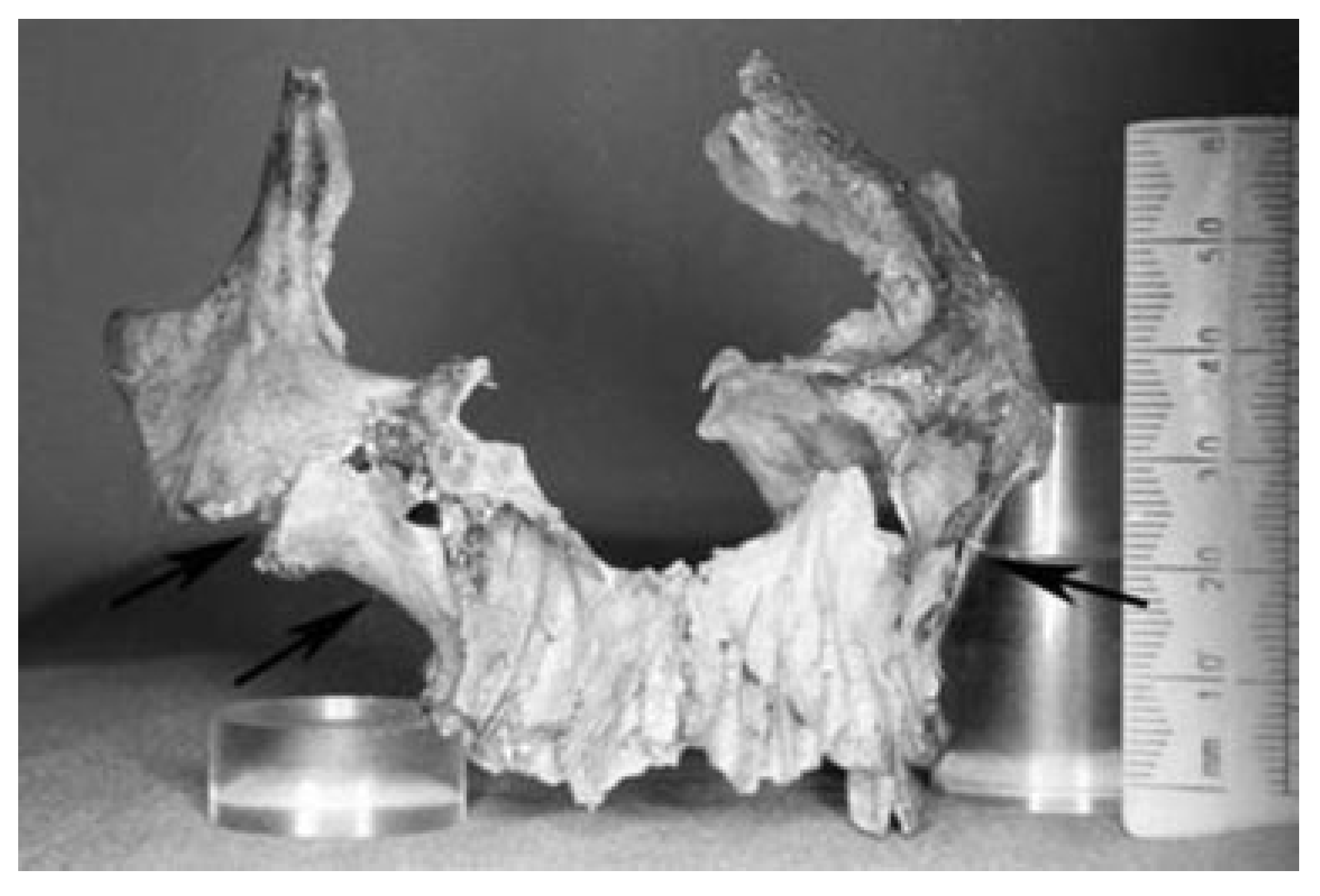
  5. Musgrave, J.H.; Neave, R.A.; Prag, A.J. The skull from tomb II at Vergina: King Philip II of Macedon. J Hell Stud 1984, 104, 60–78. [Google Scholar] [CrossRef] [PubMed]
  6. Heinemann, W. (Ed.) Pliny Natural History, Vol 2; The Loeb Classical Library: Cambridge, 1969; pp. 588–589. [Google Scholar]
  7. Riginos, A.S. The wounding of Philip II of Macedon: Fact and fabrication. J Hell Stud 1994, 114, 103–119. [Google Scholar] [CrossRef] [PubMed]
  8. Heinemann, W. (Ed.) The Geography of Strabo, Vol 3; The Loeb Classical Library: Cambridge, 1954; pp. 344–345. [Google Scholar]
  9. Heinemann, W. (Ed.) The Geography of Strabo, Vol 4; The Loeb Classical Library: Cambridge, 1961; pp. 176–177. [Google Scholar]
  10. Heinemann, W. (Ed.) Diodorus of Sicily, Vol 7; The Loeb Classical Library: Cambridge, 1971; pp. 332–333. [Google Scholar]
  11. Heinemann, W. (Ed.) Plutarch’s Moralia, Vol 4; The Loeb Classical Library: Cambridge, 1962; pp. 278–281. [Google Scholar]
  12. Heinemann, W. (Ed.) Lucian, Vol 6; The Loeb Classical Library: Cambridge, 1959; pp. 52–53. [Google Scholar]
  13. Andronikos, M. Vergina. The Royal Tombs; Ekdotike Athenon: Athens, 1993; pp. 117–175. [Google Scholar]
  14. Prag, A.J. Reconstructing King Philip II: The “nice” version. Am J Archaeol 1990, 94, 237–247. [Google Scholar] [CrossRef]
  15. Prag, A.J. Reconstructing the skull of Philip of Macedon. Danien, E.C. The World of Philip and Alexander; The University Museum of Archaeology and Anthropology: Philadelphia, PA, USA, 1990; pp. 17–37. [Google Scholar]
  16. Hartle Wyman, R. The search for Alexander’s Portrait. Lindsay Adams W, Borza EN. Philip II, Alexander the Great and the Macedonian Heritage; University Press of America: New York, USA, 1982; pp. 153–176. [Google Scholar]
  17. Holton, D. The Tale of Alexander. The Rhymed Version; Byzantine and Neohellenic Library: Salonica, 1974; pp. 37–40. [Google Scholar]
  18. Lascaratos, J.; Lascaratos, G.; Kalantzis, G. The ophthalmic wound of Philip II of Macedonia (360-336 BCE). Surv Ophthalmol 2004, 49, 256–261. [Google Scholar] [CrossRef] [PubMed]
  19. Bartsiokas, A. The eye injury of King Philip II and the skeletal evidence from the royal tomb II at Vergina. Science 2000, 288, 511–514. [Google Scholar] [CrossRef] [PubMed]
  20. Xirotiris, N.I.; Langenscheidt, F. The cremations from the Royal Macedonian tombs of Vergina. Achaeological Newspaper 1983, 1981, 142–160. [Google Scholar]
  21. Lane Fox, R. Introduction. In Brill’s Companion to Ancient Macedon; 2011; pp. 21–22. [Google Scholar]
  22. Musgrave, J.; Prag, W.; Neave, R.; Fox, R.; White, H. The occupants of Tomb II at Vergina: Why Arrhidaios and Eurydice must be excluded. Int J Med Sci 2010, 7, s1–s15. [Google Scholar]
  23. Fiedler, S.; Graw, M. Decomposition of buried corpses, with special reference to the formation of adipocere. Naturwissenschaften 2003, 90, 291–300. [Google Scholar] [CrossRef] [PubMed]
Figure 1. Map of Ancient Macedonia at the time of King Philip’s II death (336 BC) (Map_Macedonia_336_BC-es.svg: Marsyas).
Cmtr 10 00029 g001
Figure 2. Drawing of the spoon of Diocles. King Philip’s physician aimed to insert the arrow’s point into the hole of the instrument and then remove the arrow by pooling it out avoiding any additional tissue damage [18].
Cmtr 10 00029 g002
Figure 3. The reconstructed skull of King Philip II. The model realistically represents Philip’s zygomatico-orbital injury. The right supraorbital rim is traversed by an oblique scar (red arrow) while there is an osseous defect of the zygomatico-frontal suture (yellow arrow) and asymmetry of the zygomatic bones (green arrow) [15].
Cmtr 10 00029 g003
Figure 4. Frontal bone from Royal Tomb II in Vergina, arrow 1 showing the trace of the injury and arrow 2 showing the temporal bone [18].
Cmtr 10 00029 g004
Figure 5. Photo of the anterior aspect of the facial skeleton showing traces of a healed zygomatic bone fracture (right upper arrow), the other two arrows show the asymmetry of the two zygomas [22].
Cmtr 10 00029 g005
Figure 6. Photo of the posterior aspect of the facial skeleton showing asymmetry of the maxillary bones and reduced alveolar bone height on the right side [22].
Cmtr 10 00029 g006
Figure 7. Reconstruction of the face of King Philip II (source: http://alchetron.com/Philip-II-of-Macedon-806068-W#).
Cmtr 10 00029 g007

