Next Article in Journal
A Case of Glenoid Fossa Fracture, Progressive Ankylosis, Total Joint Reconstruction with Alloplastic Prosthesis to Normalized Function Including Evaluation with F18-PET/CT—A Four Year Follow-up
Previous Article in Journal
Patterns of Maxillofacial Fractures in Uttar Pradesh, India
 
 
Craniomaxillofacial Trauma & Reconstruction is published by MDPI from Volume 18 Issue 1 (2025). Previous articles were published by another publisher in Open Access under a CC-BY (or CC-BY-NC-ND) licence, and they are hosted by MDPI on mdpi.com as a courtesy and upon agreement with Sage.
Case Report

Primary Leiomyosarcoma of the Mandibular Alveolar Mucosa of a 12-Year-Old Child from Ethiopia: A Case Report

by
Tewodros Tefera Kenea
1,*,
Betel Abebe Kebede
2,
Fekadu Mesele Gozjuze
3,
Hagos Kiros
4 and
Frank Wilde
5
1
Department of Cranio-Maxillofacial and Oral Surgery, Yekatit 12 Hospital Medical College, Addis Ababa 251, Ethiopia
2
Department of Pediatrics and Child Health, Addis Ababa University, Addis Ababa, Ethiopia
3
Dental and Stomatology, Haramaya University, Harar, Ethiopia
4
Department of Pathology, Addis Ababa University, Addis Ababa, Ethiopia
5
Department of Oral and Maxillofacial Surgery, Military Hospital Ulm, Academic Hospital Ulm University, Ulm, Germany
*
Author to whom correspondence should be addressed.
Craniomaxillofac. Trauma Reconstr. 2017, 10(1), 56-59; https://doi.org/10.1055/s-0036-1582459
Submission received: 9 August 2015 / Revised: 1 November 2015 / Accepted: 6 December 2015 / Published: 27 April 2016

Abstract

:
Leiomyosarcomas (LMSs) are rare malignant mesenchymal tumors which show smooth-muscle differentiation. Most LMSs involving the oral tissues primarily affect the maxillary sinus, the maxillary or mandibular bone. We present a case of LMS of the mandibular alveolar mucosa, arising in a 12-year-old male child from Ethiopia. A malignant spindle cell-like neoplasm was diagnosed on clinical and radiographic findings as well as on incisional biopsy. The tumor was resected with wide margins. The following histopathologic examination with additional immunohistochemical studies secured the diagnosis LMS. Microscopically, the spindle-shaped tumor cells were arranged in an interlacing fascicular pattern and contained oval to elongated, blunt-ended (cigar-shaped) nuclei. The immunohistochemical examination showed immunoreactive tumor cells for vimentin, actin, desmin, and H-caldesmon, which is pathognomonic for LMS. Immunohistochemical studies are mandatory to differentiate the LMS from other similar spindle cell neoplastic lesions. Radical resection with safety margins and a lifelong periodic follow-up has to be recommended.

Leiomyosarcoma (LMS) is a very rare mesenchymal tumor that exhibits smooth-muscle differentiation [1]. Soft-tissue sarcomas account for approximately 0.7% of all malignant neoplasms, and LMSs have been reported to account for 3 to 7% of soft-tissue sarcomas [2,3]. LMS is a neoplasm that rarely affects the oral cavity because of the scarcity of smooth-muscle fibers in this region. This type of fiber is generally present in blood vessels, circumvallate papillae of the tongue, and certain primitive mesenchymal tissue [4,5,6,7].
Primary lesions are usually found in the soft tissues or in a visceral location, including the gastrointestinal, urinary, and female genital tracts [8]. Only 3 to 10% of LMSs arise in the head and neck area—the nose and paranasal sinuses, the skin and subcutaneous tissue, and the cervical esophagus are the most commonly affected sites [9]. The occurrence of LMS in an intraoral location is very rare. However, when it occurs intraorally, it occurs most frequently in the maxilla, followed by the mandible, tongue, cheek, soft palate, upper lip, and mouth floor [10,11]. In general, these lesions are well defined and symptomatic, firmly adhering to the adjacent tissues [4,6,7,11,12,13].
LMSs occurring in regions with scanty or absent smoothmuscle structures, such as the jawbones and oral tissues, possibly arise from the tunica media of blood vessels, 14 circumvallate papillae [15], myoepithelial cells [16], or from pluripotential, undifferentiated, mesenchymal cells [17,18,19,20].
LMSs usually show distinctive morphologic and architectural features: they are composed of spindle-shaped cells with elongated nuclei and eosinophilic cytoplasm, sometimes showing longitudinal fibrils [21]. The tumor cells are arranged in an interlacing fascicular pattern and contain oval to elongated, blunt-ended nuclei (cigar-shaped). The nuclei may show hyperchromatism and varying degrees of atypia and pleo-morphism [11,22]. More rarely, these tumors consist of rounded epithelioid cells with eosinophilic or clear cytoplasm (epithelioid LMS) [21]. The presence of frequent and atypical mitotic figures and necrotic foci indicates aggressive behavior. The prognosis of LMS is usually poor, due to high recurrence and metastatic rates [23,24].
This article reports on a case of primary mandibular alveolar mucosa LMS and its morphologic and immunophenotypic features.

Case Report

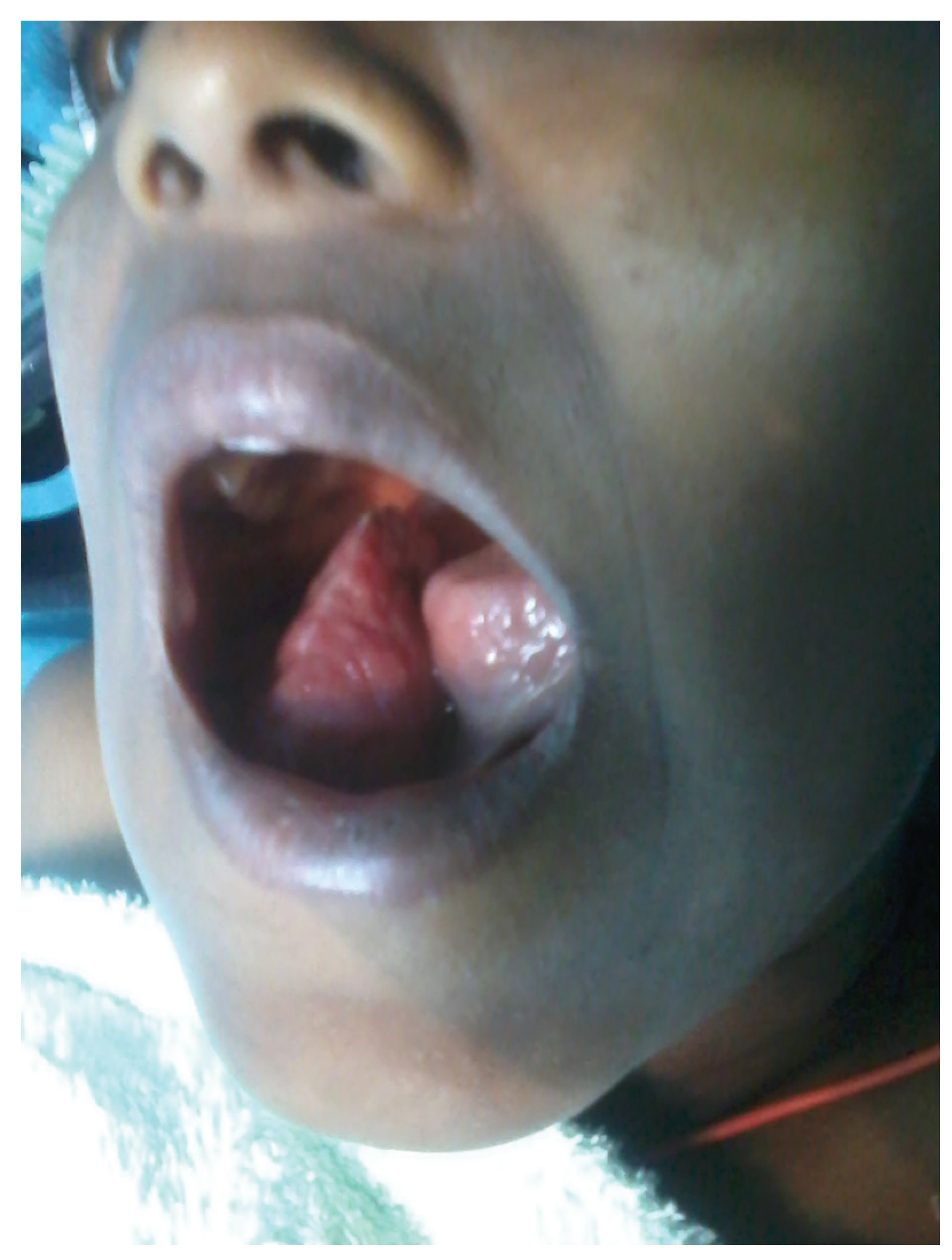
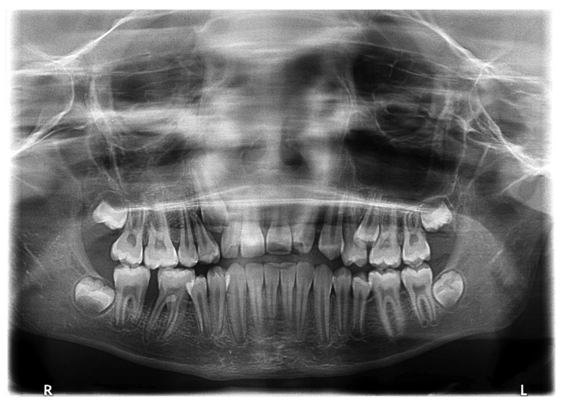
We present a 12-year-old male child presented with progressively growing soft-tissue mass of 3 months duration in the right lower jaw from mandibular alveolar mucosa between tooth 46 and tooth 47 (Federation Dentaire Internationale (FDI) tooth numbering system) (Figure 1). Clinical examination disclosed a reddish lump of soft-tissue consistency with a maximum diameter of 4.5 cm in the right mandibular vesti-bule. The tumor was pedunculated at the base, covering the complete occlusal surface of teeth 47 and 46 and partially 45. The neoplasm shows occlusal impressions of the opponent teeth. It was slightly tender on palpation and score II mobility of tooth 46 according to Miller index [25]. The extraoral examination revealed 1-week duration of right submandibular lymphadenopathy which was slightly tender to palpation. The medical and surgical history of the child was unremarkable. He and his parents denied any previous lesion or local trauma. The panoramic X-ray showed an inter-radicular osteolytic lesion between tooth 47 and tooth 46 with slight periodontal space widening of the distal root of tooth 46 (Figure 2).
Figure 1. Vestibular and lingual swelling in the right mandibular molar region. The tumor was slightly tender to palpation and pedunculated at the base, reddish in color, and measured 4.5 cm in maximum diameter covering the occlusal surface of tooth 47, tooth 46, and partially tooth 45. The neoplasm showed occlusal impressions of the opponent teeth.
Cmtr 10 00009 g001
Figure 2. The panoramic X-ray is demonstrating an osteolytic interradicular alveolar bone lesion between the teeth 47 and 46 with slight periodontal space widening of the distal root of tooth 46.
Cmtr 10 00009 g002
Based on these features, an incisional biopsy was performed. The primary microscopic examination revealed an impression of an inflammatory myofibroblastic tumor lesion in which tumor cells in form of proliferating spindle cells were imbedded in a predominantly variable amount of acute and chronic inflammatory cells along with a rich vascular network. Intersecting bundles of spindle cells were irregularly infiltrating the oral mucosa without involving the adjacent alveolar bone. Unfortunately, at that time, it was not possible to do immunohistochemistry studies (IHC) due to low resources of reagents. On the basis of the radiological, clinical, and histological findings, a glandular neoplasm, a non-neoplastic proliferative process, or a malignant tumor was considered as differential diagnosis.
The child was advised to take amoxicillin with clavulanic acid first. However, there was no improvement of the right submandibular lymphadenopathy as reaction of the antibiotic treatment. Based on the available evidence from radiographic, clinical, and histologic finding, a malignant tumor was considered.
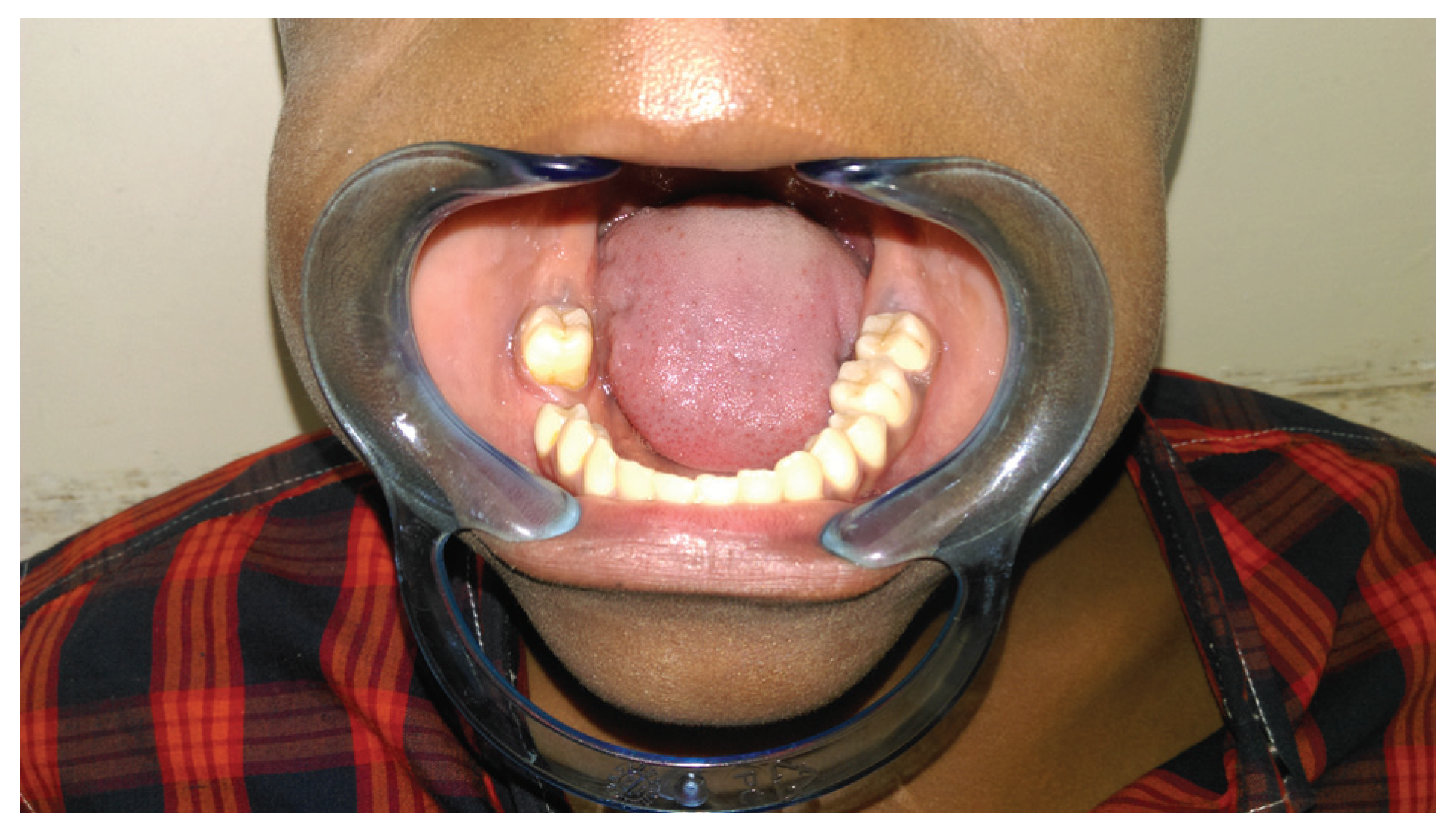
After informed consent of the parents of the child, level I selective neck dissection was performed, followed by radical intraoral tumor resection with a safety margin of approximately 1 cm in combination with extraction of tooth 46. The microscopic examination of the tumor showed spindleshaped tumor cells which were arranged in an interlacing fascicular pattern and contained oval to elongated, bluntended (cigar-shaped) nuclei. The nuclei were characterized by hyperchromatism and varying degrees of atypia and pleomorphism. Cross striation was not evident. Mitotic figures, both typical and atypical, were frequently detected (12–20/10 high power field (HPF)). Necrotic foci were scattered throughout the tumor (Figure 3a). The immunohistochemical study showed that tumor cells were strongly immunoreactive for vimentin, actin (smooth muscle or muscle specific), desmin, and h-caldesmon (Figure 3b). Based on these IHC studies, the final diagnosis was a primary LMS of the mandible alveolar mucosa. Fortunately, no lymph node metastases were identified in the neck dissection specimen. Based on the final definitive IHC diagnosis, the patient underwent staging using chest X-ray, abdominal ultrasound, and CT scan of brain and organ functions test showed no distant metastases. The patient has been kept under periodic follow-up since the date of surgery. No recurrence had been reported by the time of presenting this article. The patient and his parents are satisfied with his quality of life and remained disease free from the time of his last follow-up, 27 months after surgery (Figure 4).

Discussion

The case reported here differs in its clinical presentation from the other documented cases of intraoral LMSs due to the younger age at presentation (12-year-old male child) and of the indolent clinical course. LMS is an extremely uncommon tumor in children [26]. In children, soft-tissue sarcomas are classified as rhabdomyosarcoma or non-rhabdomyosarcoma (NRMS) [27]. Among the NRMSs of childhood, the most common are synovial sarcomas (20–25%), malignant fibrous histiocytomas (10–20%), and malignant peripheral nerve sheath tumors (10–20%) [27].
Figure 3. Histopathologic and immunohistochemistry studies of the leiomyosarcoma. (a) Short, “cigar-like” nuclei with small cytoplasmic clearing adjacent to the nucleus. There are atypical mitotic figures. (Hematoxylin and eosin, ×250). (b) Desmin reactivity in the remark-ably pleomorphic tumor cells helps confirm the smooth-muscle dif-ferentiation (ABC anti-alpha smooth-muscle actin, ×250).
Cmtr 10 00009 g003
Figure 4. Postoperative intraoral view without any signs of local recurrence after the 27th month follow-up.
Cmtr 10 00009 g004
Histopathologically, LMS is a neoplasm characterized by sheets of sweeping, alternating bundles and fascicles of densely packed spindles cells with abundant fibrillar eosinophilic cytoplasm and indistinct cytoplasmic borders, aligned in palisade pattern [28]. This malignancy is distinguished from other spindle cell sarcomas with the help of Masson trichrome staining, ICH analysis, and ultrastructural examina-tion [27,28]. Our patient was positive for vimentin, desmin, and smooth-muscle actin, thus confirming our diagnosis.
The prognosis of intraoral LMS is uncertain. Many reported intraoral LMSs recurred or metastasized repeatedly. Local recurrence occurs in 20.6% of patients and distant metastases (pulmonary or hepatic) develop in 20%. The variable outcome of LMSs may be related to different therapeutic approaches: excision, enucleation, and curettage or radical surgery have been advocated as treatment options [29,30,31]. However, from our point of view, radical surgical removal with a wide surgical safety margin of at least 1 cm has to be recommended.
Radiotherapy can be used as adjunctive treatment, postoperatively [32], even if some authors reported that it does not influence recurrence-free or overall survival rates for intrao-ral LMSs [14,29,31,32]. Chemotherapy seems ineffective and should be reserved for the palliation of patients with metastatic disease or inoperable tumors [29,31,32]. In view of the indolent course of some of these neoplasms and of possible late recurrences or metastases, prolonged or life-long followup has been suggested [17]. The patient should be closely followed up at least during the first 5 years, when LMSs are more likely to recur or metastasize [17,32].

Conclusion

Primary LMS in the pediatric age group is extremely rare. Owing to its innocuous clinical presentation, the possibility of occurrence of this tumor should be considered in differential diagnosis of any oral cavity growth in children. Appropriate clinical and histopathological evaluation supplemented with ICH is a must. Early and accurate diagnosis followed by radical treatment is of utmost importance for improving the prognosis of these tumors.

References

  1. Ezinger, F.M.; Weiss, S. Soft Tissue Tumors, 2nd ed.; C. V. Mosby Company: St. Louis, 1988; pp. 402–421. [Google Scholar]
  2. Gustafson, P.; Willén, H.; Baldetorp, B.; Fernö, M.; Åkerman, M.; Rydholm, A. Soft tissue leiomyosarcoma. A population-based epidemiologic and prognostic study of 48 patients, including cellular DNA content. Cancer 1992, 70, 114–119. [Google Scholar] [CrossRef] [PubMed]
  3. Skoulakis, C.; Chimona, T.S.; Tsirevelou, P.; Papadakis, C.E. Subcutaneus leiomyosarcoma of the neck: A case report. Cases J 2010, 3, 52. [Google Scholar] [CrossRef] [PubMed]
  4. Farman, A.G.; Kay, S. Oral leiomyosarcoma. Report of a case and review of the literature pertaining to smooth-muscle tumors of the oral cavity. Oral Surg Oral Med Oral Pathol 1977, 43, 402–409. [Google Scholar] [CrossRef] [PubMed]
  5. Freedman, P.D.; Jones, A.C.; Kerpel, S.M. Epithelioid leiomyosarcoma of the oral cavity: Report of two cases and review of the literature. J Oral Maxillofac Surg 1993, 51, 928–932. [Google Scholar] [CrossRef]
  6. Miles, A.E.; Waterhouse, J.P. A leiomyosarcoma of the oral cavity with metastasis to lymph-glands. J Pathol Bacteriol 1962, 83, 551–555. [Google Scholar] [CrossRef]
  7. Poon, C.K.; Kwan, P.C.; Yin, N.T.; Chao, S.Y. Leiomyosarcoma of gingiva: Report of a case and review of the literature. J Oral Maxillofac Surg 1987, 45, 888–892. [Google Scholar] [CrossRef]
  8. Kissane, J.M. Anderson’s Pathology, 9th ed.; C. V. Mosby Company: St. Louis, 1861. [Google Scholar]
  9. Piattelli, A.; Artese, L. Leiomyosarcoma of the tongue: A case report. J Oral Maxillofac Surg 1995, 53, 698–701. [Google Scholar] [CrossRef]
  10. Ethunandan, M.; Stokes, C.; Higgins, B.; Spedding, A.; Way, C.; Brennan, P. Primary oral leiomyosarcoma: A clinico-pathologic study and analysis of prognostic factors. Int J Oral Maxillofac Surg 2007, 36, 409–416. [Google Scholar] [CrossRef]
  11. Vilos, G.A.; Rapidis, A.D.; Lagogiannis, G.D.; Apostolidis, C. Leiomyosarcomas of the oral tissues: Clinicopathologic analysis of 50 cases. J Oral Maxillofac Surg 2005, 63, 1461–1477. [Google Scholar] [CrossRef]
  12. Stout, A.P.; Hill, W.T. Leiomyosarcoma of the superficial soft tissues. Cancer 1958, 11, 844–854. [Google Scholar] [CrossRef]
  13. Fu, Y.S.; Perzin, K.H. Nonepithelial tumors of the nasal cavity, paranasal sinuses, and nasopharynx: A clinicopathologic study. IV. Smooth muscle tumors (leiomyoma, leiomyosarcoma). Cancer 1975, 35, 1300–1308. [Google Scholar] [CrossRef] [PubMed]
  14. Phillips, H.; Brown, A. Leiomyosarcoma: Report of case. J Oral Surg 1971, 29, 194–195. [Google Scholar] [PubMed]
  15. Izumi, K.; Maeda, T.; Cheng, J.; Saku, T. Primary leiomyosarcoma of the maxilla with regional lymph node metastasis. Report of a case and review of the literature. Oral Surg Oral Med Oral Pathol Oral Radiol Endod 1995, 80, 310–319. [Google Scholar] [CrossRef] [PubMed]
  16. Kratochvil, F.J.D., III; MacGregor, S.D.; Budnick, S.D.; Hewan-Lowe, K.; Allsup, H.W. Leiomyosarcoma of the maxilla. Report of a case and review of the literature. Oral Surg Oral Med Oral Pathol 1982, 54, 647–655. [Google Scholar] [CrossRef]
  17. Martin-Hirsch, D.P.; Habashi, S.; Benbow, E.W.; Farrington, W.T. Postirradiation leiomyosarcoma of the maxilla. J Laryngol Otol 1991, 105, 1068–1071. [Google Scholar] [CrossRef]
  18. Sanerkin, N.G. Primary leiomyosarcoma of the bone and its comparison with fibrosarcoma. Cancer 1979, 44, 1375–1387. [Google Scholar] [CrossRef]
  19. Neville, B.W.; Damm, D.D.; Allen, C.M.; Bouquout, J.E. Oral and Maxillofacial Pathology; W. B. Saunders: Philadelphia, PA, 1995; pp. 406–407. [Google Scholar]
  20. Rodini, C.O.; Pontes, F.S.C.; Pontes, H.A.R.; Santos, P.S.S.; Magalhães, M.G.; Pinto, D.S., Jr. Oral leiomyosarcomas: Report of two cases with immunohistochemical profile. Oral Surg Oral Med Oral Pathol Oral Radiol Endod 2007, 104, e50–e55. [Google Scholar] [CrossRef]
  21. Sandruck, J.; Escobar, P.; Lurain, J.; Fishman, D. Uterine leiomyosarcoma metastatic to the sphenoid sinus: A case report and review of the literature. Gynecol Oncol 2004, 92, 701–704. [Google Scholar] [CrossRef]
  22. Fields, J.P.; Helwig, E.B. Leiomyosarcoma of the skin and subcutaneous tissue. Cancer 1981, 47, 156–169. [Google Scholar] [CrossRef]
  23. Martis, C. Leiomyosarcoma of the maxilla: Report of two cases. J Oral Surg 1978, 36, 62–65. [Google Scholar]
  24. Wile, A.G.; Evans, H.L.; Romsdahl, M.M. Leiomyosarcoma of soft tissue: A clinicopathologic study. Cancer 1981, 48, 1022–1032. [Google Scholar] [CrossRef] [PubMed]
  25. Miller, P.D., Jr. A classification of marginal tissue recession. Int J Periodontics Restorative Dent 1985, 5, 8–13. [Google Scholar] [PubMed]
  26. Sumida, T.; Hamakawa, H.; Otsuka, K.; Tanioka, H. Leiomyosarcoma of the maxillary sinus with cervical lymph node metastasis. J Oral Maxillofac Surg 2001, 59, 568–571. [Google Scholar] [CrossRef]
  27. Izumi, K.; Maeda, T.; Cheng, J.; Saku, T. Primary leiomyosarcoma of the maxilla with regional lymph node metastasis. Report of a case and review of the literature. Oral Surg Oral Med Oral Pathol Oral Radiol Endod 1995, 80, 310–319. [Google Scholar] [CrossRef]
  28. Gabbiani, G.; Kapanci, Y.; Barazzone, P.; Franke, W.W. Immunochemical identification of intermediate-sized filaments in human neoplastic cells. A diagnostic aid for the surgical pathologist. Am J Pathol 1981, 104, 206–216. [Google Scholar] [PubMed]
  29. Schenberg, M.E.; Slootweg, P.J.; Koole, R. Leiomyosarcomas of the oral cavity. Report of four cases and review of the literature. J Craniomaxillofac Surg 1993, 21, 342–347. [Google Scholar] [CrossRef]
  30. Kuruvilla, A.; Wenig, B.M.; Humphrey, D.M.; Heffner, D.K. Leiomyosarcoma of the sinonasal tract. A clinicopathologic study of nine cases. Arch Otolaryngol Head Neck Surg 1990, 116, 1278–1286. [Google Scholar] [CrossRef]
  31. Chang, A.E.; Rosenberg, S.A.; Glatstein, E.; Antman, K.H. Sarcomas of soft tissues. In Cancer Principles and Practice of Oncology; DeVita, V.T., Hellmann, S., Rosenberg, S.A., Eds.; Lippincott: Philadelphia, PA, 1989; pp. 1345–1398. [Google Scholar]
  32. Josephson, R.L.; Blair, R.L.; Bedard, Y.C. Leiomyosarcoma of the nose and paranasal sinuses. Otolaryngol Head Neck Surg 1985, 93, 270–274. [Google Scholar] [CrossRef]