Sesamoid bones are small osseous elements present in some tendons, where they wrap around bony prominences. [
1,
2] Although sesamoid bones of the first metatarsophalangeal joint are considered a normal part of the skeleton, sesamoids of the lesser toes are seen rarely. At the end of the 19th century and early in the 20th century, investigators began to study the frequency and distribution of the foot sesamoids concurrently with the development of radiologic imaging. More recent studies [
1,
3–
5] documented the frequency of sesamoid bones in the foot and hand in large series. On the other hand, sporadic studies [
2,
6–
10] have appeared regarding the possible association between fabellas and foot sesamoids, traumatic conditions of the lateral toe sesamoids, and unusual distributions of the sesamoids in the hand and foot.
No detailed information appears in the orthopedic literature, however, about foot sesamoids and genetic homogeneity of the study population in studies with large sample sizes. Frequencies and divisions of the lesser-toe sesamoids and bilaterality of distributions also are not well documented, with adequate statistical confirmation. In the present study, we evaluated the frequency and distribution of the metatarsophalangeal joint sesamoids in Turkish subjects.
Materials and Methods
Foot radiographs from 423 adults taken between January 18, 2000, and December 10, 2003, were examined. Patients with deformity and pain at the lesser toes were excluded from the study. Because the pathologies of the hallux sesamoid are well defined, patients with hallux complaints were included in the study population. A total of 371 patients (255 women and 116 men) and 602 foot radiographs were available for study; 222 foot radiographs were bilateral. The average patient age was 44 years (range, 19–80 years).
All of the radiographs were taken at the standard distance of 100 cm and were evaluated by two orthopedic surgeons. The presence and shape of the sesamoid bones in various digits and their distribution were recorded. In addition, the presence of one of the most common accessory bones in the foot, the accessory navicular, was recorded. (All types of accessory navicular bones were recorded: type I is true sesamoid; type II, accessory navicular with articulation; and type III, cornual accessory navicular.) Differences according to side, sex, the presence or absence of accessory bones, and bilaterality were analyzed using a software program (SPSS version 10.0; SPSS Inc, Chicago, Illinois) by applying the univariate χ2 test and performing multiple logistic regression analyses, and odds ratios and κ values were calculated. Only the bilateral radiographs (444 feet of 222 patients) were analyzed statistically to improve statistical power.
Discussion
The number of bones in the adult human skeleton can vary greatly among individuals, and this variation is mainly due to the presence or absence of sesamoid bones. [
1,
2] The reason for and pathogenesis of these ossicles in the human skeleton are still unknown. The precise number of these ossicles is also unknown. In the foot, eight sesamoid bones of the metatarsophalangeal joints have been described [
7]; to our knowledge, nine or ten sesamoids have not yet been reported. In theory, sesamoid bones protect the tendon from damage around the bony prominences and increase the mechanical advantage of the associated muscle, although the functional importance of sesamoid bones is unclear except for the patella and hallux sesamoids. The development of small sesamoids is generally thought to be due to phylogenic and mechanical factors. [
1,
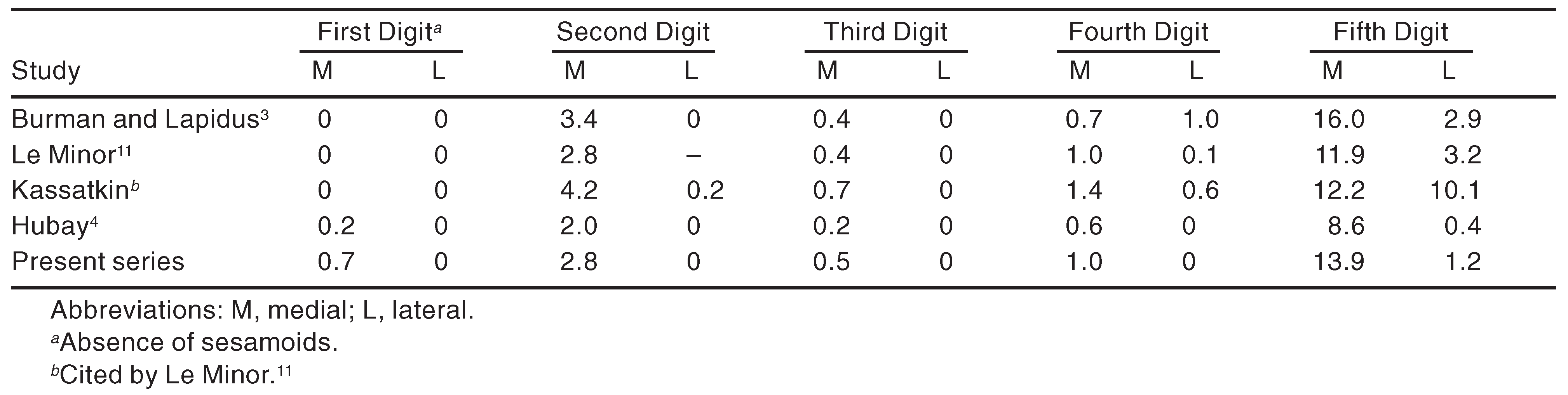
11] A mechanical stimulus could explain the common distributions of the sesamoids. In the foot, lesser-toe sesamoids are much more common on the medial side than on the lateral side, and lesser-toe sesamoids are always more predominant in the second and fifth toes than in other lesser toes. [
3–
5] Possible reasons for these differences in frequency are the asymmetry of the metatarsal head at the plantar surface of the foot and the transverse plantar arch, both of which lead to unequal stress on the metatarsal heads.
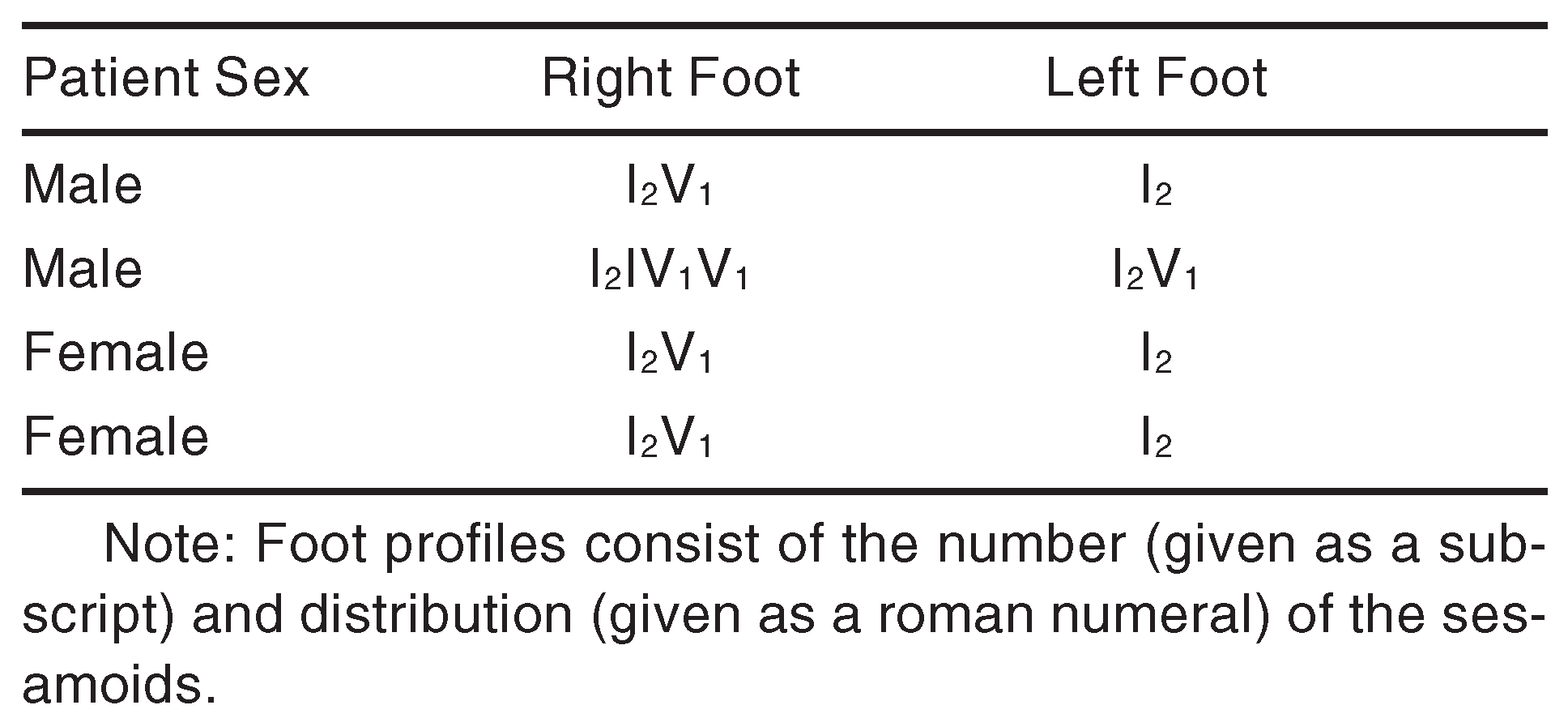
In the present study, the normal or expected foot profile, I
2, was observed in 83.2% of the feet, which is compatible with previously reported values of 81.0% to 89.9% [
3,
4,
11] and the fact that more than 15% of feet in the population could have an unusual foot profile (
Table 4). The I
1 profile (absence or hypoplasia of the hallux sesamoid) was observed bilaterally in two subjects. Congenital absence of the hallux sesamoids is an uncommon variation, and few case studies have been reported in the literature. Inge [
12] documented the first case in 1936, and since then, 15 cases have been reported. [
6,
9,
13–
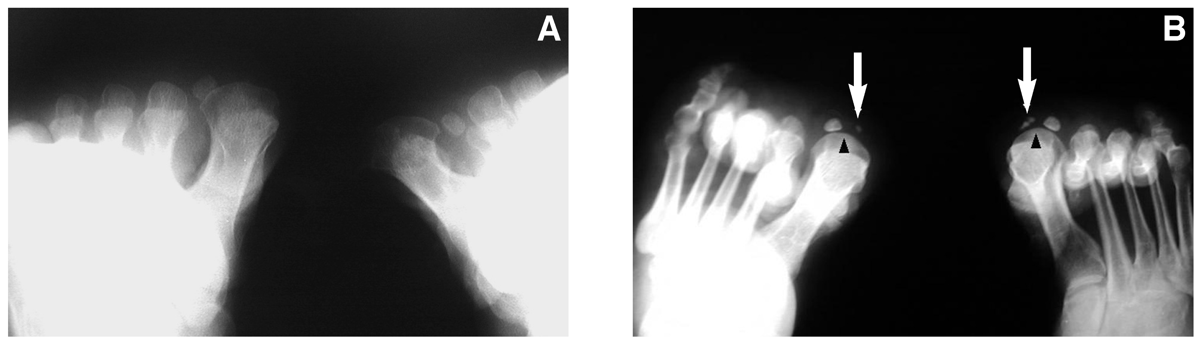
15] Absence of the lateral sesamoid and bilateral absence are rare; most cases are of the absence of medial sesamoids. Two questions should still be asked when absence of the hallux sesamoids is encountered. First, “Is this a true congenital deformity?” Previous surgery or resorption due to a destructive disease could lead to misinterpretation of the hallux sesamoids. In the radiologic examination, the physician should look for the intersesamoidal crest and both sesamoidal grooves. The absence of these structures strongly suggests a true congenital deformity (
Fig. 1). [
9] Second, “Is there any associated deformity?” As mentioned earlier, hallux sesamoids provide a variety of functions and mechanical advantages to the metatarsophalangeal joint. Although the propensity for the absence of hallux sesamoids to be symptomatic or deforming is uncertain, associations with clawing of the metatarsophalangeal joint and hallux deformity have been reported. [
14,
15] In the present study, we observed hallux valgus deformity in both cases of absence of medial sesamoids. One case had bilateral severe deformity, and only the right side was treated with proximal metatarsal osteotomy. Although no special procedure was performed for the absence of tibial sesamoids, the clinical outcome was successful (the American Orthopaedic Foot and Ankle Society Hallux Metatarsophalangeal-Interphalangeal Scale score was improved from 44 to 83). The other case with hypoplasia had bilateral mild deformity, and conservative treatment was administered. The patient was followed up for 3 years, during which she experienced mild symptoms without progression. We can speculate that the absence of medial sesamoids could lead to hallux valgus deformity owing to disrupted functional biomechanics. However, the association may be coincidental.
Divisions of the sesamoid bones are difficult for surgeons to interpret on radiographs because they can mimic sesamoid fracture. Divisions of the lesser-toe sesamoids have not been well studied. The first report was by Lapidus, [
16] who demonstrated sesamoidal bipartition of the second and third toes. Although divisions seem to be related to multiple ossification centers, [
4] trauma could lead to fragmentation. [
8,
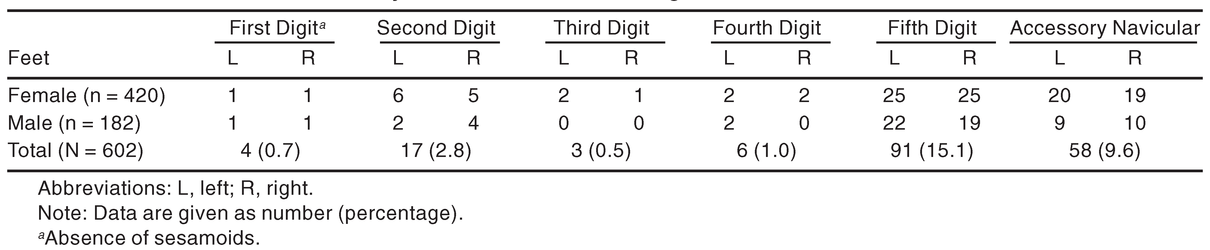
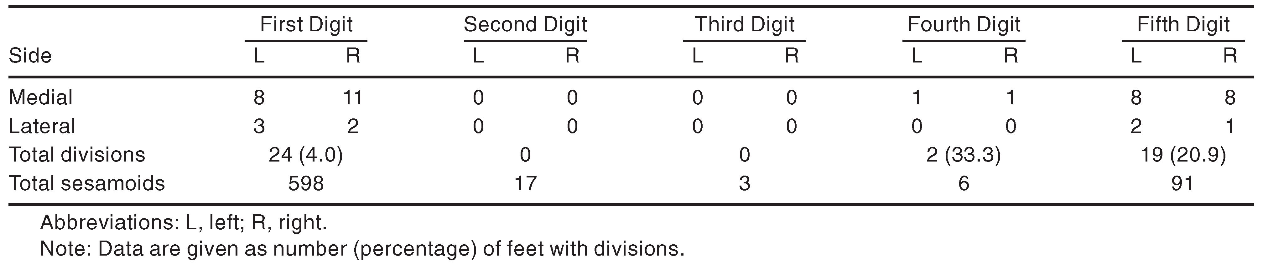
10] In the present study, sesamoidal division in the fifth digit was observed in 20.9% of asymptomatic feet (
Table 2). The incidence of sesamoidal division of the hallux has been reported to be 7.8% to 16.1%. [
3,
5,
17,
18] According to Inge and Ferguson, [
17] division of the sesamoids of the hallux is often unilateral. We found a lower rate of division of the hallux sesamoids (4.0%), and the divisions were mostly bilateral. Although previous reports [
5,
19] showed that a bilateral, symmetrical bone structure occurred in 78% to 82.9% of hand sesamoids, in our study, bilateral symmetrical sesamoid distribution was observed in 98.1% of the cases.
Research on the genetic aspects of and racial differences in the presence of sesamoid bones is sparse. The inheritance of sesamoid bones seems complex and cannot be expressed through simple genetic laws. This is largely due to the existence of multiple connections, little known as yet, between different sesamoid bones and between the left and right sides. [
5] Onat and Cebeci [
20] reported on distributions of the hand sesamoids in Turkish girls and investigated relationships with skeletal growth. In a relatively large series (126 feet), Msamati and Igbigbi [
21] reported distributions of the sesamoid bones of the foot and hand in Malawian (African) subjects. Contrary to previous findings, they did not observe sesamoid bones at the second, third, and fourth digits in the feet of the sample group. Previous prevalence studies have not documented a difference between males and females in the frequency of foot sesamoids. Females have been reported to show slight predominance for hand sesamoids. [
20] In the present study, the occurrence of sesamoids of the fifth digit was higher in males (2.7 times as many cases as in females). This observed sex difference is difficult to explain. In terms of the mechanical aspect of these bones, higher activity levels could be a factor, as boys generally are more active than girls because of participation in contact sports or work. On the other hand, males and females undergo ossification of the sesamoid bones at different times. As with all of the other skeletal tissues, ossification begins 2 years earlier in girls than in boys. [
4] Thus boys receive more mechanical stimulation with immature ossification centers.