Weed et al [
1] believed that extrinsic rearfoot posts added to orthotic shells improve control of foot position and provide greater resistance to motion. Blake and Ferguson [
2] agreed, suggesting that the addition of an extrinsic rearfoot post “increased the surface area of control of an orthotic.” They also described several variations in the design of extrinsic rearfoot posts. However, results of investigations of the effects of external forefoot and rearfoot posts have been inconclusive, showing variability between subjects and minimal impact on foot position [
3,
4]. Much of the research to date suggests that the kinematic changes produced by orthotic devices are small and inconsistent [
5–
11]. However, a change in the kinetics of a system may occur without a detectable change in the kinematics.
McPoil and Hunt [
12] maintain that a relationship exists between pathology in an anatomical structure and the degree of stress applied to it. The stresses around a given joint are related to the magnitude of the moment applied to the joint at any instant in time. A moment is the product of the force and lever arm, identified as the perpendicular distance from the application of the force to the joint axis. Kirby [
13–
16] suggested that the spatial orientation of a joint axis may have a profound effect on function and that the position of the axis affects the internal and external moments around a joint and, ultimately, the other joints in the kinetic chain. Fuller [
17,
18] advanced this work further by demonstrating how the interrelationship between the kinetic variable, the center of pressure (COP) on the plantar foot, and the spatial location of a joint axis may lead to abnormal stresses in the tissues.
The measurement of the COP describes the location of the pressure applied to the foot and is defined as a single point, calculated from the ground reaction components applied over the total plantar surface of the foot: the point at which there is no moment from all of the applied vertical pressures at a series of instants in time, as the ground reaction is applied over the plantar surface of the foot [
17,
19,
20]. The location of the COP can be used to determine the length of the lever arm and to calculate the moment about a given joint at any instant in time. To alter the moments acting about a joint, it is necessary to change either the magnitude of the force or the length of the lever arm. Foot orthoses should be capable of changing the axial position, the magnitude and position of the force, or both if reduction of tissue stress is the objective.
Fuller [
17,
18] speculated that orthoses work by changing the location of the COP and in so doing altering the moments applied around joints, but he provided no data to support this view. Limited research has been published relating to the effects of orthoses on the COP pathway [
19–
21]. Meaningful interpretation of these studies is difficult because the total COP path was considered for the stance phase of gait as a whole. This approach could result in the loss of vital information regarding more subtle changes that may occur at specific times in the stance phase. It seems that no research has specifically studied the effects of rearfoot post design on COP variables. This study investigated the effects of extrinsic rearfoot post design on the lateral-to-medial position of the COP during four stages of the stance phase of walking.
Methods
Subject
A single subject was used in this study. Previous studies [
22,
23] of the effectiveness of orthotic devices used this design to ensure that subject-specific information was not lost. We studied a 35-year-old woman with no history of traumatic injury or lower-limb pathology in the past 12 months. A lower-limb biomechanical assessment of the subject revealed the following: lower-extremity flexibility was unremarkable; no torsional deformities were observed; a slight limb-length discrepancy of less than 5 mm was measured (left shorter than right); no forefoot deformity was seen in the right foot, but a rigid plantarflexed fifth ray was identified on the left; the measurement for relaxed calcaneal stance position was 0° on the right and 6° everted on the left, with the total range of motion available at the subtalar joint recorded at 31° and 36°, respectively; the subject was categorized as having a bilateral fully compensated rearfoot varus; a high proportion of transverse plane motion was elicited at the midtarsal complex of both feet; during nonweight-bearing, the maximum range of motion at the first metatarsophalangeal joint was 90°; and with repeated weightbearing, no limitation in first metatarsophalangeal joint range of motion was found.
Instrumentation
Data were collected using an in-shoe pressure-measurement system (F-Scan; Tekscan, South Boston, Massachusetts) at a sampling frequency of 50 Hz. The CoM’nalysis add-on software for the F-Scan was used to measure COP displacement. The study protocol was designed to maximize as far as was practical the reliability and validity of the system. Note that the F-Scan in-shoe measurement system has limitations, particularly in terms of absolute data values; however, several studies [
26,
27] have suggested that the system is adequate for determining the rank order of data. In recognition of these limitations, movement of the COP in this study is not described in absolute terms but rather indicates relative displacement.
Procedure
A new pair of sensors was used for each data-collection session. Initially, the sensors were cut to the appropriate size and fitted into the subject’s footwear. Before calibration, the subject was asked to walk for a minimum of 14 cycles to allow for preconditioning of the sensor [
28]; once preconditioning was complete, the subject was weighed and the sensor was calibrated. The force recorded by the equipment was checked to ensure that it was within ±10% of the subject’s weight before proceeding with data collection.
The subject was instructed to walk at her normal walking velocity, and a metronome was adjusted to match the cadence. Throughout the session, to maintain a constant walking velocity, the subject kept time with the metronome. According to Cornwall and McPoil [
29], changes in walking speed between testing trials can influence stance phase durations, which in turn can affect the reliability of the COP path.
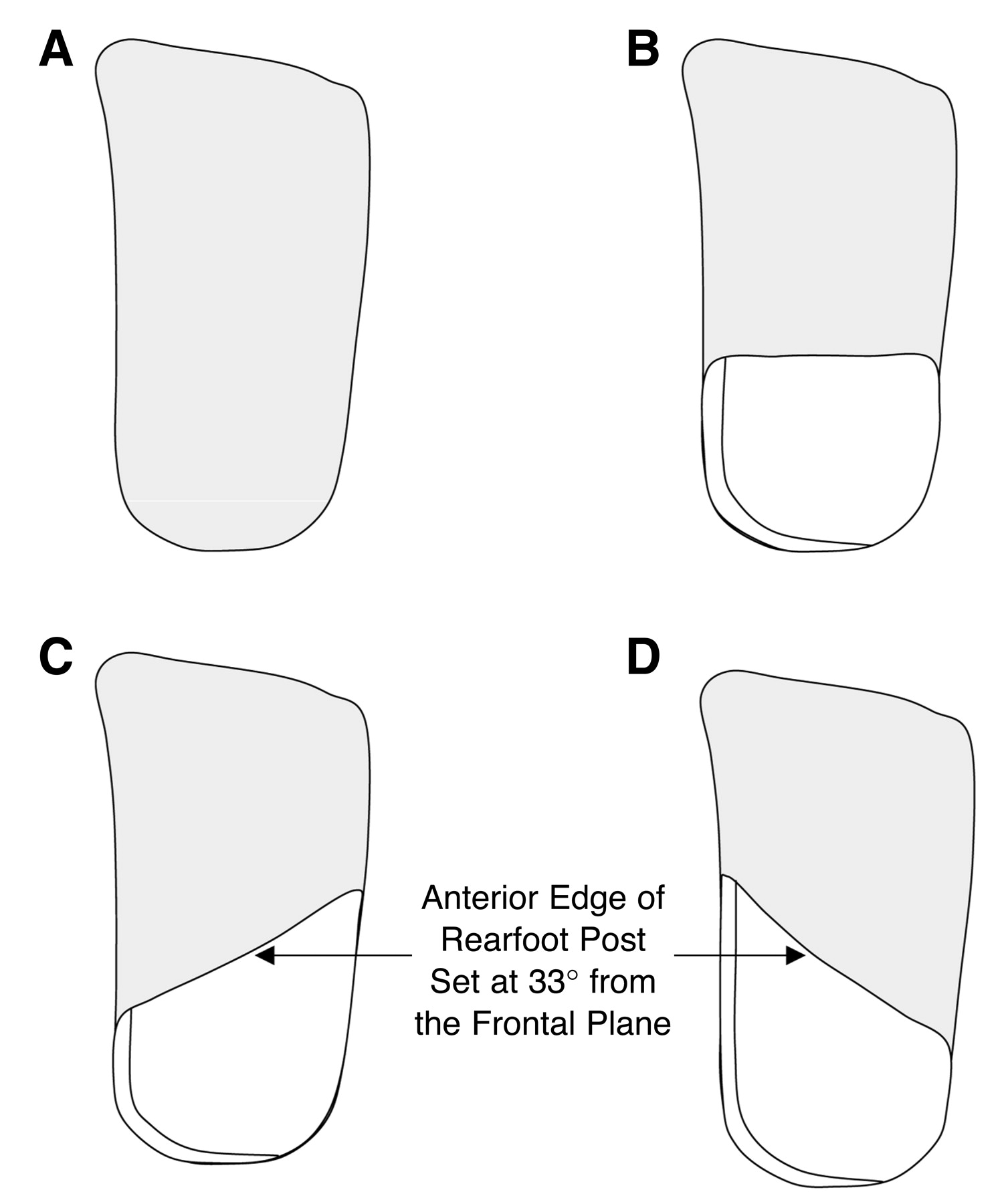
A 10-m walkway was used, which enabled the collection of four or five consecutive gait cycles. To allow for acceleration and deceleration of gait, the first and last gait cycles were disregarded. For each condition tested, three sets of data were collected, giving a total of 12 to 15 steps recorded for each foot. Four conditions were investigated: orthotic shell only, standard post, internal oblique post, and external oblique post.
The four conditions were performed at random; between conditions, the subject placed the shell-only orthotic device in the shoe and walked several lengths of the walkway. Data were collected for both feet, and the session was repeated after 3 days. Using the CoM’nalysis software, two variables were derived from the data, recording the position and velocity of the COP for each percentage of the gait cycle. The two groups of data were lateral-to-medial COP velocity versus time and lateral-to-medial COP movement versus time.
Statistical Analyses
Intraclass correlation coefficients were calculated for the left and right footsteps recorded at the two sessions to test the between-day reliability of the COP movement and velocity data. The Cronbach α was 0.9 and 0.89 for movement and velocity data, respectively, indicating good between-day reliability for the variables [
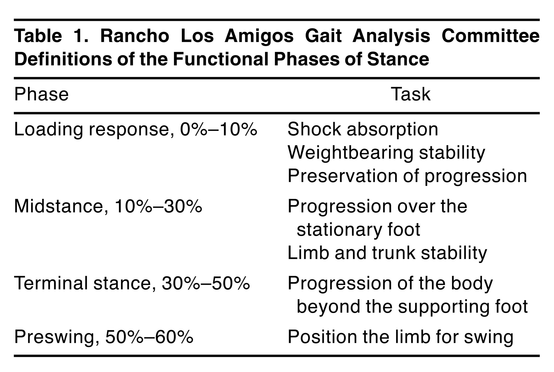
30]. The mean values of the trials for each group of data at each percentage of the gait cycle were computed. Stance period data were broken down into four functional phases according to the Rancho Los Amigos Gait Analysis Committee definitions (
Table 1) [
31].
Paired
t tests were then calculated for each phase of gait to determine whether statistically significant differences existed among the experimental conditions (
Table 2 and
Table 3). Left and right feet were examined separately. Each rearfoot post design was tested against the control (orthotic device without posting) with the assumption that the rearfoot post designs were not related to each other. Further analysis of the interaction between the various post designs was considered to be outside the scope of this study.
When using repeated t tests, the possibility of a type I error is increased. By using the Bonferroni adjustment, the α level of each test can be adjusted downward to retain a final significance level of .05. Therefore, to maintain an overall significance level of .05, each separate test should be performed with a P of .05/4 = .0125.
Results
Throughout the analyses, discrepancies in the results obtained for the right and left feet were apparent. These discrepancies may in part be accounted for by differences in the structure or function of the foot and lower limbs of the subject.
Center-of-Pressure Position
Loading Response.
The loading response period of stance can be defined as the initial double-stance period. The phase begins with initial floor contact and continues until the other foot is lifted for swing [
31]. In the left foot, the maximum medial position of the COP occurred at initial contact, the time when the foot touches the floor. This finding was consistent in the left foot for all three orthotic conditions. This may have been related to the findings from the static biomechanical examination of the left limb whereby the foot was seen to be everted during the relaxed calcaneal stance position. It seems to indicate that the subject was striking the ground with the medial side of her heel. This was followed by a rapid lateral movement of the COP, suggesting rapid supination of the foot.
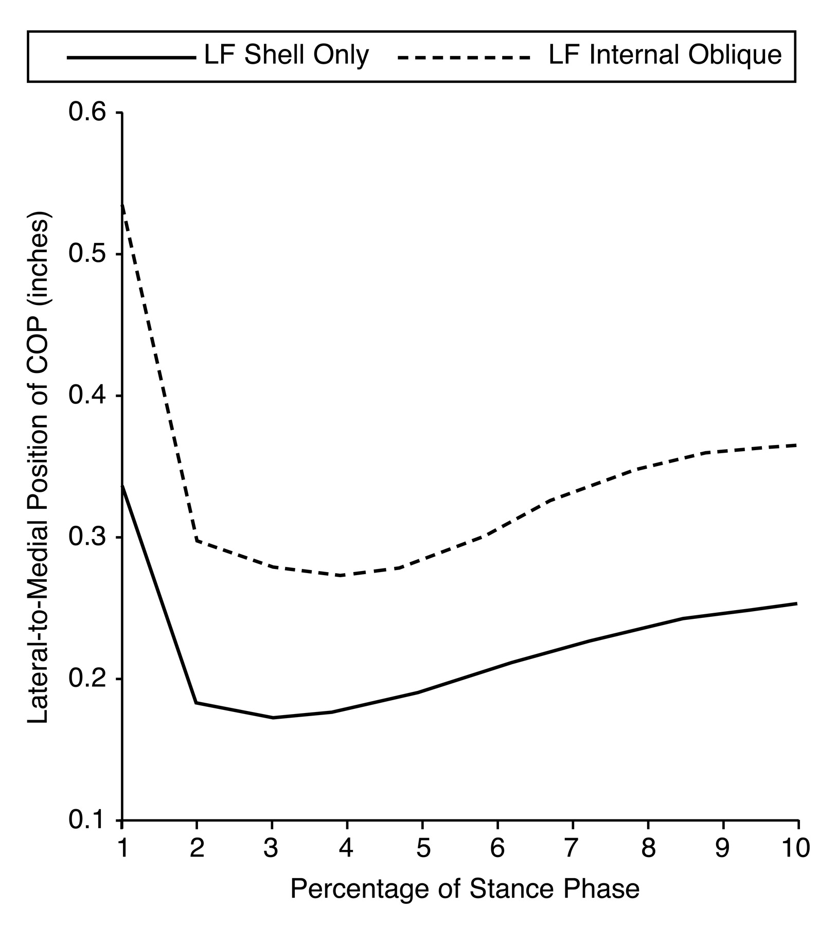
During the loading response phase of gait, significant differences were observed in the lateral-to-medial position of the COP in the left foot between two of the rearfoot posting techniques tested against the same foot functioning with the orthotic shell only (
P < .0125) (
Table 2). The effect of the rearfoot posts was to move the COP path in a medial direction. The rearfoot posting technique that seemed to have the greatest effect on the lateral-to-medial COP path was the internal oblique post (
Fig. 2).
Kirby [
16] and Fuller [
17] suggest that if the COP is positioned medial to the subtalar joint axis, it will produce a supinatory moment. The farther medial the COP is to the subtalar joint axis, the longer the lever arm and the greater the subsequent magnitude of the moment produced given a constant magnitude of force. The medial movement of the COP observed in association with the rearfoot posts suggests that the supination moment produced by the orthotic device may be increased by the addition of the rearfoot posts to the shell. However, little certainty can be accorded to the position of the axis relative to the COP at this time or to the magnitude of the force. It is possible that the shift in the COP position was also accompanied by a shift in the axial position, which may have either enhanced the effect or negated it. The magnitude of the force may also have changed, again either enhancing or negating the effect of the shift in COP in terms of the moments produced about the subtalar joint axis. However, if the medially positioned COP observed at initial contact was indicative of the subject striking the ground with the medial side of her heel, it is possible that the addition of the rearfoot posts made the subject strike the ground even more medially. Further research incorporating kinematic data is required to resolve this issue.
At initial contact of the right foot, the COP was laterally located; as the loading response progressed, a rapid medial movement of the COP occurred. This loading pattern was true of all of the orthotic conditions tested. The lateral position of the COP at initial contact is suggestive of the subject striking the floor with the lateral aspect of her heel; the medial movement of the COP that followed represents immediate and rapid pronation of the foot. The maximum medial position of the COP of the right foot occurred at 10% of the gait cycle (the point at which the opposite foot should leave the ground) for all of the orthotic conditions except the external oblique post, where the maximum medial position of the COP path was reached slightly earlier in the gait cycle. It is reasonable to assume that the maximal medial position of the COP will correspond to the point during the stance phase of gait at which maximal subtalar joint pronation is reached. If this is the case, then the loading pattern demonstrated by the right foot of this subject when wearing an orthosis with and without posting reflects the expected motion occurring in the foot during the loading response [
31] and may reflect the static biomechanical examination that showed a relaxed calcaneal stance position of 0°.
During the loading response, the lateral-to-medial COP position in the right foot failed to show a significant difference for any of the pairings tested (
P < .05) (
Table 2), indicating that the addition of a rearfoot post had no predictable effect on the COP position in the right foot of this subject. This might be explained by the comparatively large standard deviation, produced as a consequence of an outlying data point in this data set.
Midstance.
Midstance is defined as the first half of the single-limb support interval. The phase begins as the other foot is lifted and continues until the body weight is aligned over the forefoot [
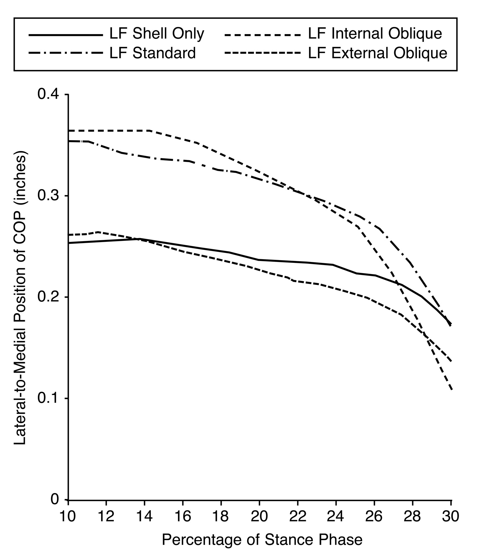
31]. During midstance, a significant difference in the lateral-to-medial position of the COP in the left foot was identified for all of the conditions tested (
P < .001), whereas in the right foot, significant differences were observed in the lateral-to-medial position of the COP in just one of the three comparisons (
P < .0125) (
Table 2).
During this phase of gait, the internal oblique post in both feet and the standard post on the left foot moved the COP path in a medial direction (
Fig. 3). As discussed previously here, the farther medially positioned the COP becomes with respect to the subtalar joint axis, the longer the lever arm and consequently the greater the magnitude of the moment attempting to supinate the subtalar joint, assuming that the subtalar joint axial position and the magnitude of the ground reaction force remain constant. The external oblique post, in contrast, seemed to have the opposite effect at midstance, moving the COP path in a lateral direction (
Fig. 3). These findings suggest that the position of the COP can be adjusted by the addition of a rearfoot post during the midstance phase of gait; furthermore, it seems that the design of the rearfoot post can translate the COP path in either a medial or a lateral direction as desired. In conclusion, if the aim of the clinician is to increase the moment attempting to supinate the subtalar joint at midstance by moving the COP in a medial direction, then an internal oblique post would be the design of choice; however, if the clinician wants to increase the pronation moment acting on the subtalar joint by moving the COP in a lateral direction during midstance, then an external oblique post should be considered, presuming all other variables remain unchanged.
Terminal Stance.
Terminal stance is the phase that completes single-limb support. It begins with heel rise and continues until the other foot strikes the ground [
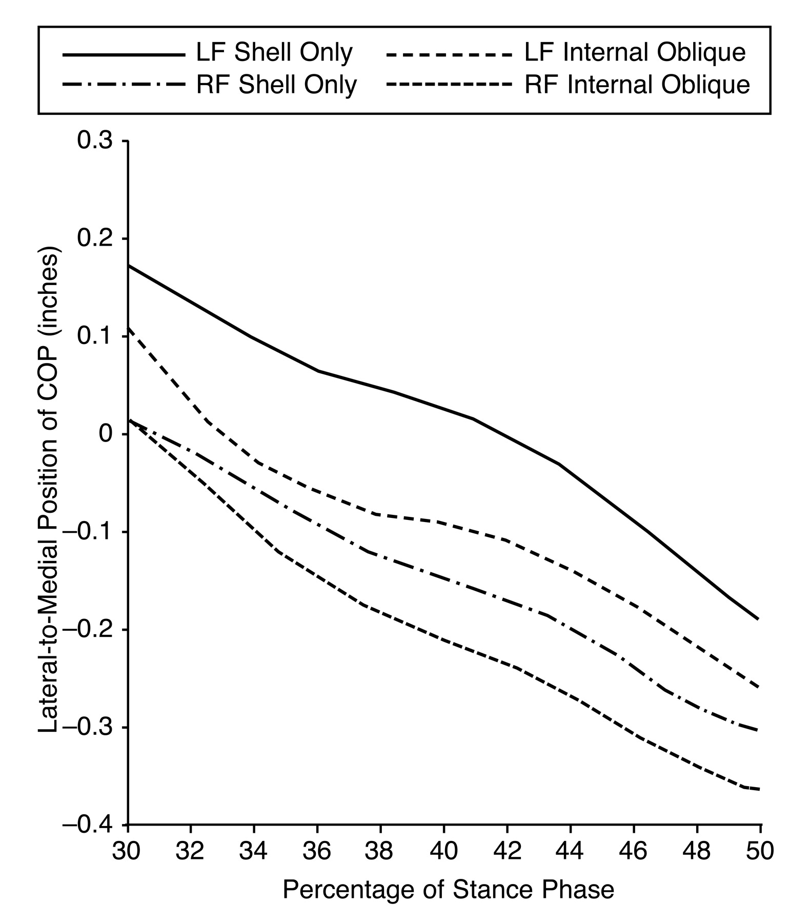
31]. During terminal stance, all but one of the comparisons tested showed a significant difference in the lateral-to-medial COP position (
P < .001 for the left and right feet) (
Table 2). All of the rearfoot post designs tested, except one, moved the COP in a lateral direction compared with the same foot functioning with the shell only. The internal oblique post seemed to have the greatest effect on the COP path at terminal stance (
Fig. 4). The exception was the external oblique post on the right side, where the change recorded was inconsistent.
Preswing.
Preswing is the final phase of stance and the second double-stance interval in the gait cycle. The phase begins with initial contact of the opposite limb and ends with ipsilateral toe-off [
31]. The lateral-to-medial position of the COP in the left foot at the preswing phase of gait failed to show a significant difference (
P < .0125), whereas all of the posting conditions when tested against the shell-only condition on the right revealed a significant effect (
P < .005) (
Table 2). In all of the pairings, the lateral-to-medial position of the COP path moved laterally compared with the shell-only condition.
Lateral movement of the COP position in both feet at terminal stance and preswing implies that the addition of a rearfoot post impacts the COP position, even at a point in the gait cycle when the heel and, consequently, the rearfoot post are no longer in contact with the supporting surface.
A lateral positional shift of the COP with respect to the plantar aspect of the forefoot at terminal stance and preswing is indicative of one or a combination of several of the following changes occurring: an associated increase in the counteracting internal supination moments created by structures intrinsic to the limb, a lateral shift in subtalar joint axial position, and a subsequent lateral shift in vertical body weight. The result will be a foot that functions about a relatively supinated arc of motion and a reduction in body weight medial to the foot. Fuller [
18] predicts that placing more weight on the lateral forefoot would decrease the load on the medial forefoot, thereby reducing tension in the plantar fascia and increasing the ease of first-ray motion and windlass mechanism activation.
No other study to date, to our knowledge, has reported on the effect of rearfoot post design on the lateral-to-medial COP position, although limited research exists relating to the effects of foot orthoses on the total COP path. Scherer and Sobiesk [
19] reported that 66% of their 18 study participants showed a lateral shift in the total COP path when they wore a pair of polypropylene casted insoles, compared with the no-insole condition. However, the study did not determine whether the effect of the orthotic device on the COP path was constant but rather provided the overall average effect, omitting important information relating to the timing of the changes recorded during the stance phase of gait and preventing useful comparisons between the two studies.
A second study by McPoil et al [
20] found that the density of material from which the orthotic device was manufactured had no influence on COP position. In contrast, it has been found that the addition of a rearfoot post has an influence on COP position, indicating that the effect of the rearfoot post might be greater than the effect of the density of the orthotic shell material. Further research using lateral-to-medial COP position and greater subject numbers is necessary to substantiate this conclusion. In the same article, McPoil et al [
20] reported that the total COP path was altered by orthotic therapy only when the subject presented with a forefoot valgus deformity, signifying that the effects of orthoses on gait depend on a large range of interacting variables and undermining the possibility of developing the generic mode of action of the foot orthosis.
Center-of-Pressure Velocity
Loading Response.
At initial contact (the moment the foot touches the floor) in both feet the rearfoot post increased the COP velocity compared with the same foot functioning with the orthotic shell alone. During the loading response phase of gait, no significant difference was identified between any of the comparisons tested in either foot (
P < .0125), except for the left foot shell only and external oblique posts. External oblique posting seemed to increase the velocity of the COP (
Table 3).
Midstance.
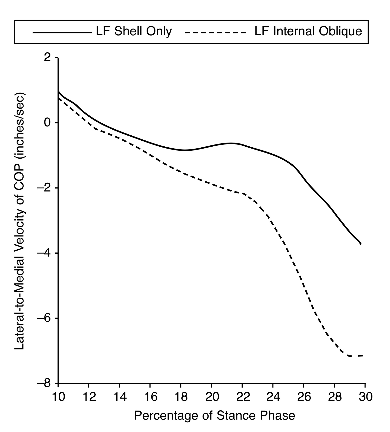
At midstance in the left foot, a significant difference was shown for all of the comparisons tested (
P < .001) (
Table 3). The tested rearfoot posts increased the COP velocity during the midstance phase of the gait cycle compared with the shell-only condition. The internal oblique post seemed to have the greatest effect (
Fig. 5). The COP velocity of the right foot at midstance was not affected (
Table 3).
Terminal Stance.
When exploring the lateral-to-medial velocity of the COP at terminal stance, just two of the six rearfoot post comparisons when tested against the shell-only condition revealed a significant difference (
P < .0125) (
Table 3). The two were shell only
versus standard in the left foot and shell only
versus internal oblique in the right foot. The effect of the different rearfoot posts in these instants seemed small and inconsistent.
Preswing.
The lateral-to-medial velocity of the COP at the preswing phase of gait showed no significant difference in any of the comparisons (
P < .0125) (
Table 3). In the right foot at toe-off in all of the comparisons, the rearfoot post increased the velocity of the COP; however, in the left foot, the rearfoot post reduced the velocity of the COP at toe-off.
The effect of adding a rearfoot post to the shell of an orthotic device on the COP velocity was found to be inconsistent, suggesting that the effect of the rearfoot post during the stance phase of gait on the velocity of the lateral-to-medial COP cannot easily be predicted or controlled. At initial contact, in all of the cases, application of the rearfoot post seemed to increase the velocity of the COP. An increase in the lateral-to-medial velocity of the COP would be consistent with an increase in the velocity of the pronation or supination occurring at that time. In the past, clinicians have attempted to use the rearfoot post to reduce the velocity of subtalar joint motion at initial contact and during the loading response, believing that reducing the velocity of the movement occurring will extend impulse time and thus reduce the magnitude of force applied to the system. With this in mind, where the aim of the clinician is to reduce the impact at initial contact, addition of the rearfoot post on the basis of findings from this study would fail to have the desired effect.
Reducing the velocity of subtalar joint pronation at initial contact may reduce the magnitude of the force applied to the system, but this positive benefit must be balanced by the possible negative effect on the mechanical response of biological tissues to increased load rate. Biological tissues, including all of the musculoskeletal components, exhibit viscoelastic characteristics, one being that the material’s response to loading (strain) is time-dependent (strain rate–dependent). The higher the rate of strain, the stiffer the material becomes, resulting in increased strength and toughness of that material (Garrett et al [
32] and Taylor et al [
33]). Therefore, reducing the velocity of initial contact and consequently its strain rate may have a detrimental effect on the strength of the tissue and its ability to withstand the strain applied to it. From this it can be surmised that the increase in velocity of the COP at initial contact resulting from the introduction of the rearfoot post may be beneficial by increasing the capability of the system to withstand force before injury. However, it would be imprudent to change current practice on the basis of this supposition until further research has been conducted.
The only other study to date to report on COP velocity in relation to orthotic therapy is by McPoil and Hunt [
12]. In their study, ten asymptomatic volunteers were used to determine the effects of two types of orthoses
versus no orthosis on the velocity of the COP path. The findings of the study showed that the orthosis had no effect on the COP velocity.
Discussion
The results of this study support those of McPoil and Hunt [
12], indicating that the lateral-to-medial COP velocity cannot easily be controlled by orthotic therapy and that it is not a suitable measurement to assess the effects of orthotic therapy on gait. However, further research using the lateral-to-medial velocity of the COP is required to determine whether the findings of these studies are representative of a general trend before a final conclusion can be drawn.
The validity of the data collected for this study requires some consideration. It is impossible to differentiate between the natural step-to-step variations in the lateral-to-medial COP path occurring during gait and variability in the F-Scan equipment from changes in the COP pattern secondary to the rearfoot post design. However, in the context of this study it is reasonable to assume that any small variation other than that associated with the rearfoot post design would be constant throughout the trial and for all of the orthotic conditions tested; therefore, although the lateral-to-medial COP data may not be entirely valid, any variation recorded between test conditions remains applicable and is demonstrated by an intraclass correlation coefficient recording good reliability.
A possible limitation of the research method is the single-subject design. A single-subject design prevents the assumption that the findings are representative of the general population. A disadvantage of the single-subject design is that it cannot recognize any general population trends relating to the mode of action of rearfoot post design on the COP path; thus it cannot be postulated that this study is representative of a generic pattern. Other studies [
9,
34] of the effects of the foot orthotic device on foot kinetics and kinematics have concluded that because of the subject-specific nature of orthotic therapy and its effects, the development of a generic model of action is not possible; therefore, it could be argued that a single-subject design is more appropriate for the study of custom orthoses. However, it is necessary for this research design to be developed using an increased sample size to establish whether the findings of this study translate to a larger population and thus can be applied to clinical practice.
The subject recruited for the study was an asymptomatic woman with a relatively unremarkable biomechanical status. It is unlikely that her response to the addition of a rearfoot post is comparable to that of a symptomatic patient, a subject with gross mechanical malalignment, or a subject with an unusual COP path. Because the current research on the COP path is scant, the introduction of additional confounding variables that could affect the outcome was avoided. However, further research that uses a larger sample and considers the use of a subpopulation of matched subjects is warranted.
The orthotic devices were posted to the vertical heel position (0° post) for this study; however, it is also common clinical practice to grind a rearfoot post into varus or valgus by a significant number of degrees. It would be reasonable to envisage that an increased degree of posting incorporated in the rearfoot post would enhance the effect of the orthotic device on the lateral-to-medial COP path, and, therefore, the changes reported in this study may represent a minimal expected effect.
The data analyzed in this study are representative only of the immediate effect of the rearfoot post design on lateral-to-medial COP position and velocity. It is possible that the maximal effect of prescription variation may become apparent only after weeks or months of wearing the orthotic device.
The implication that the COP pattern is symbolic of foot motion is too simplistic; the COP path is a result of the entire body’s kinetic and kinematic activity during gait. The actual interrelationship between COP position and the magnitude and direction of joint motion cannot be made with certainty without the relative position of the joint axis and the magnitude and direction of the force vector also being known. Therefore, the ability of this technique to reflect rearfoot motion should be approached with extreme caution. The only study [
35] to date to test this association used a single value to denote the COP path and concluded that no association was found between this and frontal plane motion of the rearfoot during gait. Further research is warranted to investigate kinetic and kinematic correlation during gait.
Conclusion
During the loading response phase of gait in the left foot, the effect of the rearfoot post was to move the COP path in a medial direction; the internal oblique post provided the greatest effect. At midstance, the internal oblique post and the standard post moved the COP path in a medial direction, whereas the external oblique post had the opposite effect of moving the COP path laterally. During the terminal stance and preswing phases of stance, the influence of all of the posting designs was to move the COP path in a lateral direction.
The velocity of the COP lateral-to-medial path was increased at initial contact for all of the orthotic conditions tested. Only the left foot at midstance showed a consistent and significant increase in COP velocity with addition of the rearfoot post.
The addition of a rearfoot post to an orthotic shell has a significant effect on COP lateral-to-medial position and velocity. Whereas the effect of the post designs seemed to provide reasonably predictable changes in COP position, the effect of the post designs on COP velocity was variable and inconsistent. The effect of the orthotic post was dependent on design and phase of gait. It seems that addition of the rearfoot post had an effect on the lateral-to-medial COP position even at the point in the gait cycle when the heel was no longer in contact with the floor. It seems that the addition of a rearfoot post and specifically the design of the post can be used by the clinician to alter the COP position and velocity. The F-Scan CoM’nalysis software is an appropriate measurement tool for assessing the effect of orthotic devices on the COP path.
Table 1.
Rancho Los Amigos Gait Analysis Committee Definitions of the Functional Phases of Stance.
Table 1.
Rancho Los Amigos Gait Analysis Committee Definitions of the Functional Phases of Stance.
Table 2.
Lateral-to-Medial Position t Tests in the Left and Right Feet.
Table 2.
Lateral-to-Medial Position t Tests in the Left and Right Feet.
Table 3.
Lateral-to-Medial Velocity t Tests in the Left and Right Feet.
Table 3.
Lateral-to-Medial Velocity t Tests in the Left and Right Feet.
Figure 1.
The four orthotic conditions used in this study: shell only (A), standard post (B), external oblique post (C), and internal oblique post (D).
Figure 1.
The four orthotic conditions used in this study: shell only (A), standard post (B), external oblique post (C), and internal oblique post (D).
Figure 2.
Lateral-to-medial center-of-pressure (COP) position curves for the left foot (LF) at the loading response phase of gait.
Figure 2.
Lateral-to-medial center-of-pressure (COP) position curves for the left foot (LF) at the loading response phase of gait.
Figure 3.
Lateral-to-medial center-of-pressure (COP) position curves for the left foot (LF) at the midstance phase of gait.
Figure 3.
Lateral-to-medial center-of-pressure (COP) position curves for the left foot (LF) at the midstance phase of gait.
Figure 4.
Lateral-to-medial center-of-pressure (COP) position curves for the left (LF) and right (RF) feet at the terminal stance phase of gait. Positive coordinates indicate the medial position; negative coordinates, the lateral position.
Figure 4.
Lateral-to-medial center-of-pressure (COP) position curves for the left (LF) and right (RF) feet at the terminal stance phase of gait. Positive coordinates indicate the medial position; negative coordinates, the lateral position.
Figure 5.
Lateral-to-medial center-of-pressure (COP) velocity curves for the left foot (LF) at the midstance phase of gait.
Figure 5.
Lateral-to-medial center-of-pressure (COP) velocity curves for the left foot (LF) at the midstance phase of gait.