To achieve effective forward progression of the center of mass during the propulsive phase of gait, approximately 65° to 75° of hallux dorsiflexion is necessary. [
1,
2,
3,
4,
5] This occurs in two stages. The first 20° to 30° is a hinge-type rotation about the axis of the first metatarsophalangeal joint. [
6,
7] The second stage of hallux dorsiflexion is achieved through plantarflexion of the first ray and activation of the windlass mechanism. [
1,
6] The windlass mechanism was first described by Hicks, [
8] who likened its effect to a cable being wound onequarter of a turn onto the drum of a windlass, with the drum of the windlass being the head of the metatarsal, the handle that does the winding being the proximal phalanx, and the cable that is wound onto the drum being the plantar aponeurosis.
Several theories have been postulated to explain how the second stage of hallux dorsiflexion might be restricted and to determine the mechanism of injury and subsequent foot pathology. One possible explanation considers an association between foot pronation and hallux dorsiflexion capacity, [
8,
9,
10,
11,
12] generated from the common understanding that excessive foot pronation will result in increased tension in the plantar fascia, increased dorsiflexion force under the first metatarsal head, and thus a reduced ability of the hallux to dorsiflex.
The only study to date investigating this relationship was conducted by Harradine and Bevan. [
10] In their study, the maximum available hallux dorsiflexion was recorded using a digital goniometer in static stance, with the subjects wearing shoes. The procedure was repeated with 3°, 5°, and 8° valgus wedges placed under the rearfoot within the shoe in an attempt to replicate the function of an excessively pronated foot. It was concluded from the results that an initial increase in rearfoot pronation produced a statistically significant decrease in hallux dorsiflexion (
P < .05). This study used valgus wedging to artificially replicate three magnitudes of pronation; therefore, the findings may not be representative of the full continuum of foot pronation seen in the general population.
To my knowledge, there is no study quantifying the association between navicular drop and hallux dorsiflexion in subjects displaying differing degrees of pronation. Therefore, the purpose of this study was to determine whether navicular drop (as a measurement of subtalar and midtarsal joint pronation) affects first metatarsophalangeal joint motion. The null hypothesis of this study was that first metatarsophalangeal joint dorsiflexion in stance is independent of navicular drop.
Methods
Subjects
A convenience sample of 24 subjects (5 men and 19 women) was recruited for the study (mean age, 33 years; age range, 21–40 years). Subjects were excluded from the study if they presented with any of the following: a history of surgery or serious injury to the foot, an inflammatory joint disorder, a neuromuscular disorder, current foot pain, limited range of motion in the first metatarsophalangeal joint during nonweightbearing, or increased flexibility in the ankle and subtalar joints. Ethical approval was provided by the Staffordshire University ethics committee, Stoke-on-Trent, England. The nature and purpose of the measurement procedure were fully explained to each subject before written consent was obtained.
Methods and Procedure
All of the measurements were performed on the same day. The environment in which the measurements were taken was standardized throughout the session, the heating and lighting were set at comfortable constant levels, and noise was kept to a minimum. The examining table was positioned at eye level for the clinician, and it was maintained at this level for all of the measurements. A single clinician with 10 years’ experience performed all of the measurements; the readings were shielded and read by an independent observer.
A standardized procedure was performed for each subject that included positioning of the subject, clinician, and independent observer; alignment and use of the instruments; and the order in which the reference marks were made and the measurements taken. The order in which the subjects attended the measurement session was unspecified. The subject stood in a relaxed bipedal stance while the clinician measured the distance between the medial malleolus using a tape measure; the subject then stood on a small elevated platform while the clinician located the navicular and drew the reference mark. Only the right foot was used, and each measurement was repeated four times, with the mean and range values calculated for each set of four measurements.
Navicular Drop
Navicular drop measures the distance the navicular tuberosity moves in stance as the subtalar joint moves from its neutral position to a relaxed position. The most common method of taking the measurement was first described by Brody [
17] and involves making a mark on the skin to identify the tuberosity of the navicular. Then, using a blank card held at right angles to the foot against the navicular, with the base of the card flat on the supporting surface, the height of the navicular is marked on the card. The participant is asked to stand in a relaxed position; again, the height of the navicular is marked on the card. The difference between the two marks is recorded as the navicular drop value.
In an effort to improve repeatability, the technique described by Brody [
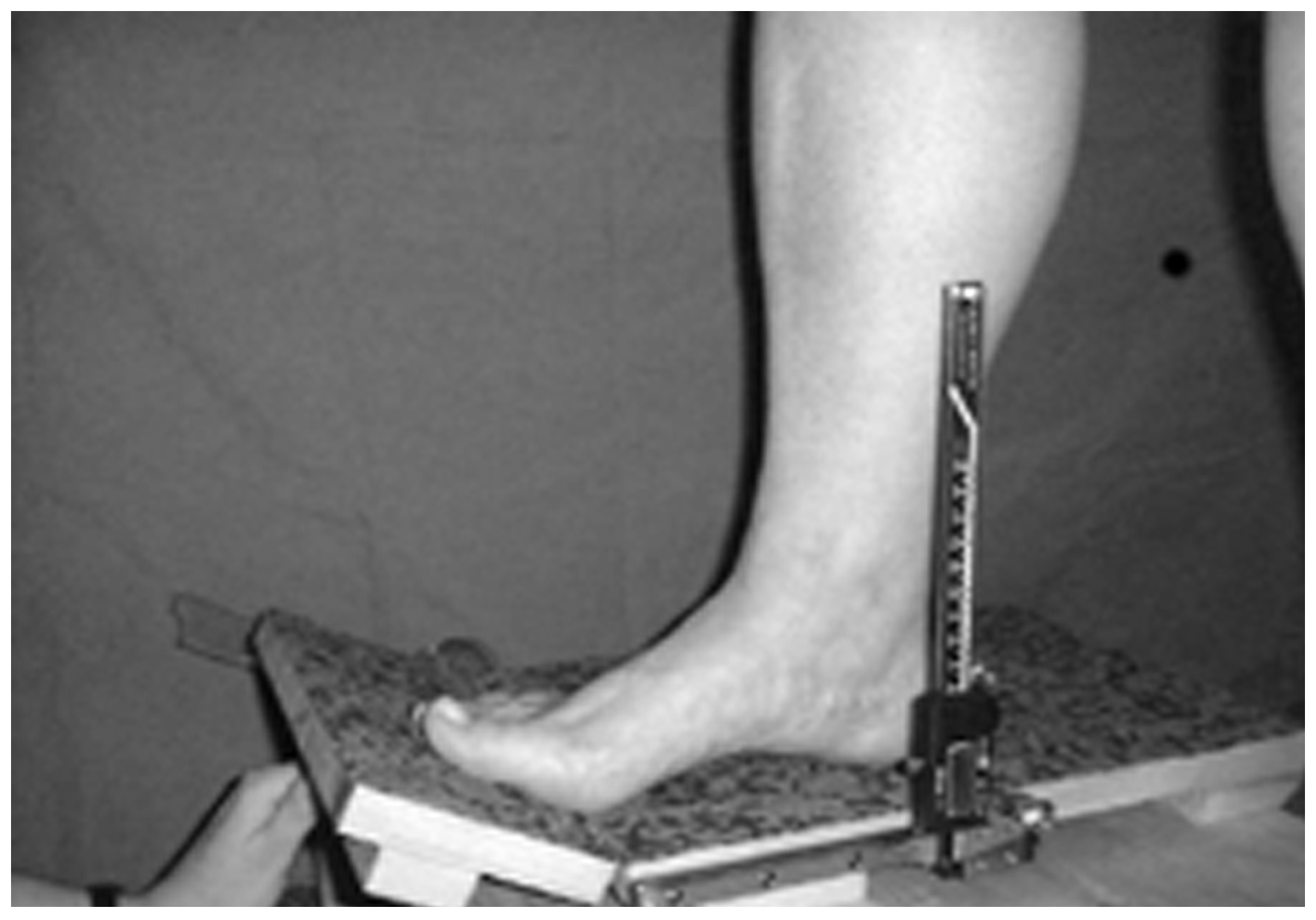
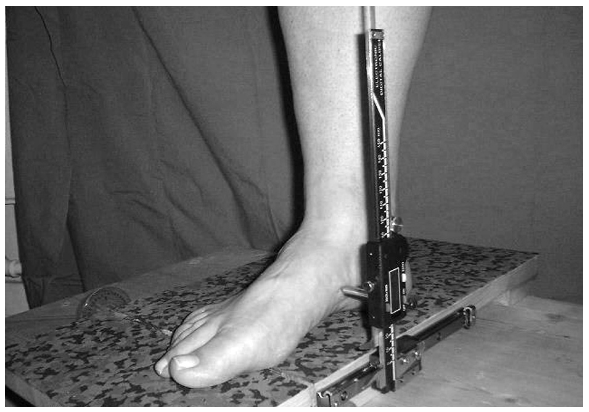
17] was adapted for the study. The subject was asked to stand in the relaxed calcaneal stance position, and a dot was marked on the skin corresponding to the navicular tuberosity. A modified digital caliper was then used to measure navicular drop (
Figure 2). With the subject standing in the subtalar joint neutral position, the caliper arm was placed over the mark and the caliper was set to zero. Subtalar joint neutral was located by palpating for talonavicular congruency and by observing when the curves superior and inferior to the lateral malleolus were equal. The subject then assumed his relaxed calcaneal stance position, and the caliper arm was again placed over the mark. The difference between the two measurements could then be recorded from the digital scale and was defined as the navicular drop value. During the procedure the subject was maintained in the base of stance on the measurement rig and was asked to relax and look straight ahead. The method used to measure navicular drop was described and demonstrated to the subject before the recording took place.
Results
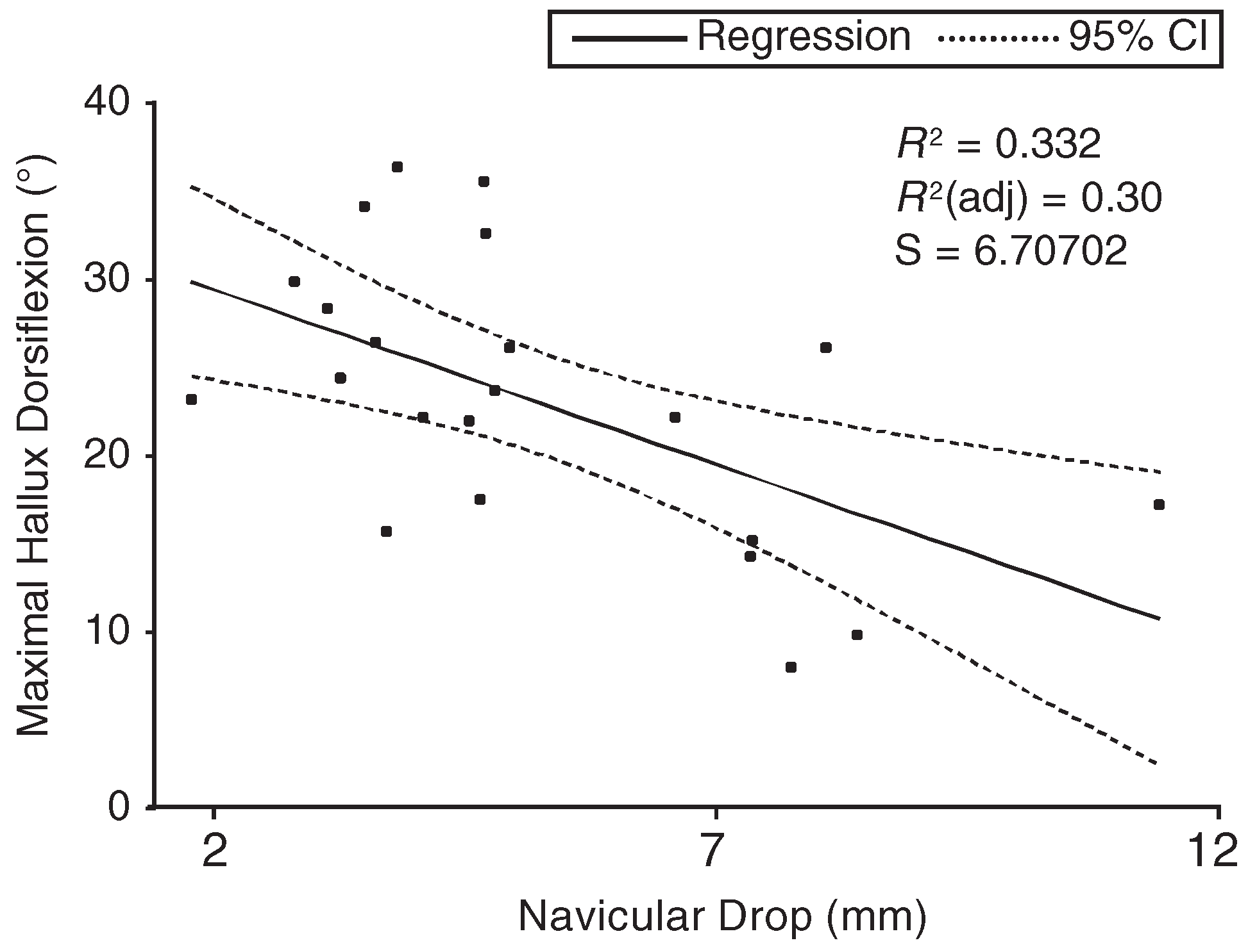
The navicular drop measurement for the 24 subjects ranged from 1.68 mm to 11.50 mm (mean, 5.48 mm). The maximum hallux dorsiflexion measurement for the same group of subjects ranged from 8° to 36° (mean, 22.78°). Analysis of the 24 subjects using the Pearson product moment correlation identified a statistically significant negative correlation at P < .05 between maximum hallux dorsiflexion and navicular drop (Pearson r = –0.474; P = .02). The relationship between the two variables was investigated further in 23 patients using simple regression analysis.
The relationship between navicular drop and maximal hallux dorsiflexion is presented graphically, and the
R2 value is displayed for the pair of variables, in
Figure 3. The null hypothesis that navicular drop has no effect on maximal hallux dorsiflexion can be rejected at the 5% level of significance (
P = .004). An examination of
R2 reinforces the view that navicular drop provides part of the explanation for subject variation in maximal hallux dorsiflexion (
R2 = 0.332). Using simple regression analysis, 33.2% of the variation in maximal hallux dorsiflexion can be explained by different navicular drop values. The
P value for the F statistic is smaller than .05, indicating
R2 at the 5% level of significance (F = 10.421;
P = .004).
Discussion
Navicular drop is negatively correlated with hallux dorsiflexion at the 5% level of significance. It is highly probable, therefore, that the measurement of navicular drop has an effect on hallux dorsiflexion because navicular drop is a measure of sagittal plane and frontal plane movement of the midfoot. As the navicular drop displayed by a subject increases, the angle of hallux dorsiflexion decreases.
These findings are in agreement with most of the related research. Jack (1953), cited in an article by Payne, [
18] found that the windlass mechanism failed to operate in unstable flat feet displaying joint collapse at the talonavicular and naviculocuneiform joints. Roukis et al [
16] demonstrated that a proportionate decrease in the range of motion of the first metatarsophalangeal joint in stance occurs with dorsiflexion of the first ray, and Harradine and Bevan [
10] state that eversion of the rearfoot lowers the maximal hallux dorsiflexion available when measured in the static setting. All agree that lowering of the height of the navicular or, conversely, dorsiflexion of the first ray decreases the passive range of motion of the hallux in stance.
Because a small subject group was used, this is a preliminary study. The information presented shows only a trend that deserves further investigation. Although there is no evidence to suggest that the measures under investigation are sex-related, any generalization of results made to the larger population must take into consideration the nearly 4:1 ratio of women to men in the sample population and acknowledge that this imbalance may have introduced bias to the results.
Although the study design aimed to maximize the repeatability of the measurements used, limitations persist. The repeatability of the navicular drop measurement depends on the examiner’s ability to reproduce subtalar joint neutral as the zero measuring position. Another potential source of error in the study was the end position of hallux dorsiflexion, which was subjectively determined by the examiner to occur with an increase in resistance to dorsiflexion and with first-ray plantarflexion. In future studies, a precisely measured dorsiflexion force at a precisely measured distance from the first metatarsophalangeal joint axis on the Roukis goniometer should further improve the accuracy of measurement of the dorsiflexion moment required to dorsiflex the hallux at the first metatarsophalangeal joint.
Conclusion
The aim of this study was to investigate the suspected association between navicular drop as a measure of foot pronation and hallux dorsiflexion in stance. The results indicate that navicular drop is negatively correlated with hallux dorsiflexion at the 5% level of significance. Although causation cannot be inferred, the results suggest that navicular drop has a linear effect on hallux dorsiflexion. As navicular drop increases, the range of hallux dorsiflexion seems to decrease. The results of this study seem to support the theory that excessive pronation results in increased tension in the plantar fascia and increased dorsiflexion force under the metatarsal head and thus reduces the ability of the hallux to dorsiflex. [
19]