Next Article in Journal
Proximal Oblique Crescentic Osteotomy in Hallux Valgus
Previous Article in Journal
Criterion Validation of Four Criteria of the Foot Posture Index
 
 
Journal of the American Podiatric Medical Association is published by MDPI from Volume 116 Issue 1 (2026). Previous articles were published by another publisher in Open Access under a CC-BY (or CC-BY-NC-ND) licence, and they are hosted by MDPI on mdpi.com as a courtesy and upon agreement with American Podiatric Medical Association.
Article

Plantar Soft-Tissue Thickness Predicts High Peak Plantar Pressure in the Diabetic Foot

by
Frag Abouaesha
1,
Carine H. M. van Schie
1,
David G. Armstrong
1,2 and
Andrew J. M. Boulton
1
1
Department of Medicine, Manchester Royal Infirmary, University of Manchester, Oxford Rd, Manchester, M13 9WL, England
2
Department of Surgery, Podiatry Section, Southern Arizona Veterans Affairs Medical Center, Tucson
J. Am. Podiatr. Med. Assoc. 2004, 94(1), 39-42; https://doi.org/10.7547/87507315-94-1-39
Published: 1 January 2004

Abstract

The aim of this study was to evaluate whether high plantar foot pressures can be predicted from measurements of plantar soft-tissue thickness in the forefoot of diabetic patients with neuropathy. A total of 157 diabetic patients with neuropathy and at least one palpable foot pulse but without a history of foot ulceration were invited to participate in the study. Plantar tissue thickness was measured bilaterally at each metatarsal head, with patients standing on the same standardized platform. Plantar pressures were measured during barefoot walking using the optical pedobarograph. Receiver operating characteristic analysis was used to determine the plantar tissue thickness predictive of elevated peak plantar pressure. Tissue thickness cutoff values of 11.05, 7.85, 6.65, 6.55, and 5.05 mm for metatarsal heads 1 through 5, respectively, predict plantar pressure at each respective site greater than 700 kPa, with sensitivity between 73% and 97% and specificity between 52% and 84%. When tissue thickness was used to predict pressure greater than 1,000 kPa, similar results were observed, indicating that high pressure at different levels could be predicted from similar tissue thickness cutoff values. The results of the study indicate that high plantar pressure can be predicted from plantar tissue thickness with high sensitivity and specificity.

Diabetic foot ulceration remains one of the most serious complications of diabetes mellitus. The recurrence rate of diabetic foot ulcers is extremely high, and these ulcers are associated with a significantly increased mortality rate.[1,2] Several contributing factors lead to diabetic foot ulceration, such as peripheral neuropathy, foot trauma, foot deformity, increased foot pressures, and callus.[3-7] Foot deformities such as Charcot’s foot deformity and claw toes have been shown to increase plantar foot pressures.[8] Claw toes are believed to be related to atrophy of the small muscles in the foot, affecting the balance between toe extensors and flexors. Although there is no direct evidence to support this hypothesis, recent studies have demonstrated that there is evidence of plantar muscle atrophy and change in plantar tissue characteristics in diabetic neuropathic patients.[9-11] Abouaesha et al[12] showed that plantar subcutaneous tissue thickness in the forefoot was significantly related to peak plantar pressure in a large group of diabetic neuropathic patients. However, it is not clear whether there is a threshold of plantar tissue thickness above which high foot pressures are highly likely. Therefore, the aims of this study were to investigate whether plantar tissue thickness could predict high plantar foot pressure and to determine the thickness threshold for high plantar pressure at each metatarsal head site.

Materials and Methods

This study was approved by the Central Manchester ethics committee, and written informed consent was obtained from all participants. A total of 157 diabetic neuropathic patients were invited to take part in the study. Inclusion criteria were evidence of neuropathy, no history of ulceration, and the presence of at least one palpable foot pulse. Neuropathy was defined as a vibration perception threshold greater than 25 V.[13-16] Vibration perception threshold was measured at the tip of the hallux of both feet using the Neurothesiometer (Horwell, London, England).[14] Plantar tissue thickness was measured during weightbearing at each metatarsal head bilaterally using the Planscan (Department of Medical Physics and Clinical Engineering, Royal Hallamshire Hospital, Sheffield, England).[17] The Planscan is a scanning platform that holds a high-resolution probe. The Toshiba SSA-240A ultrasound scanner with a 3.75-MHz curvilinear array transducer (Toshiba Medical Systems Europe, Zoetermeer, the Netherlands) was used for the assessment. Patients stood barefoot on the plastic surface of the scanning platform, which had an ultrasound probe located underneath the upper surface. The metatarsal head of interest was positioned directly above the transducer, and the distance between the most prominent part of the metatarsal head and the skin was determined as the plantar tissue thickness. Three measurements obtained in the longitudinal plane of the metatarsal head were averaged for subsequent analysis. For each patient, measurements were taken of all metatarsal heads. All callus tissue was debrided to normal skin lines before tissue thickness measurements. Dynamic plantar pressures were measured during barefoot walking using the optical pedobarograph (Department of Medical Physics and Clinical Engineering, Royal Hallamshire Hospital).[18] Five steps were analyzed for each foot; any steps regarded as atypical by the investigator, such as tripping, substantially altered gait, or aiming for the pressure plate during data collection, were not saved and thus were not used for analysis. Peak pressure was analyzed at each individual metatarsal head.

Analysis

A receiver operating characteristic analysis was used to determine the optimal combination of sensitivity and specificity for plantar tissue thickness to predict elevated plantar pressure at each metatarsal head. Sensitivity is the ability of a test (in this case, plantar tissue thickness cutoff point) to detect a positive result when the target condition is present (in this case, elevated foot pressures), and specificity is the ability of a test to correctly identify the true negative result when the condition (in this case, elevated foot pressures) is absent. The thickness and peak pressure from both feet were used for the analysis. We used 700 kPa of peak plantar forefoot pressure because this level has been identified as a conservative threshold pressure for defining risk of diabetic foot ulceration.[19] In addition, the receiver operating characteristic analysis was repeated using a cutoff value of 1,000 kPa because this threshold has previously been suggested to be the upper limit of normal pressure levels.[20] A nonparametric analysis with 95% confidence intervals was used to estimate the SE of the area under the sensitivity/specificity curve and to compare the receiver operating characteristic curve’s area to the null hypothesis area of 0.5. For all calculations, an α of .05 was used. Data are presented as mean ± SD.

Results

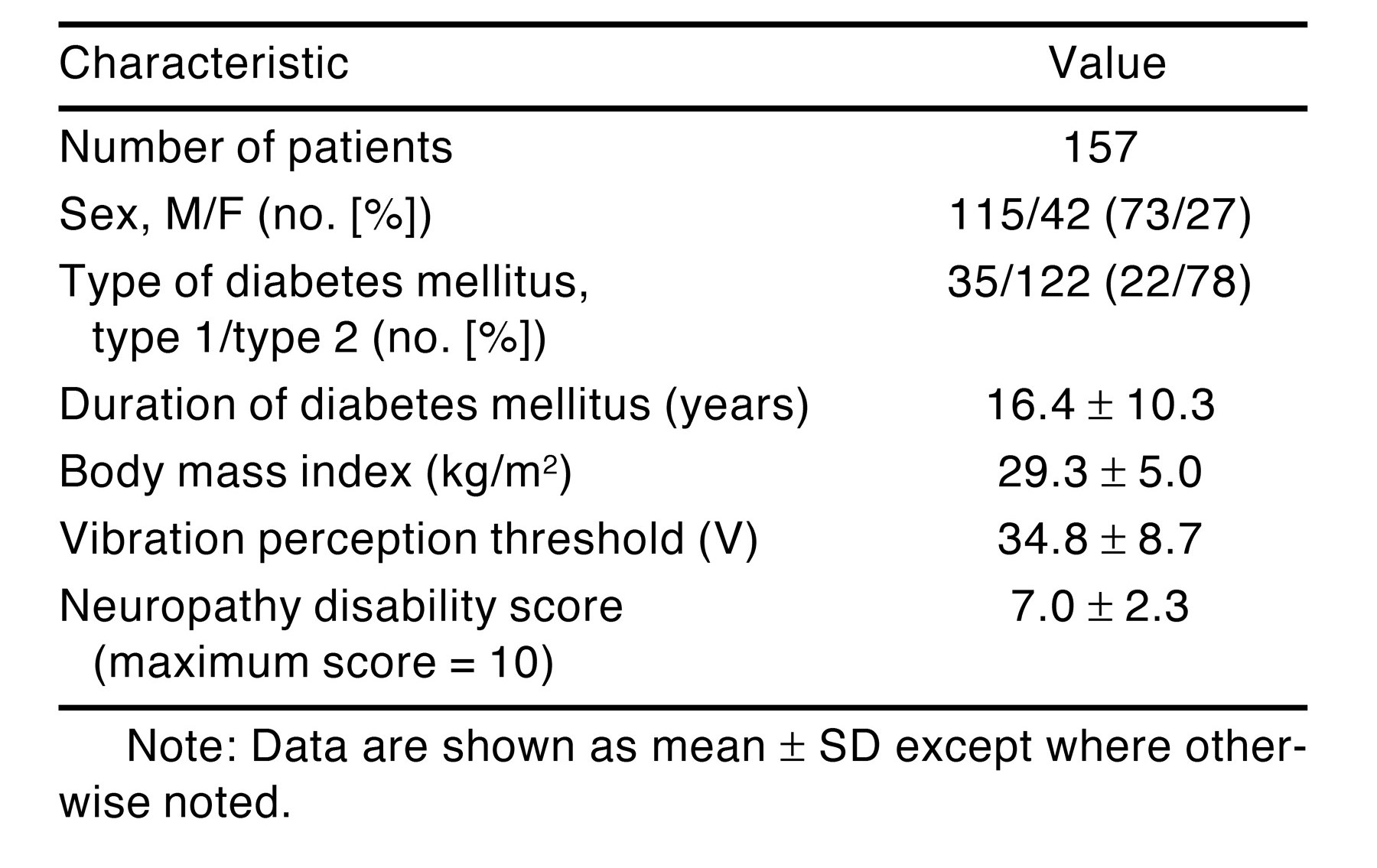
Descriptive characteristics of the study population are listed in Table 1. Tables 2 and 3 show the results of the receiver operating characteristic analysis at each metatarsal head for the pressure thresholds of 700 and 1,000 kPa. Little difference was found between the optimal tissue thickness cutoff values to predict high plantar pressure, as determined by a balance of sensitivity and specificity using the two different pressure thresholds. The area under the receiver operating characteristic curve was significantly greater than the null hypothesis area at each metatarsal head (Tables 2 and 3) using both pressure thresholds.
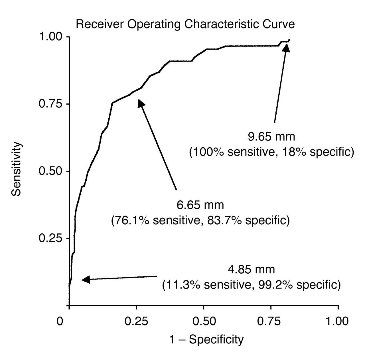
As expected, cutoff values defined at relatively high plantar tissue thicknesses were very sensitive for detecting risk of high foot pressure but very nonspecific (eg, at the third metatarsal head: 9.65 mm = 100% sensitivity and 18% specificity). Conversely, cutoff values made at low plantar tissue thicknesses were very specific for predicting high peak plantar pressure but not very sensitive (eg, at the third metatarsal head: 4.85 mm = 11.3% sensitivity and 99.2% specificity) (Fig. 1).

Discussion

The results of this study indicate that there is a tissue thickness cutoff value below which high foot pressures are highly likely. High foot pressures have been shown to be predictive of future foot ulceration. Although reduced plantar tissue thickness has not yet been shown to be predictive of future foot ulceration, it has been strongly associated with a history of ulceration.[21,22] This is clinically important, indicating that patients with thin plantar tissue pads are at increased risk of foot ulcer development. In addition, it has recently been shown that plantar tissue under the second metatarsal head undergoes compression of 40% during weightbearing,[23] indicating that it is important to have sufficient tissue under the ball of the foot to allow this soft-tissue compression under load bearing for pressure distribution.
In the present study, optimal tissue thickness cutoff values were determined for predicting foot pressures greater than 700 and 1,000 kPa. Two levels were chosen to investigate whether a conservative and a more realistic high pressure level would provide the same results. Because the difference between the optimal cutoff values to predict pressures greater than 700 or 1,000 kPa is small, it is suggested to take the most conservative approach by using the tissue thickness cutoff values that most optimally predict pressure greater than 700 kPa. Different cutoff values are indicated for each metatarsal head because there are natural differences in tissue thickness along the metatarsal heads.[12] For example, the natural tissue thickness at the first metatarsal head is more than twice that at the fifth metatarsal head.
In patients with a thin plantar tissue pad, it is extremely important to protect the skin from harmful high levels of pressure. Recent evidence has shown that injecting silicone is effective in increasing plantar tissue cushioning and subsequently reduces plantar pressure at the injected sites.[24] The finding that high plantar pressure can be easily predicted from plantar tissue thickness indicates that this new treatment might be a promising prophylactic technique for reducing repetitive pressure on the plantar tissue and skin surface, thereby reducing the likelihood of tissue breakdown.

Acknowledgments

This study was funded by a grant from the NHS Executive North West Research and Development Directorate, Manchester, England.
  1. Apelqvist J, Larsson J, Agardh CD: Long-term prognosis for diabetic patients with foot ulcers. J Intern Med 233: 485, 1993.
  2. Boyko EJ, Ahroni JH, Davignon D, et al: Increased mortality associated with diabetic foot ulcer. Diabetes Care 13: 967, 1996.
  3. Veves A, Murray HJ, Boulton AJM, et al: The risk of foot ulceration in diabetic patients with high foot pressure: a prospective study. Diabetologia 35: 660, 1992.
  4. Murray HJ, Young MJ, Boulton AJM, et al: The association between callus formation, high pressures and neuropathy in diabetic foot ulceration. Diabet Med 13:: 979, 1996.
  5. Boyko EJ, Ahroni JH, Smith DG, et al: A prospective study of risk factors for diabetic foot ulcer. Diabetes Care 22: 1036, 1999.
  6. Reiber GE, Vileikyte L, Boulton AJM, et al: Causal pathways for incident lower-extremity ulcers in patients with diabetes from two settings. Diabetes Care 22: 157, 1999.
  7. Pham H, Armstrong DG, Veves A, et al: Screening techniques to identify people at risk for diabetic foot ulceration: a prospective multicenter trial. Diabetes Care 23: 606, 2000.
  8. Ahroni JH, Boyko EJ, Forsberg RC: Clinical correlates of plantar pressure among diabetic veterans. Diabetes Care 22: 965, 1999.
  9. Suzuki E, Kashiwagi A, Kikkawa R, et al: 1H- and 31P-magnetic resonance spectroscopy and imaging as a new diagnostic tool to evaluate neuropathic foot ulcers in Type II diabetic patients. Diabetologia 43: 165, 2000.
  10. Brash PD, Foster JE, Vennart W, et al: Magnetic resonance imaging reveals micro-haemorrhage in the feet of diabetic patients with a history of ulceration. Diabet Med 13: 973, 1996.
  11. Brash PD, Foster J, Vennart W, et al: Magnetic resonance imaging techniques demonstrate soft tissue damage in the diabetic foot. Diabet Med 16: 55, 1999.
  12. Abouaesha F, van Schie CHM, Boulton AJM: Plantar tissue thickness is related to peak plantar pressure in the high-risk diabetic foot. Diabetes Care 24: 1270, 2001.
  13. Armstrong DG, Lavery LA, Fleischli JG, et al: Choosing a practical screening instrument to identify patients at risk for diabetic foot ulceration. Arch Intern Med 158: 289, 1998.
  14. Young MJ, Breddy JL, Veves A, et al: The prediction of diabetic neuropathic foot ulceration using vibration perception thresholds. Diabetes Care 16: 557, 1994.
  15. Young MJ, Boulton AJ, Sonksen PH, et al: A multicentre study of the prevalence of diabetic peripheral neuropathy in the United Kingdom hospital clinic population. Diabetologia 36: 150, 1993.
  16. Abbott CA, Vileikyte L, Boulton AJM, et al: Multicenter study of the incidence of and predictive risk factors for diabetic neuropathic foot ulceration. Diabetes Care 21: 1071, 1998.
  17. Bygrave CJ, Betts RP: The plantar tissue thickness in the foot: a new ultrasound technique for loadbearing measurements and a metatarsal head depth study. The Foot 2: 71, 1992.
  18. Veves A, Boulton AJM: The optical pedobarograph. Clin Podiatr Med Surg 10: 463, 1993.
  19. Armstrong DG, Peters EJ, Lavery LA, et al: Is there a critical level of plantar foot pressures to identify patients at risk for neuropathic foot ulceration?. J Foot Ankle Surg 37: 303, 1998.
  20. Duckworth T, Betts RP, Burke J, et al: The measurement of pressures under the foot. Foot Ankle 3: 130, 1982.
  21. Brink T: Induration of the diabetic foot pad: another risk factor for recurrent neuropathic plantar ulcers. Biomed Tech 40: 205, 1995.
  22. Gooding GA, Stess RM, Grunfeld C, et al: Sonography of the sole of the foot: evidence for loss of foot pad thickness in diabetes and its relationship to ulceration of the foot. Invest Radiol 21: 45, 1986.
  23. Cavanagh PR: Plantar soft tissue thickness during ground contact in walking. J Biomech 32: 623, 1997.
  24. Van Schie CHM, Whalley A, Boulton AJM, et al: Efficacy of injected liquid silicone in the diabetic foot to reduce risk factors for ulceration: a randomized double-blind placebo-controlled trial. Diabetes Care 23:: 634, 2000.
Figure 1. The ability of plantar tissue thickness to predict elevated plantar pressure at the third metatarsal head.
Japma 94 00039 g001
Table 1. Descriptive Characteristics of the Study Population
Japma 94 00039 i001
Table 2. Optimal Tissue Thickness Cutoff Value at Each Metatarsal Head to Predict Peak Plantar Pressure Greater Than 700 kPa
Japma 94 00039 i002
Table 3. Optimal Tissue Thickness Cutoff Value at Each Metatarsal Head to Predict Peak Plantar Pressure Greater Than 1,000 kPa
Japma 94 00039 i003