Acute ruptures and lacerations of the extensor hallucis longus tendon have been previously reported in the literature.[
1-
8] Treatment of these injuries has varied widely. Griffiths[
1] stated that repair of the extensor hallucis longus tendon was unnecessary because “its natural tendency to spontaneously repair after tenotomy is well-known.” His conclusion, however, was based on a single patient who had a satisfactory result without primary repair. Floyd et al[
2] reported on 13 patients with extensor hallucis longus tendon lacerations: 11 underwent primary repair, 1 underwent secondary repair following an infection, and 1 was not repaired. Floyd et al recommended primary repair for extensor hallucis longus tendon lacerations and noted that failure to repair the tendon could not be justified. Current literature[
3-
5] recommends that extensor hallucis longus tendon injuries, both ruptures and lacerations, be repaired primarily. Conservative treatment should be considered only if the injury is distal to the extensor expansion, where dorsiflexion of the hallux would be maintained owing to the structure and function of the extensor expansion and the extensor hallucis brevis tendon.[
3]
Potential complications of not repairing an extensor hallucis longus tendon injury include a hammered hallux, dorsal bunion, hallux flexus, hallux malleus, and mal perforans ulceration.[
3] Disadvantages of repair are postsurgical scarring and adhesion formation, which can result in loss of motion and persistent pain in the great toe.[
3,
7]
Primary repair of the extensor hallucis longus tendon is performed when the severed ends of the tendon can be drawn into close approximation.[
6] Tendon grafting may be used in conditions where the severed ends are unable to be reapproximated owing to tendon degeneration and retraction. Potential forms of autogenous grafts from the foot include the plantaris, the peroneus tertius, slips from the extensor digitorum longus or extensor digitorum brevis, or the extensor hallucis brevis. The ideal position of the tendon after repair should be neutral, and the tension across the tendon should be zero.[
3,
6]
In addition to autografts, tendon allografts are widely available. Several studies[
9-
11] have described the use of tendon allografts to reconstruct the anterior cruciate ligament, patellar tendon, lateral ankle ligaments, and Achilles tendon. Allografts provide the advantages of no donor site morbidity, shorter operative time, smaller incisions, and large graft size availability.[
12] A disadvantage of allograft use is the potential spread of the human immunodeficiency virus. Buck et al[
13] estimated the risk of transmission from an infected bone allograft to be less than one in a million with proper precautions and adequate laboratory studies.
The following case report describes a patient in whom a fascia lata allograft was used to repair a ruptured extensor hallucis longus tendon with a 6-cm defect. The tendon graft was used to stabilize the hallux against the first metatarsal in a traumatically induced flail, floppy great toe.
Case Report
A 61-year-old woman presented to the Temple University School of Podiatric Medicine in Philadelphia, Pennsylvania, with primary complaints of bunion pain, sub–second metatarsal head pain, and a contracted second digit on her right foot. The patient stated that she had had the deformities for several years and was unable to wear shoes without significant discomfort. She had tried various forms of conservative therapy, including footwear modifications, injections, and padding, without relief of pain.
Her medical history was significant for hypertension, which was controlled with atenolol administration. The patient also took estrogen, calcium, and a daily vitamin supplement. She had undergone two previous surgeries—surgery on her left foot for a bunion and second-digit hammer toe correction in 1991 and a hysterectomy in 1986—both without complications. She had never smoked and consumed alcohol socially. Her other medical history was unremarkable.
Physical examination revealed an intact vascular status with no neurologic deficits. The right foot demonstrated a moderate bunion deformity, a second-digit hammer toe that was rigidly dorsiflexed at the metatarsophalangeal joint, and severe pain elicited with palpation of the sub–second metatarsal head. Radiographs were consistent with the clinical findings, confirming a moderate hallux valgus deformity and a second digit that was dorsally dislocated at the metatarsophalangeal joint. Conservative and surgical options were discussed with the patient. Owing to the lack of relief of discomfort from conservative care, she elected to have surgical intervention.
The patient’s first surgery on her right foot consisted of a modified Austin osteotomy with a long dorsal wing, fixated with a single 0.062 Kirschner wire. The extensor hallucis longus tendon was noted to be thin and atrophic but required lengthening. To strengthen the extensor hallucis longus tendon, after an open Z-lengthening, the extensor hallucis brevis tendon was harvested and used as an onlay graft secured with 3-0 absorbable sutures. Procedures on the second digit consisted of proximal interphalangeal joint fusion, metatarsophalangeal joint release, and flexor digitorum longus to proximal phalanx tendon transfer. A partial second metatarsal head resection, with full soft-tissue metatarsophalangeal joint release, was also performed to relocate the toe. The second digit was then fixated with a 0.045 Kirschner wire crossing the proximal interphalangeal joint fusion site and extending proximally across the metatarsophalangeal joint. The patient underwent the surgery without any complications. She was placed in a posterior splint with instructions to remain nonweightbearing and was discharged from the hospital after a short stay in the recovery room.
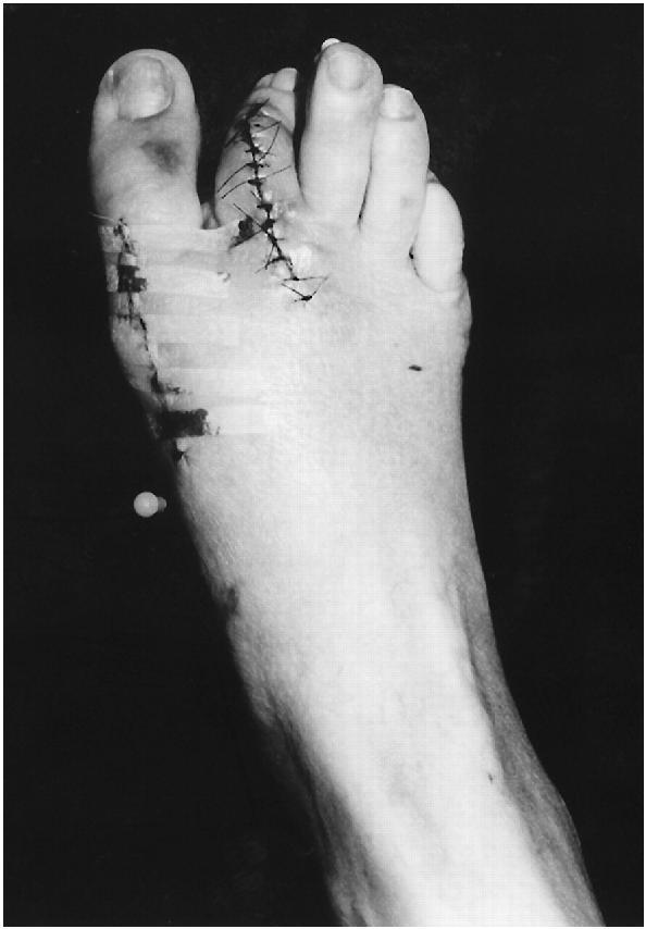
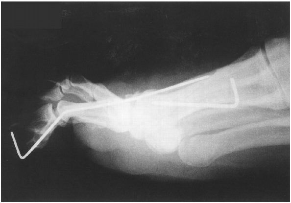
The patient was seen at the clinic 4 days later. She stated that on her second postoperative day she fell, injuring her right foot. Her neurovascular status remained intact. Clinical and radiographic examination demonstrated a second digit that was angled laterally approximately 45° to the proximal interphalangeal joint (
Figure 1 and
Figure 2). The second digit was anesthetized with lidocaine and manually manipulated into correct anatomical alignment. No angular or other deformity of the hallux was noted; however, muscle testing revealed a severe weakness of hallux dorsiflexion, although the extensor hallucis longus tendon could be felt tensing under the skin. A partial extensor hallucis longus tendon rupture was suspected at this time. Because of the patient’s age, the sustained trauma, and the close proximity of the surgery and trauma dates, it was decided to first attempt conservative care. The hallux was splinted in dorsiflexion, and the patient remained nonweightbearing.
During the next 4 weeks the patient did not regain strength in her hallux, and the toe became progressively flail at both the metatarsophalangeal and interphalangeal joints. The patient had increased difficulty walking owing to the lack of stability of the hallux. A complete extensor hallucis longus tendon rupture was now suspected. The patient was scheduled for surgical exploration and repair of the tendon rupture.
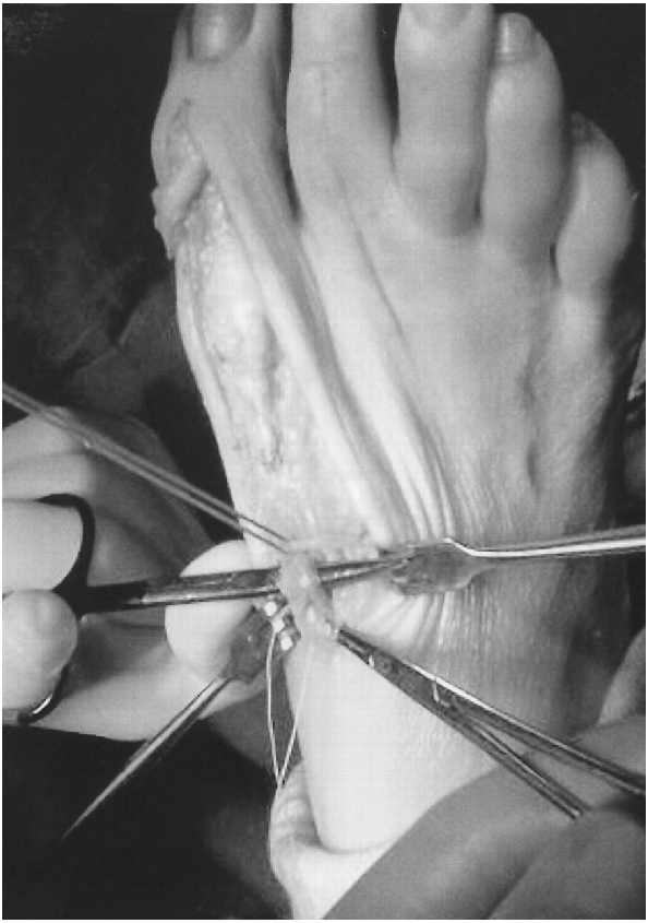
The patient underwent a second surgery on her right foot 5 weeks after the initial surgery. The extensor hallucis longus tendon was explored. A small distal portion of the tendon was identified attached at the base of the proximal phalanx of the hallux. Proximally, no tendon could be identified distal to the first metatarsocuneiform joint (
Figure 3). The extensor hallucis longus tendon was completely ruptured, with a defect of 6 cm.
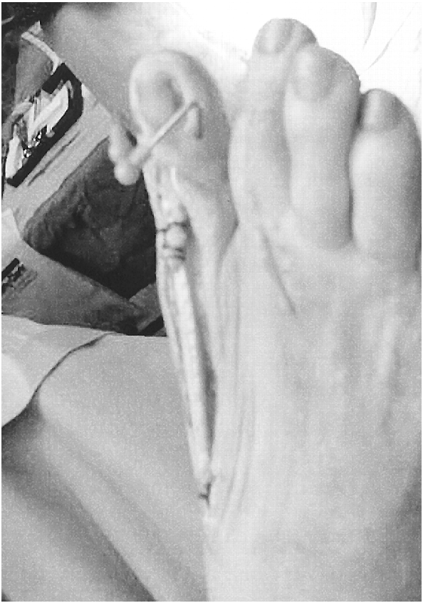
The patient underwent a hallux interphalangeal joint fusion with crossed 0.062 Kirschner wires. Next, a piece of tensor fascia lata allograft was rolled onto itself to form a linear tubelike structure. The graft was secured onto itself with 3-0 absorbable sutures. The hallux was then held in its desired anatomical position, and the tendon graft was cut to fill the extensor hallucis longus defect. The graft was then sutured to both the proximal and distal portions of the ruptured extensor hallucis longus tendon with 0 nonabsorbable sutures (
Figure 4). The graft was secured under zero tension. Deep and subcutaneous tissues were reapproximated with absorbable sutures, and the skin edges were reapproximated with nylon sutures. The patient tolerated the procedure without complications. Postoperatively she was placed in a below-the-knee cast and was nonweightbearing on her right foot.
Immobilization was continued for 6 weeks, with partial to full weightbearing as tolerated for an additional 2 weeks. Hallux dorsiflexion remained weak. The patient was referred to a physical therapist for electrical muscle stimulation and range-of-motion exercises. At the last follow-up visit (10 months), she was able to minimally dorsiflex her great toe. Despite the weakness, the hallux remained in good anatomical alignment and was stable throughout ambulation.
Discussion
This case demonstrates the use of a tendon allograft to repair a ruptured extensor hallucis longus tendon with a large defect. Tendon allografts have been well described in the literature for reconstruction of the anterior cruciate ligament, patellar tendon, lateral ankle ligaments, and Achilles tendon.[
9-
11] Several types of tendon allografts are available. At the Temple University School of Podiatric Medicine, tensor fascia lata and Achilles tendon allografts are used.
In most cases, a ruptured extensor hallucis longus tendon with a defect can be repaired using another tendon from the patient’s same foot. In this unique case, this was not a viable option. During the course of both surgeries, multiple tendons were examined, and the extensor hallucis brevis tendon was harvested during the first surgery. All examined tendons, including the extensor hallucis longus, slips from the extensor digitorum longus or the extensor digitorum brevis, and intrinsics surrounding the first and second metatarsophalangeal joints, were noted to be thin, nonhomogeneous in color, and atrophic. No palpable peroneus tertius could be appreciated.
The etiology of this patient’s degenerative, atrophic tendons is not known. Previously described potential systemic risk factors may include hypertension, obesity, diabetes mellitus, rheumatoid arthritis, and corticosteroid use.[
8,
14] Local factors include previous surgery and trauma. This patient had hypertension, which was well controlled with medication and previous corticosteroid injections into her second metatarsophalangeal joint. The only other factor contributing to her condition may have been her age.
It was the authors’ initial intent to restore complete active range of motion to the patient’s hallux. At the last follow-up visit she was able to only minimally extend her great toe, most likely because of fibrosis and adhesions to the graft. One possibility to prevent restricted motion may be the use of Silastic (Dow Corning Wright, Arlington, Tennessee) sheeting around the tendon to act as a pseudosheath.[
15]
Despite the patient’s inability to fully dorsiflex the hallux, the surgery was successful at restoring the previous flail, plantarflexed hallux to a more anatomical and functional position. The patient is able to walk without pain or difficulty and can propulse off of her medial column and hallux.
Conclusion
In this case, the patient sustained a complete extensor hallucis longus tendon rupture with a 6-cm defect. Owing to the size of the defect, the tendon could not be repaired primarily. Use of the patient’s own tendons as an autograft was not an option because of their widespread degeneration and atrophy. For this patient, repair of the extensor hallucis longus tendon using a fascia lata allograft served to stabilize the hallux onto the first metatarsal head, and fusion of the first metatarsophalangeal joint was avoided.