To the Editor:
Flattening of the top of the talus is generally considered a complication of treatment for clubfoot. It can also be secondary to trauma- or corticosteroid-induced ischemic necrosis of bone, hemophilia, or sickle cell anemia [
1-
3]. A case of bilateral flat-top talus not associated with clubfoot or any systemic disease is discussed as well as the possible causes of this deformity.
Case Report
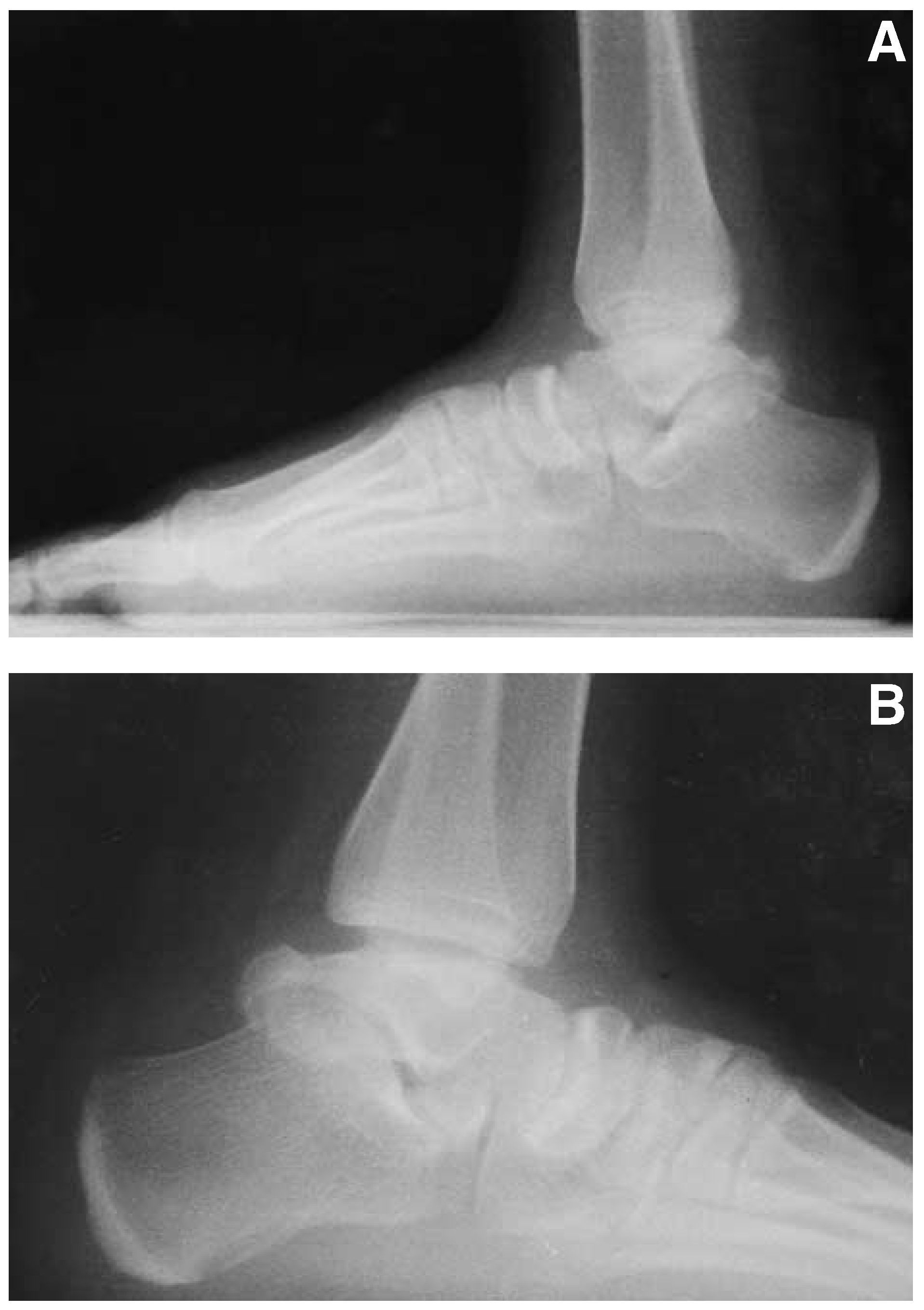
An 18-year-old woman presented to Mersin University Hospital in Mersin, Turkey, with mild left ankle pain following an ankle sprain. Mild tenderness was found on palpation of the lateral malleolus, and plain film radiographs revealed no trauma-induced lesion. Anteroposterior radiographs of the ankle were normal, but lateral radiographs showed flattening of the top of the talus. True lateral radiographs as described by Dunn and Samuelson
4]. were obtained for both ankle joints for accurate diagnosis, and flattening of the top of the talus was seen on both sides (Fig. 1). Because some rotation of the talus in the ankle mortise might be misleading, the left ankle was subjected to fluoroscopy with different degrees of rotation, from a direct anteroposterior to a direct posteroanterior view. Flat-top talus deformity was found in all positions.
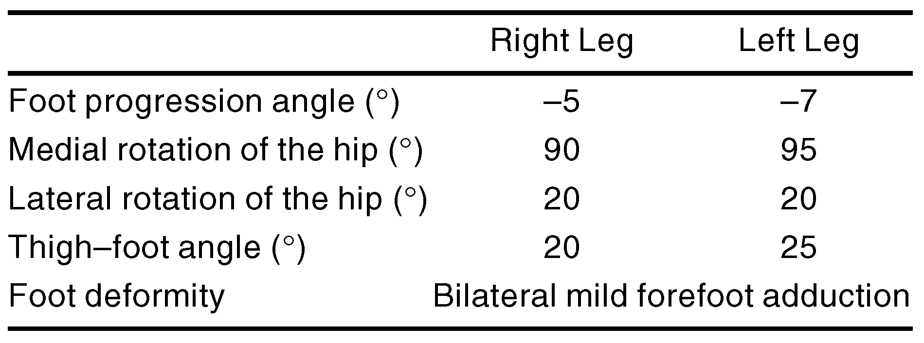
The patient reported no previous ankle injuries or problems. There was no history of corticosteroid administration and no history or findings of sickle cell anemia or hemophilia. Systemic examination was normal except for in-toeing, with the patellae rotated 45° inward. She habitually sat in a reversed tailor position with the ankles in dorsiflexion. There was no family history of any orthopedic deformities. An examination for congenital hip dysplasia was negative; however, excessive restriction of lateral rotation and increased medial rotation of both hips were detected. A torsional profile of the case, as described by Staheli et al,
5]. is displayed in Table 1. Although the patient had normal ankle range of motion, fluoroscopic examination revealed pivotal rather than gliding motion.
An anti-inflammatory drug was prescribed and an elastic bandage was dispensed to the patient. Ten days later, the patient returned for a follow-up visit, at which she reported being completely pain-free.
Discussion
Flat-top talus is generally considered a complication of forceful manipulation or prolonged casting in full dorsiflexion used in the treatment of clubfoot. It is postulated that this deformity results from excessive forces exerted on the superior aspect of the ossifying talus, which is more sensitive in the growing child [
4,
6,
7]. Kaplan,
8]. however, questioned this proposed etiology after finding flat-top talus in clubfeet that had not undergone treatment. In the case reported here, there was no history of clubfoot or previous treatment for such a condition.
Figure 1.
Left (A) and right (B) true lateral radiographs as described by Dunn and Samuelson,
4]. with the foot medially rotated to superimpose the medial and lateral malleoli, demonstrating flat-top talus.
Figure 1.
Left (A) and right (B) true lateral radiographs as described by Dunn and Samuelson,
4]. with the foot medially rotated to superimpose the medial and lateral malleoli, demonstrating flat-top talus.
Tabel 1.
Torsional Profile
Tabel 1.
Torsional Profile
Although osteonecrosis of the talus may result in flattening of the talar dome, the patient had no history of corticosteroid administration, trauma, or sickle cell anemia, which may cause osteonecrosis [
1,
3]. Furthermore, the radiographs did not show any evidence of ischemic necrosis of the talus, such as sclerosis. Patients with hemophilia may display a depression of the top of the talus, [
2]. but the case presented here had no history of hemorrhagic diathesis.
To the authors’ knowledge, this is the first case of flat-top talus reported in the literature that was not associated with clubfoot deformity or any systemic disease. There are two possible explanations for the flat-top talus in this case. The first is the patient’s habitual reversed tailor sitting position in which the ankles are dorsiflexed forcefully, exerting forces similar to those in clubfoot treatment; the second is a congenital flattening of the dome of the talus that is distinct from clubfoot.
It is commonly believed that flattening of the dome of the talus converts the normal gliding motion of the ankle joint to a pivotal motion, resulting in decreased range of motion and pain, making flat-top talus a contraindication to the treatment of clubfoot [
9]. Although pivotal motion was detected in this patient, the range of motion of the ankle joint was within normal limits and not accompanied by pain. This suggests that pain and decreased range of motion in patients with clubfoot may be due not to the shape of the talus, but to other factors associated with clubfoot or its treatment, indicating that flat-top talus may not be a contraindication to the treatment of clubfoot.
It is not possible to predict whether the shape or the pivotal motion of the talus in this case will result in early osteoarthritis, but the patient will be examined annually to assess the natural course of the condition.