Next Article in Journal
Treatment of Scleroderma Skin Ulcers Using Becaplermin Gel and Hydrocolloid Membrane
Previous Article in Journal
Relationship Between Proximal Articular Set Angle and Hallux Abducto Valgus
 
 
Journal of the American Podiatric Medical Association is published by MDPI from Volume 116 Issue 1 (2026). Previous articles were published by another publisher in Open Access under a CC-BY (or CC-BY-NC-ND) licence, and they are hosted by MDPI on mdpi.com as a courtesy and upon agreement with American Podiatric Medical Association.
Article

Diabetic Foot Wound Care Algorithms

by
Paul Han
1 and
Ruben Ezquerro
2
1
Diplomate, American Board of Podiatric Surgery; Medical Director, Diabetic Foot Care Center, Coastal Communities Hospital, Tenet Health System, Santa Ana, CA
2
Staff Podiatric Physician, Diabetic Foot Care Center, Coastal Communities Hospital, Tenet Health System, Santa Ana, CA
J. Am. Podiatr. Med. Assoc. 2002, 92(6), 336-349; https://doi.org/10.7547/87507315-92-6-336
Published: 1 June 2002

Abstract

The purpose of this article is to present reference guidelines to assist clinicians when treating diabetic patients with foot wounds. Diabetic patients with limb-threatening foot ulcers often have multiple coexisting medical conditions that frequently become impediments to the resolution of foot wounds. Each foot wound is unique and its etiology is multifactorial; therefore, each foot wound should be managed differently. The treatment algorithm presented in this article is divided into three categories: Algorithm I describes the treatment of septic foot wounds, which may be considered true podiatric surgical emergencies; Algorithm II describes the treatment of ischemic foot ulcers or gangrene with or without underlying osteomyelitis; and Algorithm III describes the treatment of neuropathic foot ulcers with or without underlying osteomyelitis.

According to a recent report by the American Diabetes Association [1], 15.7 million people, or 5.9% of the population, in the United States have diabetes mellitus, and nearly 18.4% of people 65 years and older have diabetes mellitus. Diabetic foot complications are the most common cause of nontraumatic lower-extremity amputations in the United States, and more than 50% of all nontraumatic amputations in the United States occur among people with diabetes mellitus. Twenty-five percent of diabetic patients will have foot problems during their lifetime, and such foot problems are the most frequent reason for hospitalization. Foot infections in the United States account for up to 25% of all diabetic hospital admissions [1]. The number of lower-limb amputations performed annually in diabetic patients has climbed from 67,000 to 86,000 in the United States in just the past 2 years, according to the latest national health data [2].
Diabetic foot complications, one of the most common precursors to subsequent lower-limb amputation, are a major public health problem. Diabetic ulceration in the foot has been identified as a factor in 84% of lower-extremity amputations [3]. The diabetic patient has coexisting medical conditions that make resolution of foot wounds much more complicated and challenging. The most common components in the causal pathways to amputation are peripheral vascular disease, peripheral neuropathy, and ulceration. The presence of acute sepsis in the foot, chronic indolent soft-tissue infection, or osteomyelitis underlying an ulcer can further complicate the problem. Foot wounds with underlying neuropathy or ischemia manifest differently and should be managed accordingly. Septic foot wounds, true surgical emergencies, carry high rates of amputation and mortality. Delay in surgical therapy in these patients is likely to lead to major limb amputation.
Prevention of foot ulcers through patient education can be one of the most cost-effective means of curtailing the rate of diabetes mellitus–related amputations; however, once diabetic patients develop a limb-threatening foot wound, a clear, descriptive, etiology-oriented, and systematic treatment scheme is needed to direct the physicians to the most appropriate therapies in an expeditious manner. Such a system was designed by the main author of this article (P.H.) and was implemented during the past 6 years at the Diabetic Foot Care Center at Coastal Communities Hospital in Santa Ana, California, which is a 14-physician team comprising all medical and surgical specialties. The authors believe that the comprehensive medical team approach to diabetic patients with impending amputation is the most effective method of prevention. This concept is incorporated into the diabetic foot wound care algorithms presented here. These algorithms represent the clinical practice and experience of the authors. They are not meant to be construed as evidence-based, and they are not the result of any formal consensus panel discussions.
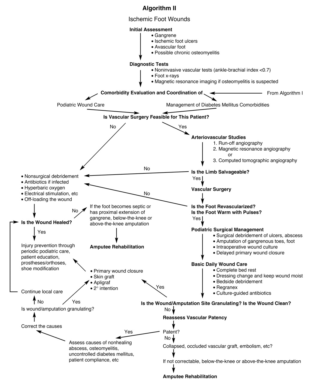
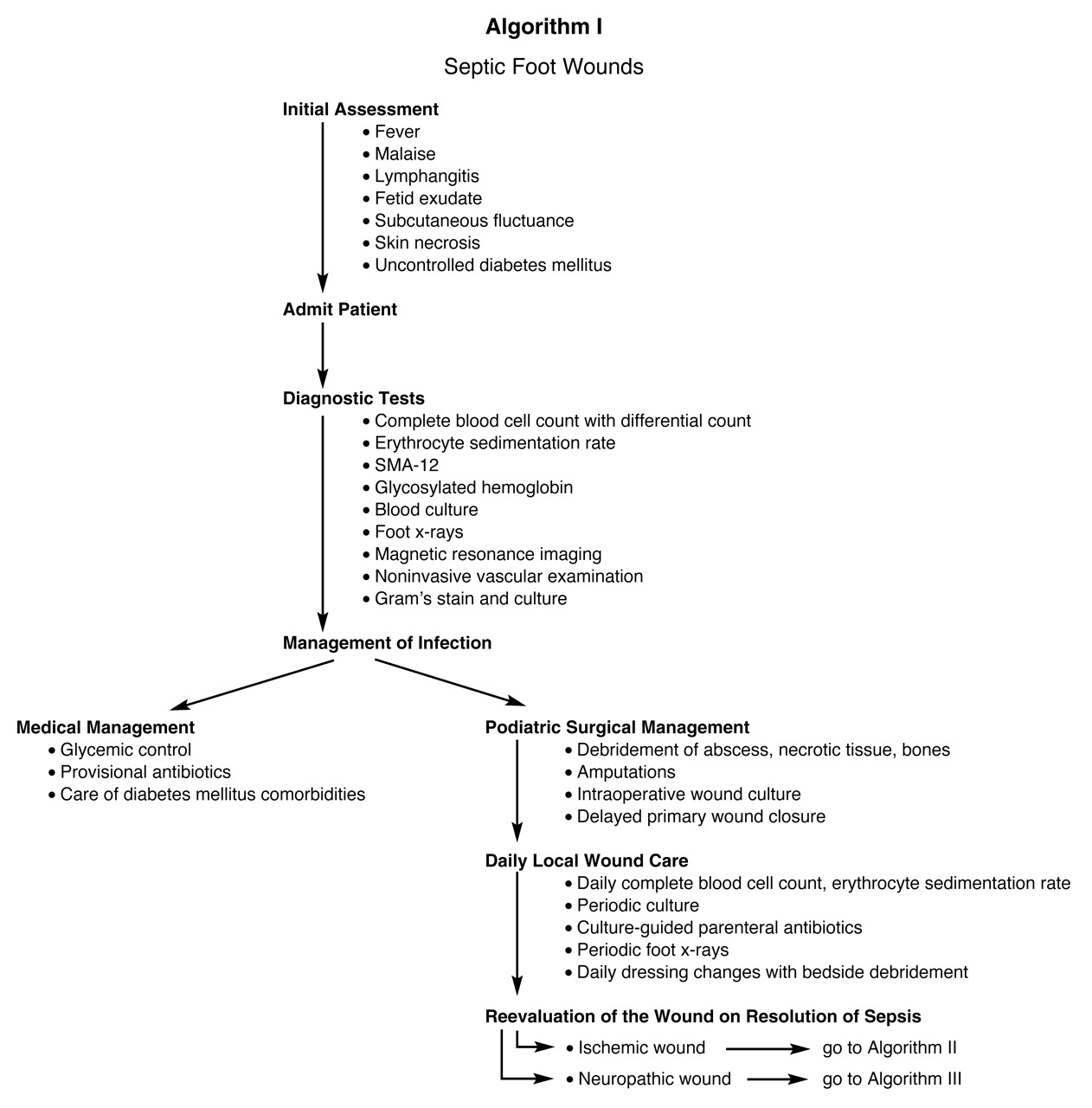
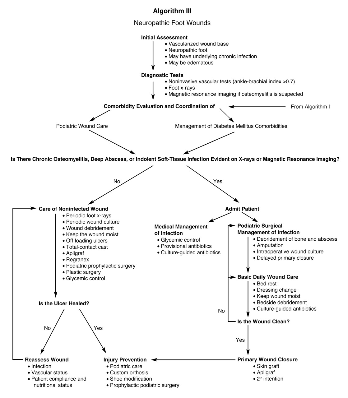
In diabetic foot wounds, both ischemia and neuropathy may coexist, with one more pronounced than the other. Wound-care algorithms for the diabetic foot are divided into three categories based on the nature of underlying pathologic mechanisms. Algorithm I describes the management guidelines for septic diabetic foot wounds (Fig. 1). Algorithm II is designed for the treatment of chronic diabetic foot wounds of which the major underlying pathologic mechanism is peripheral vascular disease that may or may not be accompanied by chronic underlying osteomyelitis (Fig. 2). Algorithm III addresses the management of chronic neuropathic diabetic foot wounds with or without chronic indolent osteomyelitis (Fig. 3).
Figure 1. Algorithm I describes the management guidelines for septic foot wounds. (Modified from Han PY: The Diabetic Foot Centers: Operational Manual and Practice Guidelines, Asian American Patient Education Advocate League, Los Angeles, 1999).
Japma 92 00336 f01
Figure 2. Algorithm II describes the management guidelines for ischemic foot wounds. (Modified from Han PY: The Diabetic Foot Centers: Operational Manual and Practice Guidelines, Asian American Patient Education Advocate League, Los Angeles, 1999).
Japma 92 00336 f02
Figure 3. Algorithm III describes the management guidelines for neuropathic foot wounds. (Modified from Han PY: The Diabetic Foot Centers: Operational Manual and Practice Guidelines, Asian American Patient Education Advocate League, Los Angeles, 1999).
Japma 92 00336 f03

Algorithm I—Management Guidelines for Septic Foot Wounds

Step 1: Initial Assessment

Systemic signs and symptoms of a septic process occur when the infection spreads to deeper tissue planes. Clinically, these patients may have signs of systemic toxic effects, including malaise, fever, lymphangitis, cellulitis, and edema. These patients may have preexisting mal perforans ulcers, wet gangrene, or dermatomycosis of the web spaces that may have served as portals for pathogenic bacteria. Acute foot infections are usually polymicrobial [4], with anaerobes contributing to significant tissue destruction. Evidence of large numbers of anaerobes may include skin necrosis, a fetid odor [5-8], soft-tissue gas [5,6], and crepitus. Pain and tenderness are frequently absent owing to neuropathy. These patients may not feel discomfort in the foot or see the problem. On the other hand, patients may experience pain and tenderness in foot infections that coexist with ischemia and minimal neuropathy.

Step 2: Hospital Admission

Diabetic patients with acute foot infection require immediate incision and drainage. Delay in surgical debridement and appropriate antibiotic therapy can result in high rates of amputation and mortality [9].

Step 3: Diagnostic Tests

Blood Tests. Initial laboratory tests should include blood culture, SMA-12, a complete blood cell count with differential count, and erythrocyte sedimentation rate, which may help monitor resolution of infection. Blood glucose level should be carefully monitored, as uncontrolled hyperglycemia is common in diabetic foot infection. Glycosylated hemoglobin level is valuable in determining the long-term control of diabetes mellitus.
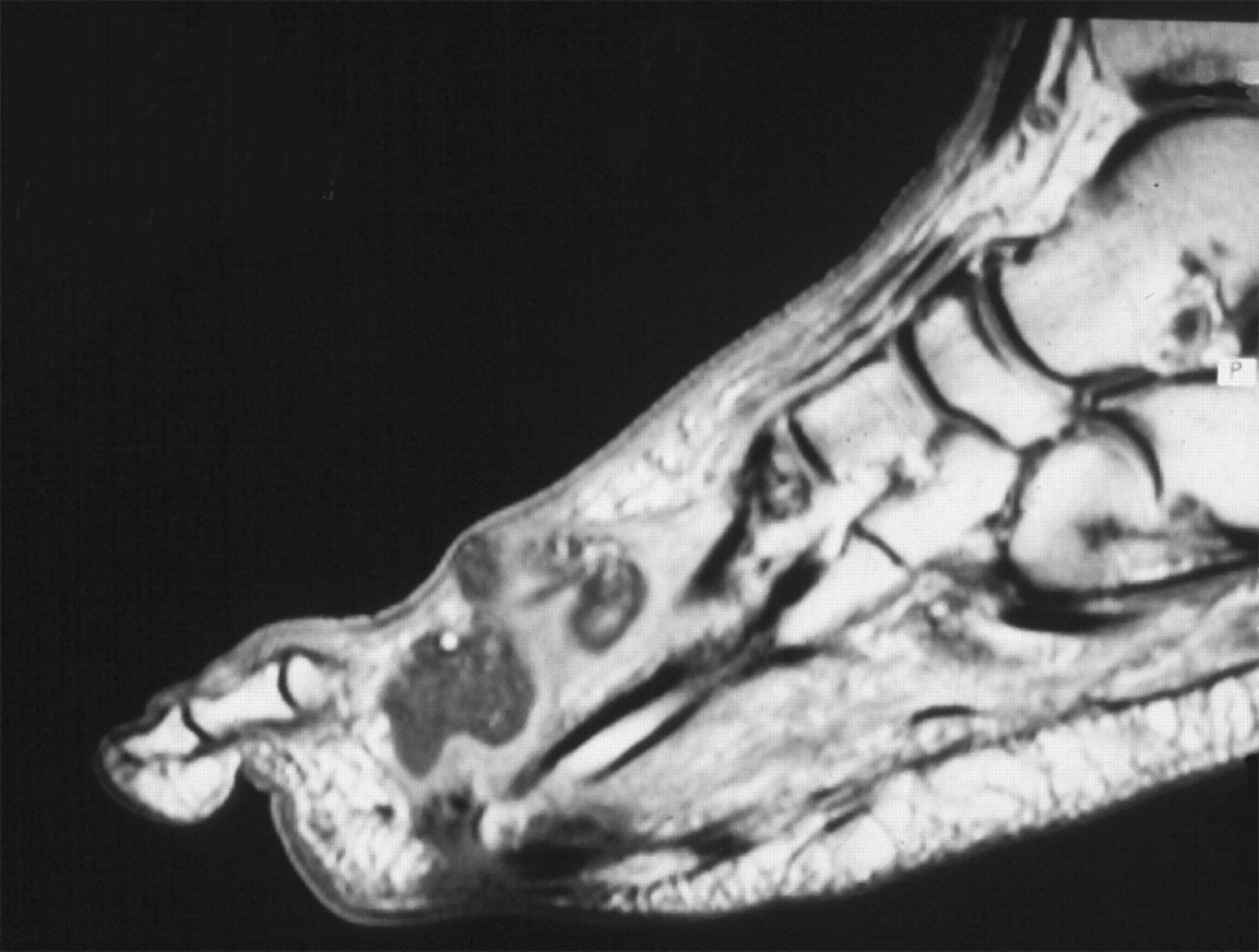
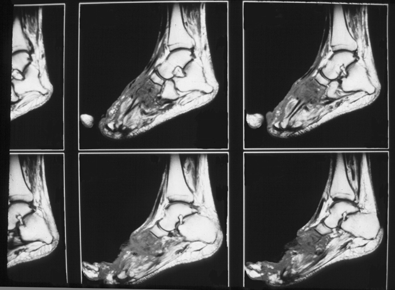
Imaging Studies. Initial radiographic evaluation for a septic foot is usually plain film foot x-rays examined for evidence of gas in the soft tissue, foreign bodies, and osteomyelitis. Soft-tissue gas is seen as a collection of radiolucent areas dissecting along soft-tissue planes as well as in the subcutaneous tissue. During the initial period of medical stabilization of the patient in preparation for surgery, if prompt magnetic resonance imaging (MRI) is available, it should be used to locate abscess formation (Fig. 4) and to evaluate the extent of osteomyelitis (Fig. 5).
Figure 4. T1-weighted magnetic resonance image showing abscess formation from plantar to dorsal with the presence of a mal perforans ulcer at the plantar aspect of the foot.
Japma 92 00336 f04
Figure 5. The sagittal plane view of a T1-weighted magnetic resonance image with low signal intensity in the bone marrow of the entire foot just distal to the midtarsal joint.
Japma 92 00336 f05
Noninvasive Vascular Examination. Various diagnostic evaluations are discussed in detail in Algorithm II. However, a simple and quick vascular evaluation can be done before taking the patient into the operating room using an arterial Doppler scan or pulse volume measurements. These tests can be omitted if the patient has pedal pulses, digital hair, and brisk capillary filling time, as these are the signs of adequate blood supply to the wound. In the care of septic foot wounds with coexisting underlying arterial occlusive disease, control of sepsis takes precedence over correction of vascular disease. Once sepsis is controlled with surgical debridement and appropriate parenteral antibiotic therapy, steps should be taken toward vascular reconstruction for limb salvage.

Step 4: Medical Management of Infection

Provisional Intravenous Antibiotics. Appropriate parenteral antibiotic therapy along with immediate surgical therapy can lower the mortality rate and save the limb. Initial antibiotic treatment should be broad spectrum, as most diabetic foot infections are mixed infections of gram-positive aerobes, gram-negative bacilli, and anaerobes [5-8,10,11]. Other factors to consider when selecting initial antibiotics are the patient’s preexisting renal and hepatic disorders, previous antibiotic therapy, if any, and the severity of the infection.
In the presence of a deep abscess, evidenced by soft-tissue crepitus, special consideration should be given to the anaerobic component of infection when choosing initial antibiotics. It is the anaerobes that are responsible for abscess formation. Once the specific bacterium is identified by culture and sensitivity analysis, antibiotic adjustments can be made if needed.
Glycemic Control. The relationship between infection and hyperglycemia has been reported in the literature [12-20]. Infection often leads to hyperglycemia. Conversely, hyperglycemia may impede resolution of infection. If clinical improvement of infection is not evident in a few days with surgical debridement and intravenous antibiotic therapy, it may be because of either an inadequately drained abscess or poor glycemic control. Diabetic patients have defects in polymorphonuclear leukocytes that correlate with the level of hyperglycemia. For these reasons, glucose control is of paramount importance as a part of the treatment of infected foot wounds [12,13,21,22].

Step 5: Surgical Management of Infection

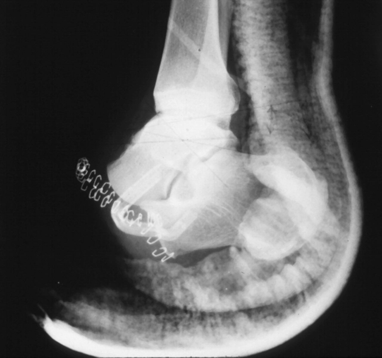
Antibiotic therapy alone will not resolve the infection. Deep abscesses and infected necrotic tissues should be surgically debrided. The goal of surgical care of the infected foot wound is to free it of necrotic and nonviable tissue, thus stimulating the formation of granulation tissue. If cellulitis originates from plantar foot ulcers and there is radiographic evidence of the bone underlying the ulcer, then the infected bone should also be resected at this time. If it is necessary to control sepsis, the infected part of the foot should be amputated at this time (Fig. 6). Soft tissue that does not bleed at the time of surgical debridement should be considered nonviable and must be excised. The wound is then loosely packed open to achieve delayed primary closure and to closely monitor for resolution of infection during daily wound care.
Figure 6. To control sepsis in the patient shown in Figure 5, it was necessary to “buy time,” during which control of the infection could be instituted. The option used for this patient was guillotine amputation through the midtarsal joint.
Japma 92 00336 f06

Step 6: Postsurgical Wound Care

Postsurgical wound care consists of bedside debridement, irrigation with sterile saline at each dressing change, and keeping the wound moist. With continuation of culture-guided antibiotic therapy and timely surgical intervention, a wound with adequate blood supply should appear clean, and healthy granulation tissue should appear in a few days. If the wound shows any residual necrotic tissue, the patient should be taken back to the operating room for further debridement and irrigation.

Step 7: Management of the Wound After Control of Sepsis

Once sepsis is controlled, the wound should be evaluated for vascular status and should be classified as either ischemic or nonischemic.
Ischemic Wound. Whether the foot wound is the result of surgical debridement in an attempt to control sepsis or a foot ulcer that has served as a portal for bacteria causing acute sepsis, ischemic wounds are devoid of bleeding granulation tissue at the base and edges of the wound. Once sepsis is controlled, the patient should be evaluated for limb salvage vascular reconstruction (go to Algorithm II).
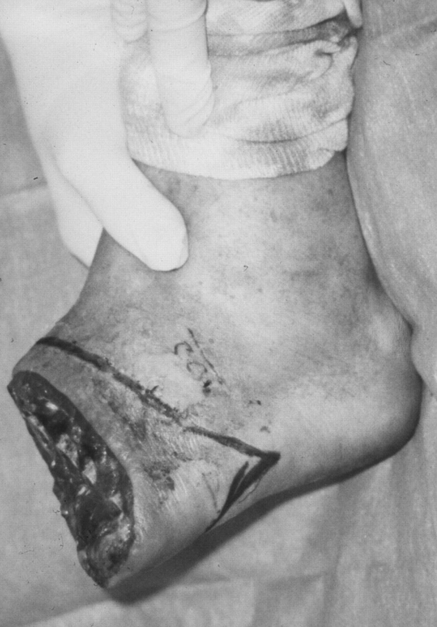
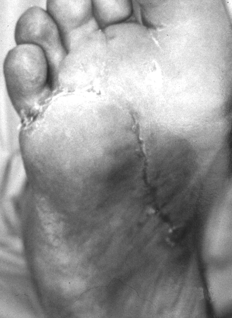
Nonischemic (Neuropathic) Wound. The surgical incision can be primarily closed once sepsis is resolved (Fig. 7). If the patient has an existing neuropathic ulcer that has served as a portal for sepsis, it can now be managed through Algorithm III.
Figure 7. Once sepsis was controlled, the guillotine amputation shown in Figure 6 was revised to a Chopart amputation with a percutaneous tenotomy of the Achilles tendon.
Japma 92 00336 f07

Algorithm II—Management Guidelines for Ischemic Foot Ulcers and Gangrene

Step 1: Initial Assessment
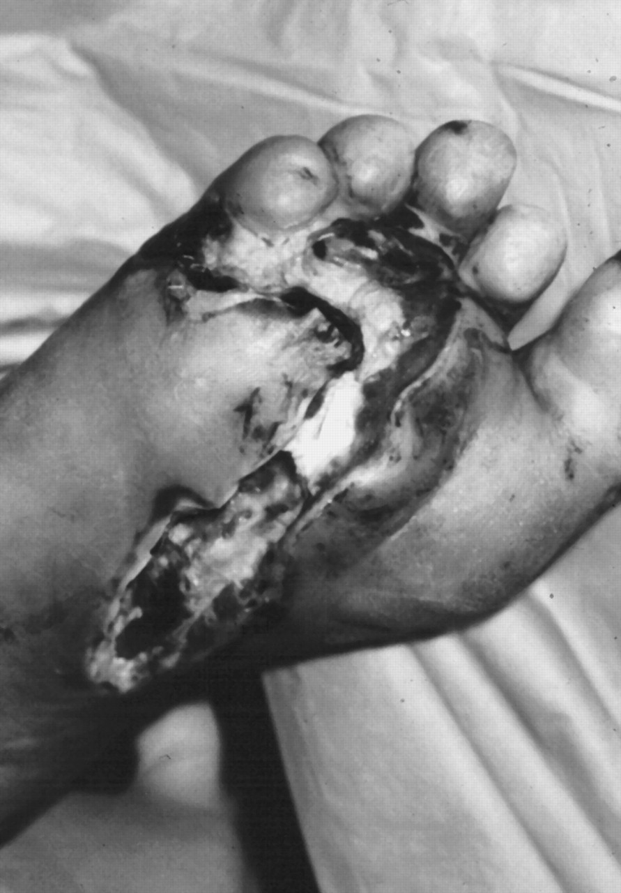
Ischemic foot wounds may be in the form of gangrene or foot ulcers with no appreciable amount of granulation tissue present at the base of the wound (Fig. 8). The base of the wound is whitish gray, unlike that of a neuropathic ulcer, which has a beefy red, granular appearance. The ischemic foot usually is characterized by brittle toenails, hairless digits, and shiny, atrophic skin and may be cold and clammy to the touch. The foot may exhibit rubor on dependency and palor when elevated above the heart. Unlike neuropathic wounds, ischemic wounds tend to be accompanied by intense pain.
Figure 8. Ischemic foot wound. This patient developed a plantar abscess through the portal of the fourth interdigital space. Swelling and erythema occurred in the region of the medial arch, with lymphangitic streaks up toward the medial leg. A longitudinal oblique plantar incision was made overlying the area of maximal fluctuation toward the porta pedis to debride the deep abscess, including part of the flexor tendons. The incision was left open for delayed primary closure. Once sepsis was controlled, limb salvage revascularization was attempted.
Japma 92 00336 f08

Step 2: Diagnostic Tests

Noninvasive Vascular Testing. Patients with an ankle-brachial index less than 0.7 or transcutaneous oxygen measurement of 40 mm Hg or less will probably not experience wound healing without vascular reconstructive surgery [3,4].
Radiographs and MRI. Initially, foot x-rays are used to detect chronic osteomyelitis of the bone adjacent to the ischemic wound or foreign bodies. Radionuclide scans may be considered if there are signs of osteomyelitis on the radiographs; however, in the presence of arterial occlusive disease, a larger amount of radiotracer is needed to reach the site of infection in order to avoid false-negative results. Therefore, MRI may be more useful for diagnosing osteomyelitis in the presence of peripheral vascular disease.

Step 3: Diabetes Mellitus Comorbidity Evaluation

Coexisting medical conditions that may interfere with wound healing should be identified at the onset of wound care. For example, peripheral vascular disease commonly coexists with cardiovascular and cerebrovascular diseases. Hypertensive diabetic patients must be screened for renal impairment, which may have a negative impact on the overall care of a foot wound.
If patients have one or more medical conditions, including renal, hepatic, hematologic, neurologic, or immunologic disease, they should be monitored and treated during the course of wound care. If the patient has severe malnutrition, as evidenced by an albumin level less than 20 g/L, a dietitian should be a member of the wound-care team. It is common for diabetic patients to be taking aspirin or coumadin; these medications should be adjusted, especially when invasive procedures and testing are considered. The use of corticosteroids, cytotoxic agents, or immunosuppressive medications should also be monitored by an internist during the course of wound care. A history of bleeding disorders or human immunodeficiency virus may also interfere with diabetic wound management.

Step 4: Is Limb Salvage Vascular Surgery Considered?

A vascular surgeon evaluates the patient for the feasibility of vascular surgery. Patients who have intractable ischemic pain, wet gangrene, a fetid wound, or underlying osteomyelitis may benefit from vascular surgery for limb salvage. Vascular reconstruction, however, is not free of risks. In general, there are concerns about the risks of performing vascular reconstruction in a population with advanced renal disease or extensive arteriosclerosis [23-25]. For example, an elderly, bedridden patient with dry, noninfected gangrene may be monitored indefinitely rather than subjected to the risks of arteriography, vascular surgery, or amputation. Another example would be a patient with advanced renal disease who might need below-the-knee or above-the-knee amputation rather than being subjected to vascular reconstruction for limb salvage.
Evaluation by a cardiologist before vascular surgery may be required, as cardiac complications from major vascular reconstruction are frequent. Diabetic patients with renal failure should be evaluated and monitored by a nephrologist during and after vascular surgery.
If vascular surgery is deemed feasible, the next step is selection of the proper arteriovascular study of the lower extremities—run-off angiography, magnetic resonance angiography, or computed tomographic angiography—to locate the occlusions or stenosis of the arteries.

Step 5: Arteriovascular Studies

Traditional run-off arteriography is not risk-free and should be used only as prevascular work-up. A major complication is renal shutdown due to the use of radiocontrast materials. Diabetic patients with creatinine levels greater than 5 mg/dL are particularly at risk [25]. In diabetic patients with abnormal renal function, the authors rely on digital subtraction angiography; magnetic resonance angiography or computed tomographic angiography can also be considered.

Step 6: Determine Limb Salvageability

Using the arteriovascular studies, limb salvageability can be determined. If occlusion or stenosis of large and medium-sized arteries is the cause of ischemia and gangrene in the foot, it can be corrected by vascular surgery to achieve limb salvage.

Step 7: Limb Salvage Vascular Procedures

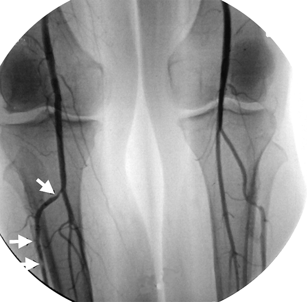
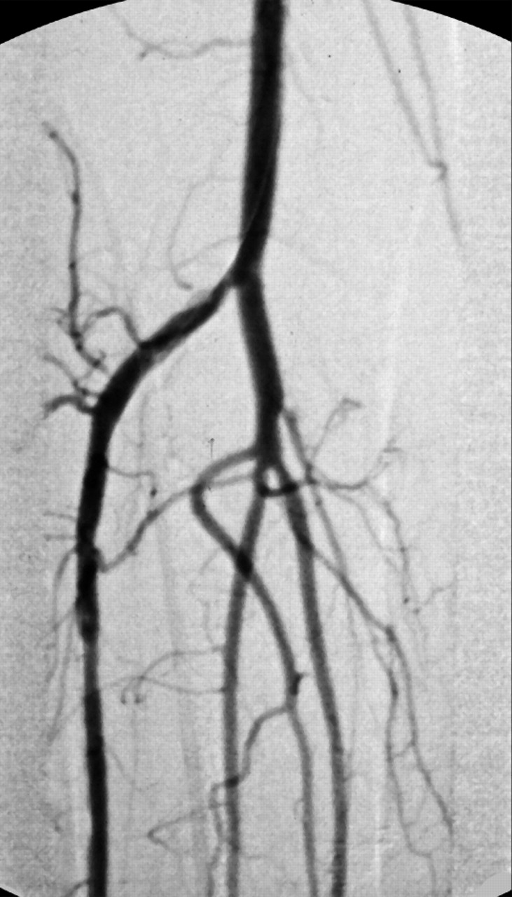
In general, vascular procedures for limb salvage include reconstructive bypass surgery and percutaneous transluminal angioplasty (Figs. 9 and 10). For detailed information regarding vascular repair for peripheral vascular disease, the reader is advised to consult a standard text on vascular surgery [26-28].
Figure 9. Arteriographic findings from the patient shown in Figure 8 were consistent with multiple stenotic vascular lesions (arrows) in the anterior tibial artery.
Japma 92 00336 f09
Figure 10. Arteriogram of the patient shown in Figure 8 immediately after dilation of the anterior tibial artery by means of percutaneous transluminal angioplasty.
Japma 92 00336 f10

Step 8: Debridement or Amputation of the Toes and Foot

Immediately after vascular reconstruction, pulses may be palpable, the foot becomes warm to the touch, and capillary filling time should be improved. The base of the ulcer should show new granulation tissue in a few days. The gangrenous toe can now be amputated, with improved potential for healing. The wound, resulting from amputation or debridement, should be left open and should be observed for a few days with standard local wound care.

Step 9: Healing of the Wound

Failure to develop granulation tissue or manifest bleeding with sharp debridement may be an indication of failed arterial reconstruction. Bypass patency and blood flow beyond the distal anastomosis should be monitored using an arterial Doppler duplex scan. The vascular surgeon should identify and correct the cause of failure as early as possible to achieve limb salvage. If the limb salvage procedure fails, more proximal amputation, such as below-the-knee or above-the-knee amputation, may be necessary.
If vascular reconstructive surgery is successful, the wound should no longer appear ischemic but should be granulating. The newly granulating wound should be carefully protected, and meticulous daily local wound care should be performed to promote healing and prevent infection. The clean wound can then be covered with a split-thickness skin graft, undergo primary closure, or be closed by secondary intention (Fig. 11).
Figure 11. After revascularization of the foot by means of angioplasty, the patient shown in Figure 8 was maintained nonweightbearing for the next 4 months, at which time the wound was healed.
Japma 92 00336 f11

Step 10: Care of the Nonsalvageable Foot

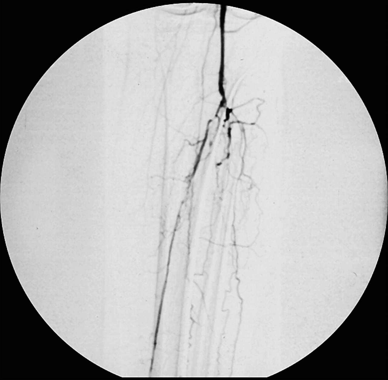
In some patients, limb salvage vascular surgery is not beneficial, especially when there is an adequate inflowing arterial source for a bypass but absence of run-off vessels to be supplied by a bypass (Fig. 12). For patients who cannot benefit from limb salvage vascular surgery or for whom surgery is contraindicated, steps should be taken for conservative wound care to prevent infection, preclude weightbearing, and protect from external irritation.
Figure 12. Arteriogram of a patient with gangrene. This patient has an adequate inflowing arterial source for a bypass proximally, but there is no available run-off bed distal to the popliteal artery to be supplied by a bypass.
Japma 92 00336 f12
Ischemic wounds in general should not be debrided, as it will lead to progression of tissue damage and further ulceration in the ischemic setting. Instead, the wound should be protected with a dressing to provide a barrier to pathogens and to maintain a moist wound environment. The ischemic foot should be protected with a foot cradle, avoiding contact with shoes or a mattress at all times.
Wet-to-dry dressings can be problematic, as the gauze becomes dry and adheres to the base of the wound, with the following dressing change disrupting the wound bed. Alternative, commercially available products such as hydrocolloid, calcium alginate, or hydrogels should be considered. For chronic and nonhealing ischemic foot wounds, where revascularization is not a viable option, systemic hyperbaric oxygen or electrical stimulation has been reported to be effective [29,30].

Step 11: Care After Amputation or Resolution of the Wound

Care after resolution of the wound includes proper prostheses, orthoses, patient education, and rehabilitation by a physiatrist. Protection of the foot is important in injury prevention. Well-constructed protective footwear and custom-molded inserts are used to prevent tissue breakdown. Preventive podiatric medical care on a regular basis should be instituted after the postoperative period to prevent recurrence of lesions.
Rehabilitation of foot or leg amputees is most successfully managed when a podiatrist, vascular surgeon, physiatrist, physical therapist, prosthetist, and pedorthist work together as a team. An important factor to consider in patients with underlying vascular disease is the risk of developing cardiovascular problems during rehabilitation. These patients should be evaluated and monitored by a cardiologist before and during the rehabilitation period.

Algorithm III—Management Guidelines for Neuropathic Foot Ulcers

Step 1: Initial Assessment
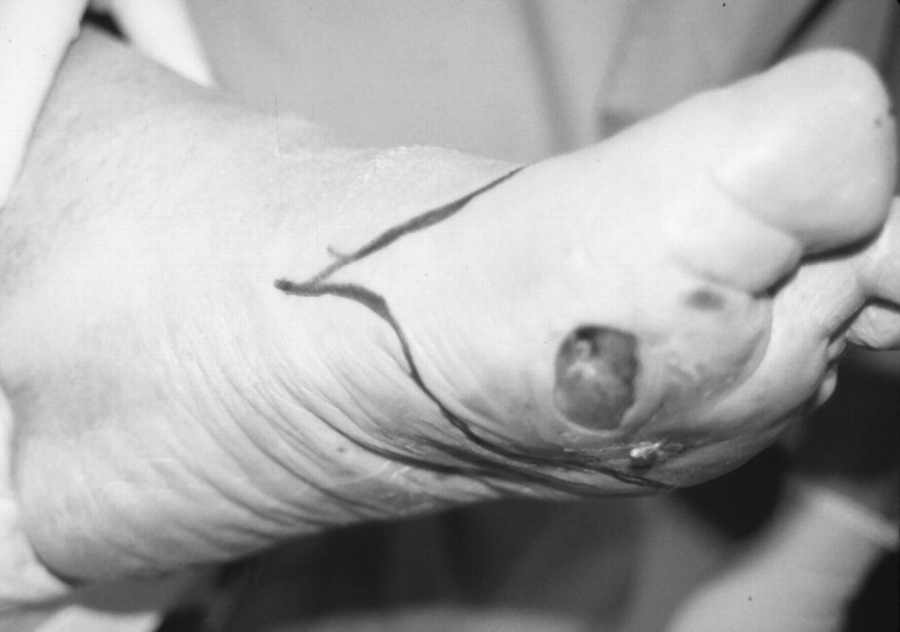
Neuropathic ulcers, by definition, occur in a well-vascularized foot, as demonstrated by palpable pedal pulses, and commonly are the result of neglected and painless calluses or corns. In the presence of neuropathy, these lesions frequently go unnoticed or untreated owing to the absence of pain, which then allows tissue breakdown and eventual ulceration, necrosis, and gangrene. These ulcers tend to be chronic as a result of continuous microtrauma with ambulation.
Chronic indolent osteomyelitis underlying the foot ulcer is common, but signs and symptoms are frequently absent. There may be minimal swelling at the site of ulceration, which is often dismissed as chronic induration of soft tissue from a long-standing ulceration. Chronic soft-tissue infection without systemic toxic effects is also common in these patients. Leukocytosis, warmth, and edema are often absent [31]. Pathogens may eventually penetrate deeply into the foot, causing a limb-threatening abscess and cellulitis (see Algorithm I). If a malodorous exudate is noticed, then the ulcer should be probed for depth and for the possibility of a sinus tract (Fig. 13). If the bone or joint is exposed, further diagnostic tests are necessary to rule out osteomyelitis or the presence of a deep abscess.
Figure 13. The left hallux was slightly indurated but not warm or erythematous. The lymph nodes were not palpable, and the patient was afebrile and leukocytosis was not present. There was a deep ulceration probing to the bone.
Japma 92 00336 f13

Step 2: Diagnostic Tests

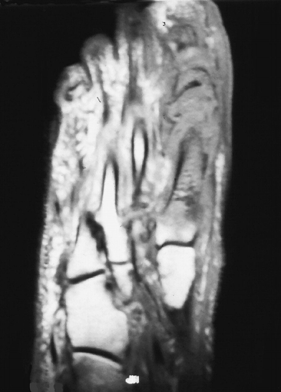
In the care of chronic neuropathic foot wounds, periodic foot radiographs and wound culture are helpful in early detection and treatment of infection. Radiographic changes of osteomyelitis may not be clearly distinguishable from those of diabetic neuroarthropathy [32]. Radionuclide scans are more sensitive than radiographs in distinguishing osteomyelitis from diabetic arthropathy, but false-negative results may occur [33], especially in diabetic patients with peripheral vascular disease, in whom the radiotracer may not be able to reach the foot in the focus of osteomyelitis. Magnetic resonance imaging is recommended when there is a strong suspicion of osteomyelitis (Fig. 14).
Figure 14. T1-weighted magnetic resonance image of the patient shown in Figure 13 clearly showing the exact demarcation of the extent of osteomyelitis, unlike in traditional bone scans. Magnetic resonance imaging can guide the surgeon to accurately determine the level of amputation or debridement of the infected bone.
Japma 92 00336 f14

Step 3: Diabetes Mellitus Comorbidity Evaluation

Patients are evaluated for significant comorbidities, as discussed in Algorithm II.

Step 4: Care of the Noninfected Neuropathic Ulcer

If the ulceration is free of underlying osteomyelitis or soft-tissue infection, the treatment regimen should be aimed at prevention of infection and closure of the wound as early as possible. To achieve these objectives, basic principles of wound care should be followed. Aggressive and periodic wound debridement of nonviable tissue is essential. Sharp debridement is preferred to using proteolytic agents or whirlpool, as these modalities do not debride neuropathic wounds adequately [34].
The causative pressure and shear friction on the wound should be removed by off-loading the site of neuropathic wounds. Removal of pressure and friction can be difficult to achieve in poorly motivated patients; therefore, patient education is important. Total-contact casts are useful in the reduction of plantar pressure [35].
In some cases, a split-thickness skin graft, tissue flaps, or a mechanical tissue expander [36] can be used for resolving the open wound. Recently, a human skin equivalent, Apligraf (Novartis Pharmaceutical Corp, East Hanover, New Jersey), has been approved for the treatment of diabetic ulcers. Topical Regranex (Ortho-McNeil Pharmaceutical, Raritan, New Jersey), which consists of platelet-derived growth factors, has been marketed for diabetic foot wounds.
The importance of meticulous glycemic control in wound healing cannot be overemphasized. Periodic culture of the wound as well as periodic foot x-rays are important to monitor infection of the bone and soft tissue.
Podiatric surgery to improve foot function or to excise bony protuberances that can lead to ulceration can be considered at this time. Foot surgery, when well planned and executed, can be effective in the treatment and prevention of recurrence.

Step 5: Care After Resolution of the Ulcer

See Step 11 of Algorithm II for injury prevention.

Management of Neuropathic Ulcers with Osteomyelitis

During the course of treatment of neuropathic foot ulcers, bones underlying the ulcers often become infected. In such cases, the management guidelines are as follows:

Step 6: Hospital Admission

Diabetic patients with foot ulcers and underlying osteomyelitis should be admitted to the hospital for parenteral antibiotic therapy, surgical management of osteomyelitis, and control of glycemia.

Step 7: Medical Management of Osteomyelitis

Glycemic Control. Meticulous control of diabetes mellitus is the initial step in the treatment of osteomyelitis, as hyperglycemia is the key pathologic factor in the development of foot infection [37,38].
Provisional Antibiotics. The choice of an initial antibiotic regimen depends on the Gram’s stain of the exudate. It should not be assumed that organisms isolated from the ulcer are the same organisms causing osteomyelitis. Bone biopsy is necessary for a definitive diagnosis of osteomyelitis.

Step 8: Surgical Management of Osteomyelitis

Intravenous antibiotics alone will not eradicate osteomyelitis in the diabetic foot. Infected bone, necrotic tissue, and abscess cavities must be surgically debrided. If necessary, the infected part of the foot should be amputated at this time (Fig. 15). After initial debridement of the infected bone and soft tissue, a deep cleft may result. The wound is loosely packed open to allow delayed primary closure.
Figure 15. The patient shown in Figure 13 had magnetic resonance imaging evidence of osteomyelitis in the entire first metatarsal bone and the head of the second metatarsal bone; therefore, the level of the most distal amputation was disarticulation at Lisfranc’s joint.
Japma 92 00336 f15

Step 9: Care of the Surgical Wound

Dressings can be changed twice a day or more frequently depending on the status of the wound and the degree of infection. Intravenous antibiotics, based on intraoperative culture of the infected bone, should be administered. Complete eradication of infected bone can be assessed by repeating foot x-rays, MRI, or cultures. If infection persists, the patient should be taken back to the operating room for additional debridement or more proximal amputation.

Step 10: Primary Closure of the Wound

When the foot is free of infection, the wound can be closed.

Step 11: Injury Prevention

See Step 11 of Algorithm II for injury prevention.

Conclusion

Foot wounds in diabetic patients are a major cause of morbidity, and their care has become a significant part of podiatric medical practice. It is important to develop systematic guidelines for the care of this complication. At the Coastal Communities Hospital in Santa Ana, California, a team of physicians from all medical and surgical disciplines was organized in 1996 and dedicated its services to the diabetic population for the prevention of amputation. This model of group practice, established as a hospital department and approved by the California State Health Department, has been effective in preventing foot complications and in caring for diabetic foot wounds. The treatment guidelines used since the inception of the program and the authors’ experiences in the team approach have been presented. It is the authors’ intention to contribute their experiences and expertise in the form of algorithms presented here for the prevention of major limb amputations in diabetic patients.

Acknowledgment

Teresa Scovil, MA, Jessica Tran, and Peggy Lee for helping with data collection and manuscript preparation; Amy Ezquerro, BA, for preparing the artwork for the manuscript. This project was funded in part by Coastal Communities Hospital, Tenet Health System, Santa Ana, CA.

References

  1. American Diabetes Association: Vital Statistics, American Diabetes Association, Alexandria, VA, 1996.
  2. American Diabetes Association: Diabetes in the News, American Diabetes Association, Alexandria, VA, November 12, 2000.
  3. Pecoraro RE, Reiber GE, Burgess EM: Causal pathway to amputation. Diabetes Care 13: 513, 1990.
  4. Armstrong DG, Liswood PJ, Todd WF: Prevalence of mixed infections in the diabetic pedal wound: a retrospective review of 112 infections. JAPMA 85: 533, 1995.
  5. Louie TJ, Bartlett JG, Tally FP, et al: Aerobic and anaerobic bacteria in diabetic foot ulcers. Ann Intern Med 85: 461, 1976.
  6. Fierer J, Daniel D, Davis C: The fetid foot: lower-extremity infections in patients with diabetes mellitus. Rev Infect Dis 1: 210, 1979.
  7. Sapico FL, Canawati HN, Witte JL, et al: Quantitative aerobic and anaerobic bacteriology of infected diabetic feet. J Clin Microbiol 12: 413, 1980.
  8. Sapico FL, Canawati HN, Witte JL, et al: The infected foot of the diabetic patient: quantitative microbiology and analysis of clinical features. Rev Infect Dis 6 (suppl 1): S171, 1984.
  9. Stamenkovic I, Lew PD: Early recognition of potential fatal necrotizing fasciitis. N Engl J Med 310: 1689, 1984.
  10. Alex DA: Microbiology of foot infections. J Foot Surg 26: S3, 1987.
  11. Sharp CS, Bessman AN, Wagner WF Jr, et al: Microbiology of deep tissue in diabetic gangrene. Diabetes Care 1: 289, 1978.
  12. Rayfield EJ, Ault MJ, Keusch GT, et al: Infection and diabetes: the case of glucose control. Am J Med 72: 439, 1982.
  13. Tan JS, Anderson JL, Watanakunakorn C, et al: Neutrophil dysfunction in diabetes mellitus. J Lab Clin Med 85: 26, 1975.
  14. Kozai K, Ogawa K, Tsubura E: Impaired granulocytes nitroblue tetrazolium reduction in patients with diabetes mellitus. Tokushima J Exp Med 30: 65, 1983.
  15. Mowat AG, Baum J: Chemotaxis of polymorphonuclear leukocytes from patients with diabetes mellitus. N Engl J Med 284: 621, 1971.
  16. Bagdade JD, Stewart M, Walters E: Impaired granulocyte adherence: a reversible defect in host defenses in patients with poorly controlled diabetes. Diabetes 27: 677, 1978.
  17. Stickle JE, Tvedten HW, Schall WD, et al: Adherence of neutrophils from dog with diabetes mellitus. Am J Vet Res 47: 541, 1986.
  18. Bagdade JD, Walters E: Impaired granulocyte adherence in mildly diabetic patients: effects of tolazamide treatment. Diabetes 29: 309, 1980.
  19. Stecher VJ, Chinea G, Greame ML, et al: The effect of anti-inflammatory drugs and diabetes on neutrophil adherence [abstract]. Federation Proc 36: 1070, 1977.
  20. Bagdade JD, Root RK, Bulger RJ: Impaired leukocyte function in patients with poorly controlled diabetes. Diabetes 23: 9, 1974.
  21. Kelly MK, Brown JM, Thong YH: Neutrophil and monocyte adherence in diabetes mellitus, alcoholic cirrhosis uraemia and elderly patients. Int Arch Allergy Appl Immunol 78: 132, 1985.
  22. Repine JE, Clawson CC, Goetz FC: Bactericidal function of neutrophils from patients with acute bacterial infections and from diabetics. J Infect Dis 142: 869, 1980.
  23. Boucher CA, Brewster DC, Darling RC, et al: Determination of cardiac risk by dipyridamole-thallium imaging before peripheral vascular surgery. N Engl J Med 312: 389, 1985.
  24. Burnham SJ, Johnson G, Gurri JA: Mortality risk for survivors of vascular reconstructive procedures. Surgery 92: 1072, 1982.
  25. Harkonen S, Kjellstrand CM: Exacerbation of diabetic renal failure following intravenous pyelography. Am J Med 63: 939, 1997.
  26. Boyd AM: Natural course of arteriosclerosis of lower extremity. Proc Roy Soc Med 53: 591, 1962.
  27. Rutherford RB: Vascular Surgery, 3rd Ed, WB Saunders, Philadelphia, 1989.
  28. Wilson SE, Williams RA: Vascular Surgery, McGraw-Hill, New York, 1986.
  29. Faglia E, Favales F, Adelgi A, et al: Adjunctive systemic hyperbaric oxygen therapy in treatment of severe prevalently ischemic diabetic foot ulcer: a randomized study. Diabetes Care 19: 1338, 1996.
  30. Baker LL, Chambers R, DeMuth SK, et al: Effects of electrical stimulation on wound healing in patients with diabetic ulcers. Diabetes Care 20: 405, 1997.
  31. Hertzer NR: Fatal myocardial infarction following lower extremity revascularization: two hundred seventy-three patients followed six to eleven postoperative years. Ann Surg 193: 492, 1981.
  32. Wheat J: Diagnostic strategies in osteomyelitis. Am J Med 78 (suppl 6B): 218, 1985.
  33. Park HM, Wheat LJ, Siddiqui AR, et al: Scintigraphic evaluation of diabetic osteomyelitis: concise communication. J Nucl Med 23: 569, 1982.
  34. Martin SJ, Corrado OJ, Kay EA: Enzymatic debridement for necrotic wounds. J Wound Care 5: 310, 1996.
  35. Armstrong DG, Lavery LA: Evidence-based options for off-loading diabetic wounds. Clin Podiatr Med Surg 15: 95, 1998.
  36. Johnson GR, Han P, Giacopelli JA: Tissue expansion as an alternative to skin grafting for closure of skin deficits. JAPMA 82: 249, 1992.
  37. Kilo C: Value of glucose control in preventing complications of diabetes. Am J Med 79 (suppl 2B): 33, 1985.
  38. Raskin P, Rosenstock J: Blood glucose control and diabetic complications. Ann Intern Med 105: 254, 1986.

Additional References

  1. Abele JE: Balloon catheters and transluminal dilatation: technical considerations. AJR Am J Roentgenol 135: 901, 1980.
  2. Alpert JR, Ring EJ, Freiman DB, et al: Balloon dilation of iliac stenosis with distal arterial surgery. Arch Surg 115: 715, 1980.
  3. Andros G, Harris RW, Dulawa LB, et al: The need for arteriography in diabetic patients with gangrene and palpable foot pulses. Arch Surg 119: 1260, 1984.
  4. Ansell G, Tweedie M, West C, et al: The current status of reactions to intravenous contrast media. Invest Radiol 15: 532, 1980.
  5. Centers for Disease Control and Prevention: Lower extremity amputations among persons with diabetes mellitus: Washington, 1988. MMWR Morb Mortal Wkly Rep 40: 737, 1991.
  6. Cruz C, Hricak H, Samhouri F, et al: Contrast media for angiography: effect on renal function. Radiology 158: 109, 1986.
  7. Cutler BS, Wheeler HB, Paraskos JA, et al: Applicability and interpretation of electrocardiographic stress testing in patients with peripheral vascular disease. Am J Surg 141: 501, 1981.
  8. Gomes A, Baker JD, Martin-Paredro V, et al: Acute renal dysfunction after major arteriography. AJR Am J Roentgenol 145: 1249, 1985.
  9. Jeffrey CC, Kunsman J, Cullen DJ, et al: A prospective evaluation of cardiac risk index. Anesthesiology 58: 462, 1983.
  10. Joseph WS: Treatment of lower extremity infections in diabetics. Drugs 42: 984, 1991.
  11. Rizzo N: Physical therapy management of diabetics undergoing lower extremity amputations. Diabetes Educator 8: 24, 1983.
  12. Sharp CS, Bessman AN, Wagner WF, et al: Microbiology of superficial and deep tissue in infected diabetic gangrene. Surg Gynecol Obstet 149: 217, 1979.
  13. Skyler JS: Diabetes and exercise: clinical implications. Diabetes Care 2: 307, 1979.
  14. Weinrauch LA, Healy RW, Leland OS, et al: Coronary angiography and acute renal failure in diabetic azotemic nephropathy. Ann Intern Med 86: 56, 1977.
  15. Wheat LJ, Allen SD, Henry M, et al: Diabetic foot infections: bacteriologic analysis. Arch Intern Med 146: 1935, 1986.