Nearly every physician dedicated to the diagnosis and treatment of disorders affecting the lower extremity has witnessed the dramatic effects of Charcot’s neuroarthropathy on the foot and ankle. This is especially true of physicians who specialize in diabetic foot care or practice in tertiary-care facilities, teaching hospitals, and wound-care centers. Initial recognition of this disorder can be quite challenging even for physicians who encounter it frequently. Early diagnosis and prompt treatment are necessary to prevent severe deformity and subsequent ulceration. The purpose of this article is to familiarize the reader with stage 0 Charcot’s neuroarthropathy and to provide information to facilitate the recognition of the condition in its earliest stage so that proper treatment can be instituted.
While there is no debate about the role of neuropathy in the development of Charcot changes, the precise neural mechanism responsible for Charcot’s neuroarthropathy remains a subject of considerable debate. Two main theories have been advanced to explain the underlying mechanisms responsible for the development of the neuropathic joint.
The first theory focused on damage to the “trophic” centers present in the anterior horn cells of the spinal cord [
1]. Charcot, the originator of this theory, believed that these centers played an essential role in protecting joints by maintaining their nutrition [
2]. Damage to these trophic centers as a consequence of trauma or systemic disease resulted in what Charcot termed ataxic neuropathy. Although the existence of Charcot’s trophic centers has never been proven, the idea that joint nutrition is directly related to blood flow has led many investigators to examine the effect of hypervascular states on the development of neuropathic joints. This theory has become known as the neurovascular or French theory and is based on the idea that bone and joint destruction occur as a result of osteoclast activation that develops from a neurally initiated increase in blood flow to the affected part [
3].
The second theory was advanced and supported by Volkmann and Virchow, who adamantly opposed Charcot’s theory, arguing that the neuropathic joint develops as a consequence of repeated trauma to an insensate foot [
3]. The authors argued that patients prone to Charcot’s joint develop progressive fractures or dislocations as a result of the abnormal mechanical stresses sustained by neuropathic patients during ambulation. This theory has become known as the neurotraumatic or German theory.
In 1917, Eloesser [
4] performed a series of experiments in which he induced a nerve lesion in one of the hind limbs of a group of cats in an attempt to produce a Charcot joint. The other hind limb in each cat was not affected by the nerve lesion and was used as a control. He then measured the number of pathologic fractures or neuropathic joints that occurred in the insensate limbs and compared the mechanical strength of the bone in the control and insensate hind limbs. Eloesser found no difference in the strength of bones in the two limbs and concluded that trauma was the causative factor in the development of the Charcot joint. Johnson [
5] repeated the study in 1967 using cats and dogs. Johnson also concluded that trauma, not underlying bone weakness, was the causative factor in the development of the Charcot joint. According to Johnson, the most beneficial finding was the realization that protection from trauma can prevent neuropathic joints from occurring in individuals who are at risk for the development of this condition.
Other investigators have questioned the ability of the neurotraumatic theory to explain the development of Charcot’s arthropathy, arguing that an underlying weakness of neuropathic bone is the key factor in the development of the Charcot joint. The rapidity with which many Charcot’s joints develop led Brower and Allman [
6] to argue that the neurotraumatic theory cannot fully explain the pathogenesis of the Charcot joint. In their study of 91 patients with neuropathic joints, 6 patients developed destructive changes within 6 weeks. Furthermore, 23% of the patients developed Charcot’s joints as a result of spontaneous or stress fractures that developed without any history of trauma or increased activity. These findings led Brower and Allman to conclude that Charcot changes occurred in a majority of their patients only after a neurally initiated vascular reflex had led to increased blood flow and subsequent weakening of bone in the injured extremity.
The concept of a hypervascular diabetic limb may seem contrary to conventional wisdom. Physicians are traditionally taught that the diabetic lower extremity is predisposed to severe peripheral vascular disease that occurs as a result of both microvascular and macrovascular disease. Adequate blood flow, however, is generally considered a prerequisite to the development of a Charcot joint [
7].
Increased blood flow in the neuropathic foot has been linked to increased arteriovenous shunting that occurs as a result of sympathetic denervation of the lower-extremity vasculature [
2,
3]. Loss of sympathetic tone allows arteriovenous shunts to open, leading to venous distention, lower-extremity edema, and an increase in skin temperature. Boulton et al [
8] measured the venous partial pressure of oxygen (P
o2) in diabetic and control groups and found that the venous P
o2 in diabetic patients with neuropathy and active foot ulcers averaged 63.0 mm Hg, while nondiabetic controls had an average venous P
o2 of 45.5 mm Hg. The authors also examined diabetic patients with neuropathy but no history of foot ulcers and diabetic patients without neuropathy. The average venous P
o2 in these groups was 53.8 and 52.8 mm Hg, respectively. The results of this study clearly indicate that increased lower-extremity perfusion was present in subjects with diabetes compared with subjects without diabetes, and lower-extremity perfusion was dramatically increased in neuropathic diabetic subjects with active foot ulcerations.
Edmonds et al [
9] used noninvasive Doppler studies to compare blood flow in the feet of diabetic subjects with neuropathy, diabetic subjects without neuropathy, and control subjects. They found markedly abnormal blood velocity profiles in diabetic patients with severe neuropathy, demonstrated by a dramatic increase in diastolic flow, compared with the diabetic subjects without neuropathy and the control group. The researchers concluded that diabetic neuropathy was associated with increased peripheral blood flow and arteriovenous shunting.
There appears to be overwhelming evidence that patients with diabetic neuropathy have increased blood flow and pooling in the feet. Furthermore, this pathologic derangement has been directly correlated with decreased bone density in the Charcot foot. Using radionuclide imaging with technetium-99m methylene diphosphonate (MDP) to study the feet of acute Charcot patients, Edmonds et al [
10] found increased bone blood flow and hypothesized that this resulted in increased osteoclastic activity within the affected part. The authors argued that these changes were the result of autonomic neuropathy. Gough et al [
11] provided direct evidence of increased osteoclastic activity in the acute Charcot foot by measuring markers for both osteoclastic and osteoblastic activity in patients with acute and chronic Charcot’s joint.
Young et al [
12] used bone densitometry to compare patients with Charcot’s joints with matched neuropathic non-Charcot patients. The authors found a statistically significant difference in the bone mineral density of the two groups of patients, with the density in the feet of neuropathic non-Charcot patients and Charcot patients averaging 1.27 g/cm
2 and 1.09 g/cm
2, respectively. The authors also found that peroneal nerve conduction velocities were directly correlated to bone mineral density: patients with higher nerve conduction velocities had a higher bone density. Cundy et al [
13] also confirmed the presence of decreased bone mineral density in Charcot patients.
While the debate over the exact neural mechanism responsible for the development of Charcot’s neuroarthropathy is likely to continue, the current authors believe that both neurotraumatic and neurovascular mechanisms are involved. Awareness of the possible mechanisms responsible for the development of the Charcot process is an integral part of understanding the natural history of Charcot’s neuroarthropathy and will help physicians to recognize the disorder in its earliest stage so that prompt treatment can be initiated.
Clinical Staging
Clinical staging is an essential step in the evaluation of the Charcot process because the stage of the deformity strongly influences the course of treatment. In his classic treatise on the Charcot joint, Eichenholtz
14 outlined three distinct stages of neuroarthropathy based on radiographic findings. Eichenholtz believed that each stage could persist for weeks to years before progressing to the next stage. He also noted that the Charcot process could end after the first or second stage and remain unchanged. Alternatively, the completed process could repeat and move through all of the stages again.
Stage 1 (stage of development) of the Eichenholtz staging process is characterized by capsular distention, fragmentation of the subchondral bone and attached articular cartilage, and debris formation at the articular margins. Stage 2 (stage of coalescence) is characterized by absorption of fine debris and fusion of large fragments, which eventually adhere to the adjacent bones. Sclerosis of the end of bones may be present, occurring as a result of a loss of vascularization that developed during the first stage. Reformation of joint architecture takes place during stage 3 (stage of reconstruction). The bone ends and major fragments become rounded and there is a decrease in sclerosis as a result of revascularization.
A weakness of the Eichenholtz staging system is the lack of correlation between radiographic findings and associated clinical findings. Staging of the Charcot deformity based solely on radiographic findings is often difficult, even for the most experienced practitioner. More accurate staging is accomplished when the associated clinical findings are correlated with radiographic findings.
In the initial stage of the deformity, the Charcot patient exhibits increased temperature, edema, and redness. Edema and warmth are profound at this time, and pedal pulses are often bounding. An increase in joint mobility may also be noted on examination. A progressive decrease in skin temperature, edema, and redness occurs as the patient progresses through stages 2 and 3. The pedal pulses return to normal when the inflammatory cycle ceases. A progressive decrease in joint mobility and increased stabilization occur as the reconstruction process progresses. The patient who has progressed through all three stages usually has significant residual deformity or joint instability.
Several authors [
15-
17] have added a fourth stage (stage 0) to the Eichenholtz classification in an effort to indicate the high risk associated with the neuropathic patient who has sustained acute fractures, dislocations, or sprains. According to these authors, radiographic evaluation of the stage 0 patient may show simple or comminuted fracture, widening of joint spaces, dislocation, or, in some cases, normal anatomy. Clinical evaluation of the patient will demonstrate swelling, warmth, and possible joint instability as a result of weakening of the periarticular structures. Differentiating stage 0 from stage 1 on the basis of radiographic evaluation can be difficult following an acute injury in the neuropathic patient. Radiographic findings of osteopenia and fracture fragmentation indicate that the condition has progressed from stage 0 to stage 1 [
16,
17].
The current authors believe that any acute injury in a neuropathic patient has the potential to develop into a full-blown Charcot process. The authors also believe that many in the medical community, especially primary-care providers, often fail to recognize the stage 0 Charcot patient. This is especially true for patients who have sustained injury without obvious radiographic evidence of fracture or dislocation. As a result, this population is at risk for progressive deformity that could ultimately lead to ulceration, infection, and lower-extremity amputation. The criteria by which these patients are identified and treated at St Vincent Charity Hospital in Cleveland, Ohio, are described below.
The Stage 0 Charcot Patient
The most common initial presenting complaint of the Charcot patient is the sudden onset of moderate or profound swelling of the foot, ankle, and leg. This edema is rarely mild. It may be accompanied by varying degrees of erythema and calor and is most commonly misdiagnosed as deep vein thrombosis or gout by physicians who are not experienced in or knowledgeable about the condition. When subsequent testing to rule out a thromboembolic phenomenon is negative, a diagnosis of gout or cellulitis is often made and treatment initiated despite the absence of laboratory findings to suggest either diagnosis.
Pain may or may not be present. Deformity is not usually evident but may be difficult to evaluate owing to edema. Patients frequently complain of the inability to wear conventional shoes and may have developed some areas of superficial irritation from shoe pressure as a result of the edema. A history of a minor injury, such as a misstep or minor ankle sprain injury, is commonly recalled; in some cases, the injury history is remote or nonexistent. Some patients are unable to recall an inciting event, indicating that they simply woke one morning and noticed significant swelling.
Physical examination confirms the presence of moderate to severe edema that is pitting in nature, mimicking severe venous insufficiency or thrombophlebitis. Unless the patient has a prior history of venous pathology, the classic skin changes associated with these disease processes are absent. The presence of erythema is variable, but it may mimic cellulitis when present. The increase in temperature may be quite significant in comparison with the contralateral extremity. The authors routinely use the Crystalline II Temperature Trend Indicator (Sharn, Inc, Tampa, Florida) to obtain an objective skin temperature measurement. This device is an adhesive, single-use, liquid crystal thermometer that is mounted in a non-latex-based paper. The thermometer reads skin temperature and displays the temperature on a scale adjusted to represent core body temperature. Using this device, the authors have found that normal skin surface temperature of the uninvolved extremity of a Charcot patient remains below 94°F. Temperature increases as high as 10° to 12° F have been noted in the involved extremity.
The pedal pulses are bounding in many cases. In other cases, the pedal pulses may be difficult to perceive because of edema, but they are readily confirmed through simple Doppler testing or other noninvasive testing modalities. Capillary rebound is usually instantaneous or normal owing to hyperemia. Light touch, proprioception, vibration, and temperature perception are usually altered in both lower extremities; the ankle joint reflexes are usually diminished as well. Deformity of the foot is generally not noted, but it can be difficult to assess without pedal radiographs if the edema is severe and hinders visualization of normal anatomical landmarks such as the fifth metatarsal base, navicular tuberosity, or malleoli. Passive range of motion of the rearfoot and ankle joints may be normal or reveal laxity or increased mobility compared with the opposite foot. Extrinsic muscle function is usually intact, while intrinsic muscle atrophy may be present to varying degrees.
In the stage 0 Charcot patient, conventional weightbearing or nonweightbearing radiographs are unremarkable in the absence of an acute fracture or dislocation. A significant increase in soft-tissue volume and density may be the most impressive finding. A few small flecks of bone adjacent to one or more joints may be noted, but these are commonly overlooked or disregarded as incidental. A subtle but discernible change in the overall alignment of the foot may be evident, especially in comparison with previous or contralateral films.
Confirmation of clinically suspected Charcot’s neuroarthropathy begins with confident exclusion of other diagnoses, including deep vein thrombosis, cellulitis or other infectious processes, and inflammatory arthritides such as gout. Laboratory studies, including a complete blood cell count with differential, erythrocyte sedimentation rate, and C-reactive protein may prove helpful, but they are not a substitute for an accurate and complete history and physical examination.
The most reliable specialized test during this earliest stage of the disease is a conventional triphasic bone scan (technetium-99m MDP). This test reveals significant and impressive uptake in all three phases with a progressive concentration of the isotope within the affected bone 3 to 6 hours following the initial injection. In cases of suspected osteomyelitis, a labeled white blood cell scan (indium-111 or technetium-99m hexamethylpropyleneamine oxime [HMPAO]) will demonstrate positive uptake 3 to 6 hours after injection, but it should not be considered positive until a 24-hour follow-up reading is obtained. In the experience of the senior author (G.V.Y), a positive indium-111 or technetium-99m HMPAO scan should be considered a false positive for osteomyelitis unless positive uptake is demonstrated at 24 hours. A negative indium-111 or technetium-99m HMPAO scan at 3 to 6 hours essentially rules out an active, underlying infectious process of bone.
Once the radionuclide studies provide convincing evidence of an osseous pathology, despite the lack of obvious bone disease on conventional radiographs, appropriate treatment can be initiated. If further assessment of the bone process is desired, a computed tomographic (CT) or magnetic resonance imaging (MRI) scan can clearly delineate the extent of bone stress or microfracture. Subtle tarsal or tarsometatarsal subluxation also may be evident. An MRI scan will often demonstrate marrow edema indicating occurrence of a stress phenomenon within the affected bone, which is consistent with the overall clinical presentation and the conventional bone scan. Magnetic resonance imaging is likely to be as sensitive as a conventional bone scan in diagnosing the underlying osseous pathology, although an MRI scan is subject to greater misinterpretation when reviewed by an inexperienced radiologist with minimal or no knowledge of the disease process. In the experience of the authors, MRI findings associated with the Charcot joint are often misinterpreted by radiologists as bone contusions, stress fractures, or nonspecific vascular abnormalities. Thus the authors do not routinely use MRI to establish or confirm the initial diagnosis of Charcot’s neuroarthropathy.
Primary treatment of the stage 0 Charcot patient consists of patient and family education regarding the disease process and its sequelae. Ensuring tight control of the patient’s blood glucose level is very important. Patients should be encouraged to pursue such control through their primary-care physician or endocrinologist. Other coexisting conditions should also receive appropriate medical attention. The importance of family and spousal support cannot be overemphasized.
The most important local treatment is the offloading of all weight from the affected extremity. Prior to immobilization of the lower extremity in a cast or splint, Jones compression bandages are applied at weekly intervals until all edema has subsided. This process usually takes no more than 2 to 3 weeks unless the patient is noncompliant. Diuretic agents may also be helpful, and the patient is encouraged to maintain elevation of the limb and avoid periods of dependency.
A short-leg, nonweightbearing, synthetic cast is applied once the edema has resolved. Multiple layers of compression material and one or two elastic bandages are applied beneath the synthetic rolls to maintain the desired position of the foot, which is usually a neutral attitude with the ankle at 90°. Offloading is typically maintained for 12 to 16 weeks. Cast changes are infrequent and rarely done more than once during this time period. Gradual protective weightbearing is initiated when the surface temperature of the skin has reached near normal. Removable cast braces, often with inflatable air bladders, are used for the transition from nonweightbearing to full weightbearing over the course of several weeks to months. Radiographs are taken every 4 to 6 weeks to monitor changes in the architectural alignment and configuration of the foot. The frequency of radiographs will be influenced by the physician’s clinical impression of whether subtle changes are occurring within the architecture of the foot or ankle.
Noninvasive bone stimulation has proven to be a valuable adjunct in the treatment of Charcot’s deformity affecting the foot and ankle [
18,
19]. The current authors use this therapy whenever possible, especially for patients considered to be at high risk of further breakdown. Financial implications continue to be the single greatest barrier to the routine use of noninvasive bone stimulation. However, once multiple or comminuted fractures and dislocations are identified, strong consideration should be given to this treatment modality.
Long-term, custom-molded, functional bracing is used for patients who demonstrate clinical or radiographic changes in the alignment and position of the foot. The specific bracing technique and device are left to the expertise of a certified orthotist, prosthetist, or pedorthist knowledgeable about Charcot’s joint disease. In higher-risk patients, functional bracing of the contralateral extremity is prescribed at the onset of treatment to help minimize breakdown of the foot, which is subject to increased stress during the prolonged course of treatment. Custom orthotic devices with or without extra-depth shoes may be sufficient once the process has completely stabilized and joint alignment has been maintained.
When difficulty is encountered and edema, warmth, and overall inflammation of the foot persist, strong consideration should be given to the administration of a bisphosphonate such as pamidronate to help arrest the process [
20-
22]. Although this drug has not been approved by the US Food and Drug Administration for the treatment of Charcot’s neuroarthropathy, the authors have found it to be a safe and efficacious tool for decreasing the pain and edema associated with this condition. In addition, this drug aids in the overall consolidation process.
The optimal dose and number of administrations of pamidronate necessary for the successful treatment of Charcot’s neuroarthropathy is still a subject of debate. At St Vincent Charity Hospital, an intravenous infusion of 60 or 90 mg is given at 2-week intervals, with a maximum of three doses. The efficacy of pamidronate was recently studied in a series of patients, and the drug was found to be an effective and safe adjunct for treatment of the acute Charcot patient [
23]. In this study, the authors found that a single dose of 90 mg of pamidronate led to a reduction of bone turnover, symptoms, and disease activity in diabetic patients with active Charcot’s neuroarthropathy.
Surgical intervention is indicated to prevent further breakdown of the foot and ankle complex in cases of acute fracture or dislocation demonstrating significant displacement. Surgical intervention is also considered for patients in whom progressive destruction of the foot or ankle has continued despite appropriate treatment. While the specific details of surgical reconstruction of the acute Charcot foot and ankle are beyond the scope of this article, the authors give consideration to either percutaneous or open stabilization of the affected joint and joint fusion with or without tendo Achillis lengthening in patients who have not responded to conservative therapy. The specific surgical procedure is influenced by a number of factors, including the patient’s overall health status, degree of control of diabetes, extent of neuropathy, number and location of joints involved, and level of patient compliance.
Case Report
A 55-year-old, insulin-dependent, hypertensive man presented to the office of the senior author complaining of swelling in his left foot of 9 months’ duration of unknown etiology. He reported sustaining an inversion ankle sprain 9 months earlier and had received treatment at an urgent-care facility. Radiographs of the foot and ankle were reported to be negative for fracture or dislocation. Minimal treatment was instituted and the patient was instructed to follow up with his primary-care physician if symptoms did not resolve. He was subsequently treated by a number of specialists including an endocrinologist, a vascular surgeon, and his primary-care physician for persistent edema. The patient was treated for presumed deep vein thrombosis even though three duplex ultrasound studies were all negative for venous abnormality. He had noticed subtle changes in his walking pattern with increased pressure to the outer border of this foot, although he denied any obvious change in the alignment of his foot or ankle.
In addition to insulin-dependent diabetes mellitus, which was well controlled, the patient was under medical care for hypothyroidism and anxiety. His current medications consisted of levothyroxine sodium and fluoxetine hydrochloride. He reported no drug allergies. Previous surgeries and hospitalization consisted of a penile implant and an unspecified sinus surgery, both without complication. The patient denied the use of alcohol, nicotine, or recreational drugs. His review of systems was remarkable for abnormal sensations in his hands and feet, which were presumed to be secondary to diabetic peripheral neuropathy. The patient was married and employed as an attorney.
Physical examination revealed a well-developed, well-nourished man who appeared anxious and frustrated. Examination of the lower extremities revealed profound pitting edema of the left foot, ankle, and lower leg. There were no areas of ulceration. Some early diffuse tyloma formation was noted along the plantar lateral aspect of the foot. Temperature was increased in the affected foot, but was not measured at the time of examination. Decreased epicritic sensation (vibratory, sharp/dull, and light touch) consistent with diabetic peripheral sensory neuropathy was noted in both lower extremities. Muscle function was intact with no weakness of the extrinsic muscle groups noted. The overall alignment and architectural configuration of both feet were equal. No equinus was present and no excessive mobility or laxity of any of the joints was noted. Active and passive motions of the foot and ankle were without pain, limitation, or crepitus.
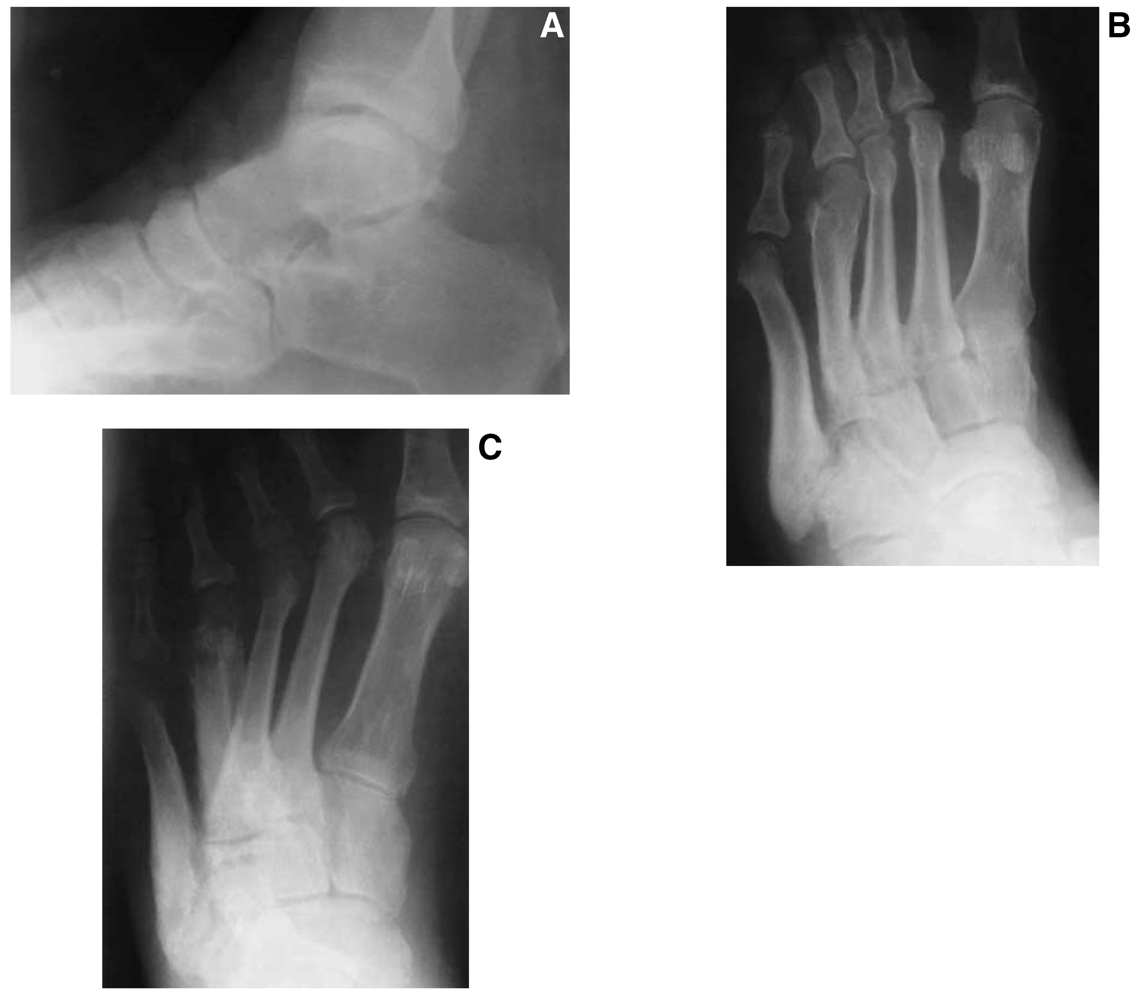
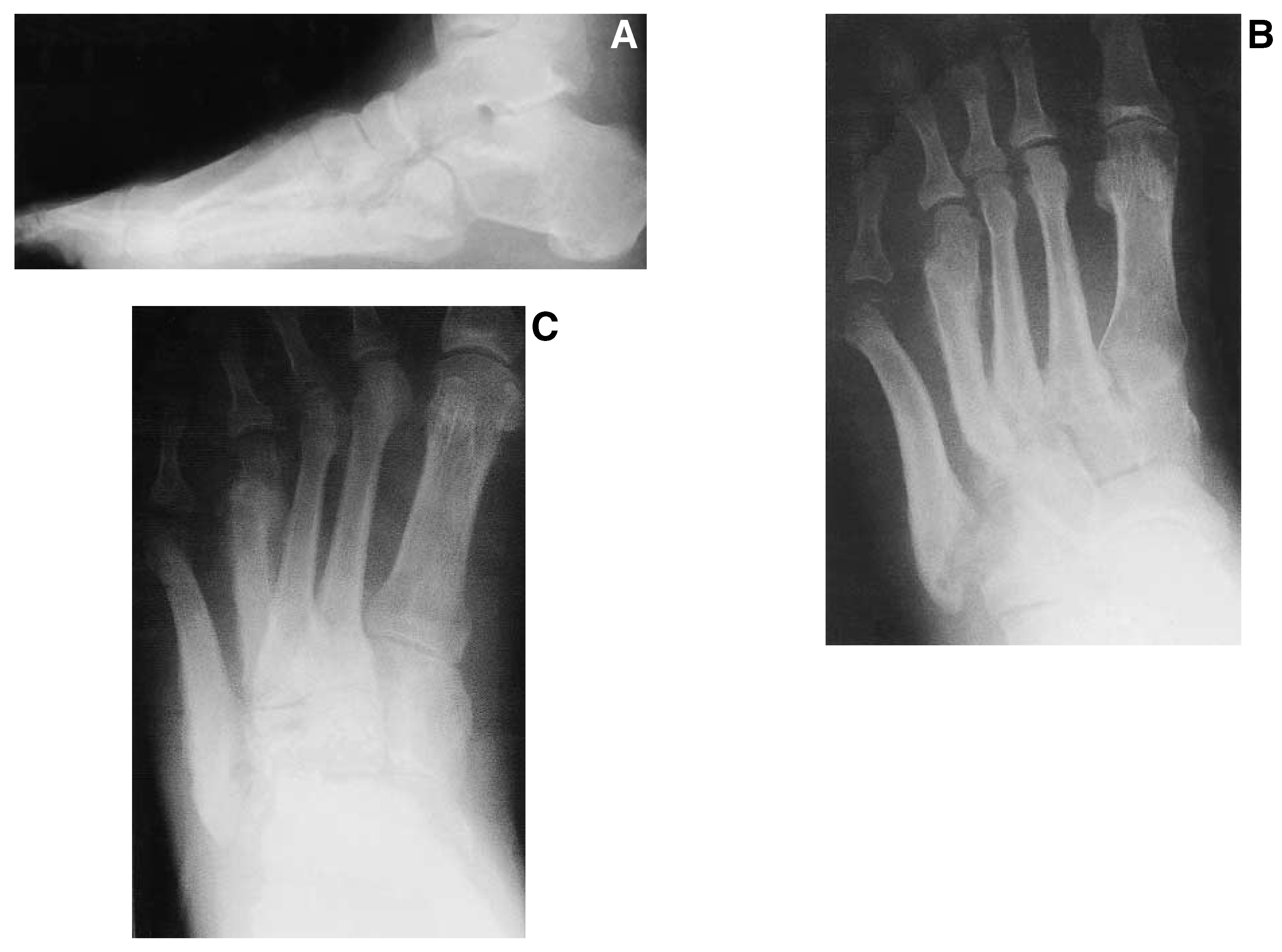
Initial radiographs of the foot and ankle taken at the time the edema developed were reviewed and found to be negative for any fractures or dislocations (
Fig. 1). No subtle subluxations were noted. An incidental os peroneum and mild cavus deformity were evident. Subsequent radiographs taken at the time of presentation to the office of the senior author 9 months later revealed significant alteration of the fourth and fifth metatarsal segments and their corresponding articulations with the cuboid (
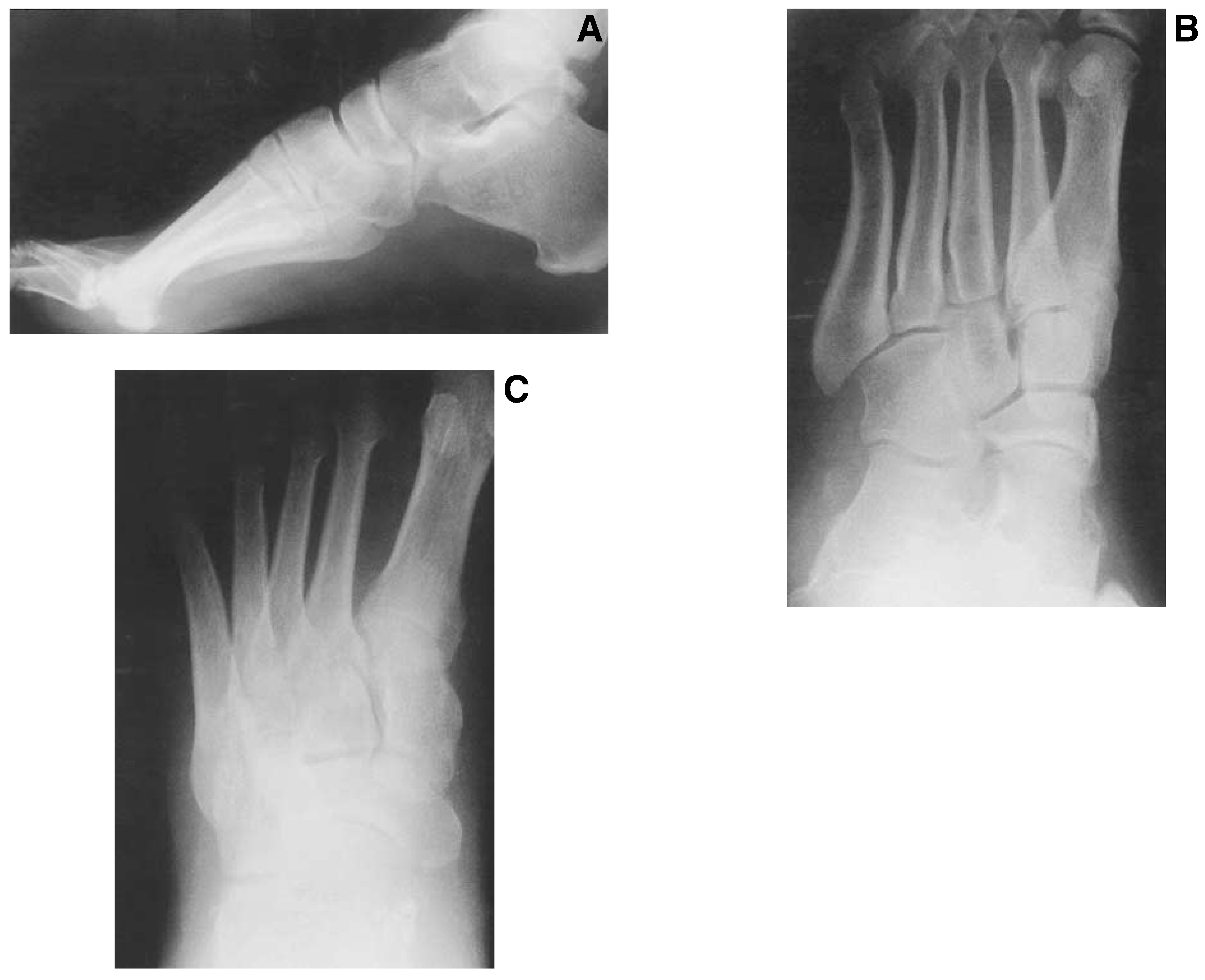
Fig. 2). Significant osseous resorption and osseous destruction were readily apparent with obvious remodeling of the area. A healing fracture of the distal aspect of the fourth metatarsal was also noted. Some angular malalignment of the fourth metatarsal was observed but was acceptable overall and did not correspond with any specific clinical findings of concern. Some early changes in the cuneiform bones and corresponding metatarsal bases were felt to be secondary to increased stress and generalized hyperemia of the foot resulting from a long-standing neuropathic process.
Figure 1.
Initial lateral (A), oblique (B), and anteroposterior (C) radiographs taken several days following the initial ankle sprain injury. Note the absence of any fractures or dislocations. An increase in soft-tissue volume and density is evident. A conventional bone scan most likely would have confirmed or excluded the diagnosis of Charcot’s neuroarthropathy and would have been the imaging study of choice in the face of continued clinical symptoms.
Figure 1.
Initial lateral (A), oblique (B), and anteroposterior (C) radiographs taken several days following the initial ankle sprain injury. Note the absence of any fractures or dislocations. An increase in soft-tissue volume and density is evident. A conventional bone scan most likely would have confirmed or excluded the diagnosis of Charcot’s neuroarthropathy and would have been the imaging study of choice in the face of continued clinical symptoms.
Figure 2.
Lateral (A), oblique (B), and anteroposterior (C) radiographs taken 9 months after the initial ankle sprain injury demonstrate obvious osseous changes of the fourth and fifth metatarsals and their corresponding articulation with the cuboid. The changes are consistent with later stages of Charcot’s neuroarthropathy. Note the destructive changes of the bones consistent with osteoclastic activity and prolonged hyperemia. A healing fracture of the distal aspect of the fourth metatarsal is also consistent with the patient’s history and a diagnosis of diabetic Charcot’s neuroarthropathy.
Figure 2.
Lateral (A), oblique (B), and anteroposterior (C) radiographs taken 9 months after the initial ankle sprain injury demonstrate obvious osseous changes of the fourth and fifth metatarsals and their corresponding articulation with the cuboid. The changes are consistent with later stages of Charcot’s neuroarthropathy. Note the destructive changes of the bones consistent with osteoclastic activity and prolonged hyperemia. A healing fracture of the distal aspect of the fourth metatarsal is also consistent with the patient’s history and a diagnosis of diabetic Charcot’s neuroarthropathy.
Initial treatment consisted of compression bandages and splints to resolve the profound edema, followed by nonweightbearing cast immobilization for 10 to 12 weeks. An external bone stimulator was applied to facilitate bone healing. The patient was instructed in proper footwear and educated regarding the diabetic Charcot process to avoid future incidents. His foot healed uneventfully; no further breakdown of the area occurred and no further deformation developed (
Fig. 3). The swelling resolved completely. The patient was seen periodically every 3 to 4 months to monitor his overall pedal status and was very compliant with his treatment regimen.
Figure 3.
Subsequent lateral (A), oblique (B), and anteroposterior (C) radiographs taken 3 months after initial presentation to the senior author’s office reveal consolidation and remodeling of the same bones and joints, indicating stabilization and resolution of the neuropathic process. No further destruction of bone is seen. The overall alignment and architecture have remained stable. This foot is now considered ready for appropriate protective bracing or possibly a custom orthotic device.
Figure 3.
Subsequent lateral (A), oblique (B), and anteroposterior (C) radiographs taken 3 months after initial presentation to the senior author’s office reveal consolidation and remodeling of the same bones and joints, indicating stabilization and resolution of the neuropathic process. No further destruction of bone is seen. The overall alignment and architecture have remained stable. This foot is now considered ready for appropriate protective bracing or possibly a custom orthotic device.
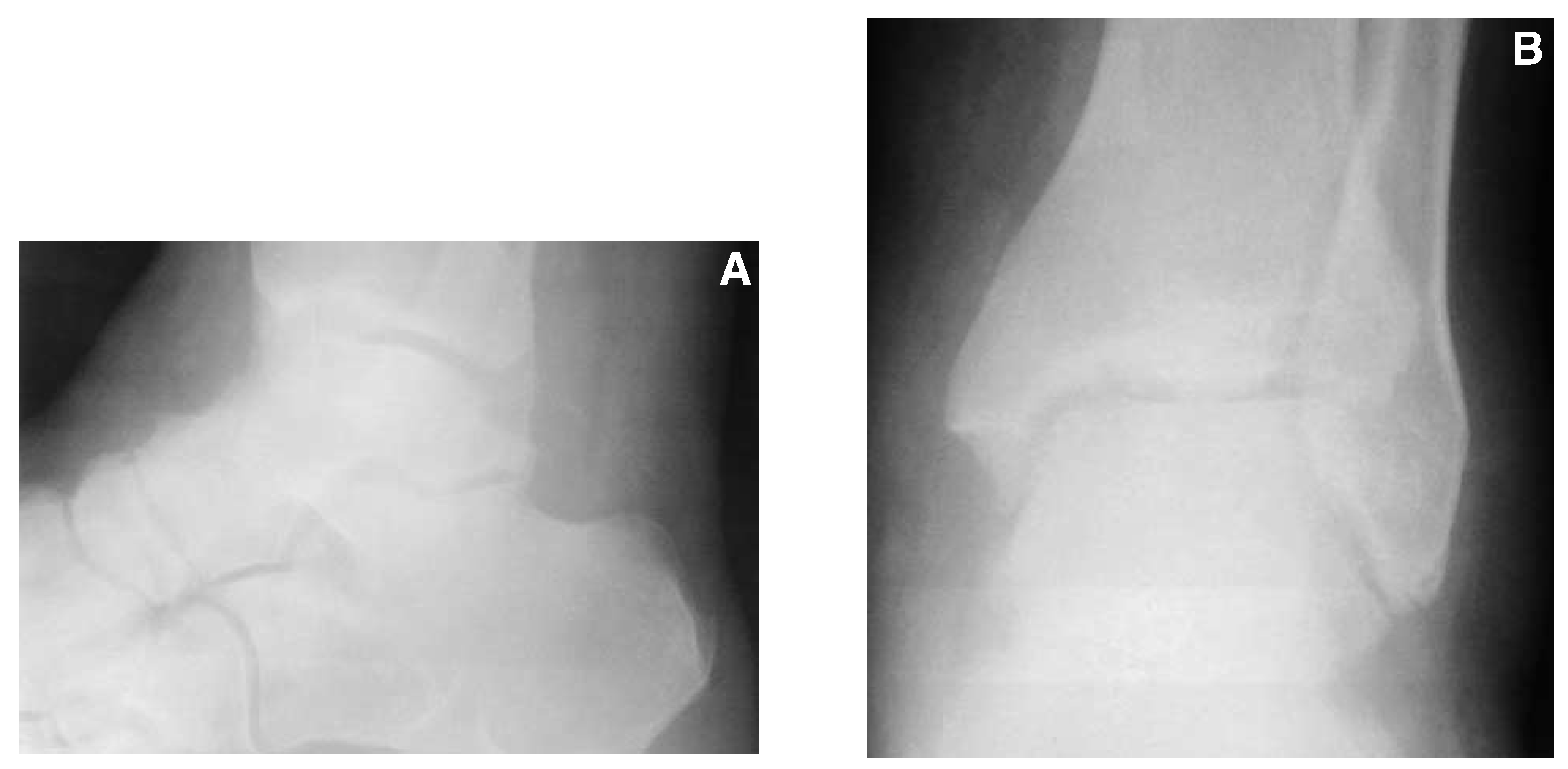
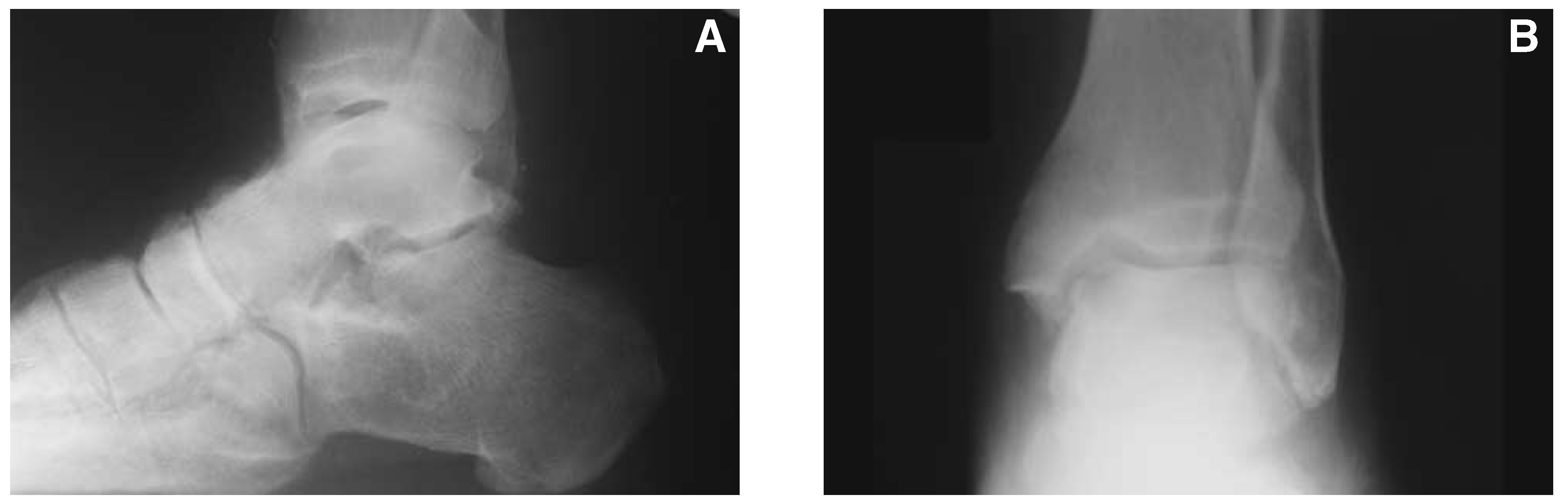
The patient returned more than 1 year later with a sudden onset of edema of his left lower leg and ankle. He denied any history of trauma to the area. Physical examination revealed significant pitting edema of the ankle with no obvious deformation. Muscle function was again intact. There was no indication of a tendon rupture or inflammation. Because there was no indication of thrombophlebitis, no work-up for a thromboembolic phenomenon was undertaken. Radiographs of the ankle were negative for acute fracture or dislocation (
Fig. 4). Some old secondary changes were noted, but they were not thought to be related to the current problem.
Figure 4.
Lateral (A) and anteroposterior (B) radiographs of the ankle taken 15 months after initial presentation (2 years following the initial ankle sprain injury) when the patient returned complaining of the sudden onset of swelling of several days’ duration involving the ankle and lower leg. An increase in soft-tissue volume and density correlated with the clinical findings. No evidence of acute fracture or dislocation is evident. Old secondary changes of the talonavicular and ankle joint are present.
Figure 4.
Lateral (A) and anteroposterior (B) radiographs of the ankle taken 15 months after initial presentation (2 years following the initial ankle sprain injury) when the patient returned complaining of the sudden onset of swelling of several days’ duration involving the ankle and lower leg. An increase in soft-tissue volume and density correlated with the clinical findings. No evidence of acute fracture or dislocation is evident. Old secondary changes of the talonavicular and ankle joint are present.
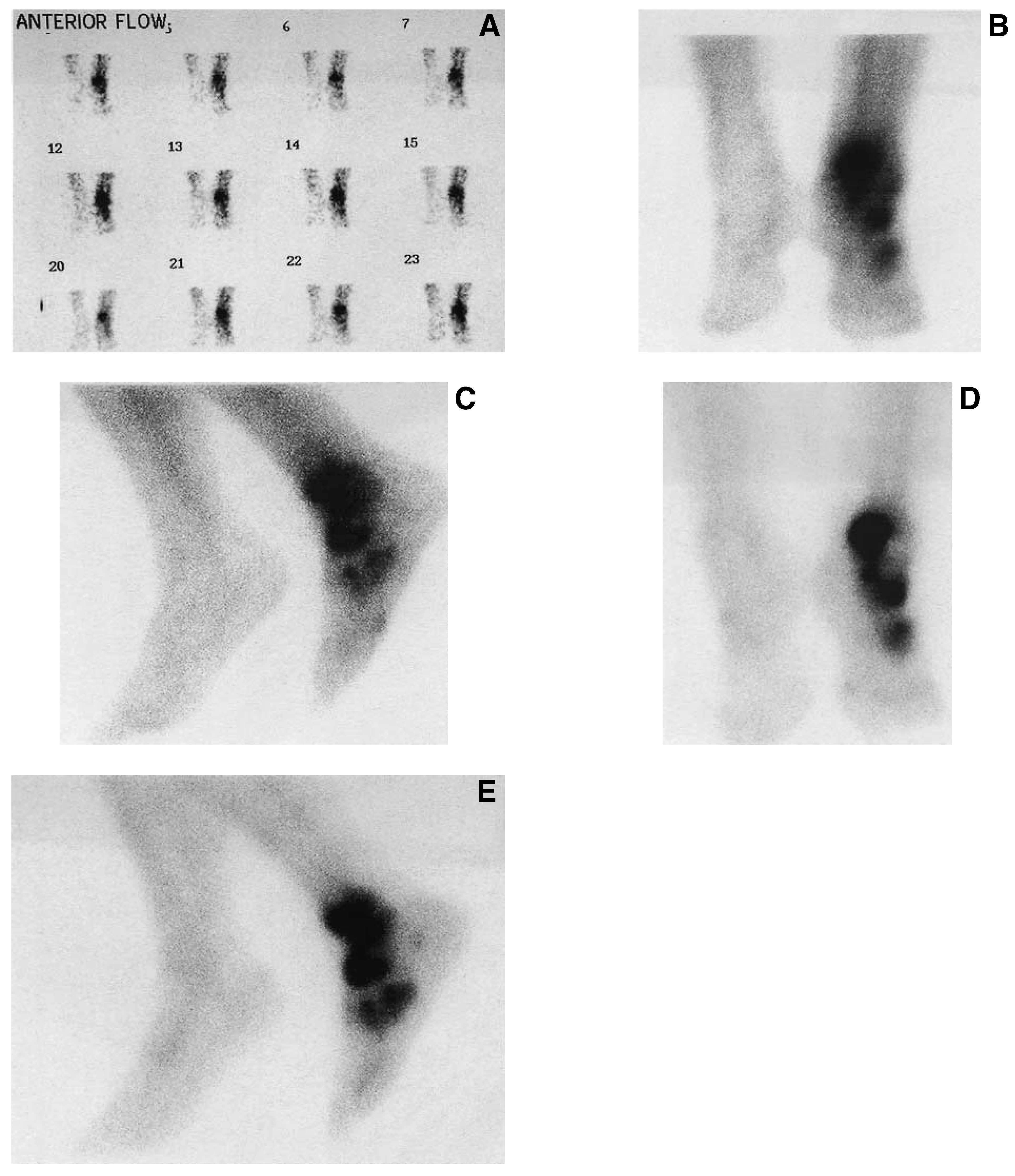
Owing to the high index of suspicion for an acute onset of Charcot’s arthropathy, a conventional triphasic (technetium-99m MDP) bone scan was obtained (
Fig. 5). A dramatic increase in perfusion to the left lower leg, ankle, and foot was found. The delayed images revealed increased focal uptake in the distal tibia, talus, and navicular. Less impressive uptake was evident in the area of the previous Charcot process of the fourth and fifth metatarsal and cuboid and corresponded well with the conventional radiographs and clinical picture.
Figure 5.
Immediate (A), blood pool (B and C), and delayed (D and E) images of a conventional triphasic bone scan (technetium-99m MDP) confirming a diagnosis of stage 0 Charcot’s neuroarthropathy. A significant increase of the radioisotope in the left lower extremity indicates significantly increased perfusion of the foot and ankle consistent with the early Charcot process in a new location. Delayed images demonstrate intense uptake in the distal tibia, talus, and navicular consistent with the clinical picture and further confirm the diagnosis. Less uptake is seen in the fourth and fifth metatarsocuboid area, where the previous Charcot process has remained stable as a result of previous treatment.
Figure 5.
Immediate (A), blood pool (B and C), and delayed (D and E) images of a conventional triphasic bone scan (technetium-99m MDP) confirming a diagnosis of stage 0 Charcot’s neuroarthropathy. A significant increase of the radioisotope in the left lower extremity indicates significantly increased perfusion of the foot and ankle consistent with the early Charcot process in a new location. Delayed images demonstrate intense uptake in the distal tibia, talus, and navicular consistent with the clinical picture and further confirm the diagnosis. Less uptake is seen in the fourth and fifth metatarsocuboid area, where the previous Charcot process has remained stable as a result of previous treatment.
Treatment consisted of serial Jones compression bandages followed by nonweightbearing bracing with a removable pneumatic walker. The patient also received three infusions of pamidronate. The patient was monitored closely and serial radiographs were obtained to monitor any architectural changes within the foot or ankle (
Fig. 6). The patient responded well to treatment and gradually returned to full weightbearing with an ankle-foot orthosis to prevent further recurrences. The swelling resolved and the patient resumed a normal lifestyle. He continues to be cautious about his activities and examines his feet regularly for any detrimental changes.
Figure 6.
Long-term follow-up lateral (A) and anteroposterior (B) radiographs of the ankle joint demonstrate no progressive breakdown of the bones or joints, indicating good response to the initial treatment and good patient compliance.
Figure 6.
Long-term follow-up lateral (A) and anteroposterior (B) radiographs of the ankle joint demonstrate no progressive breakdown of the bones or joints, indicating good response to the initial treatment and good patient compliance.
Conclusion
Charcot’s neuroarthropathy continues to present a major challenge for physicians and surgeons dedicated to the treatment of pedal disease in the diabetic patient population. Recognition of the stage 0 Charcot foot has received little attention in the podiatric and orthopedic literature. A high index of suspicion coupled with knowledge of the natural disease process can lead to the earliest possible diagnosis of this serious disease and ensure timely and proper treatment, which should decrease the incidence of serious deformity and the well-known sequelae of ulceration, soft-tissue infection, and osteomyelitis. Radionuclide imaging studies are essential to confirm the diagnosis of stage 0 Charcot’s arthropathy in the absence of radiographic findings of osseous destruction. Initial treatment of stage 0 Charcot’s arthropathy should include serial compressive dressings and prolonged nonweightbearing of the affected extremity. Consideration should be given to bisphosphonate therapy, as well as noninvasive bone stimulation, to aid in the consolidation process. Functional bracing, custom-molded inserts, or both should be employed for long-term protection following consolidation.