Phenol was discovered in 1834 by the German chemist Friedlieb Runge, who isolated it from coal tar distillate and called it carbolic acid.[
1] Phenol is an aromatic benzene ring with a hydrogen atom replaced by a hydroxyl group, resulting in its chemical formula of C
6H
6O. Because of its ionization properties, phenol has an acidity approximately 1,000 times less than that of water, making it a weak organic acid.[
2] The chemical is unique in that it is both hydrophilic and lipophilic, which contributes to its solubility characteristics. Phenol is soluble in water to an extent of 6% to 7% at room temperature and is highly soluble in nonpolar organic solvents such as isopropyl alcohol, glycerin, ketones, and esters.[
1] Owing to phenol’s solubility properties in organic solvents, nonpolar organic solvents are used as an immediate treatment for phenol burns. It has been shown that isopropyl alcohol is potentially the most effective treatment for these burns when applied to the skin soon after contact with phenol.[
3]
Liquefied phenol is very stable when stored in an airtight amber bottle at room temperature. The chemical tends to assume a pinkish or reddish color when it is exposed to room air and light; however, this has little effect on its potency or concentration. Because of phenol’s stability, the US Food and Drug Administration does not require chemical manufacturers to supply an expiration date on the bottle (J Fowler, Mallinkrodt Inc, personal communication, 2002). The senior author (J.S.B.) had a rarely used bottle of 89% phenol solution in his office that had the date of manufacture written on the product label. This 23-year-old bottle of phenol solution was sent for an independent chemical assay, and the concentration was 93.0%. However, this does not reflect typical office usage, in which bottles are opened and closed on a daily basis. This exposure to the environment could reduce the effective concentration.
Phenol is a protoplasmic poison with the potential to cause serious local and systemic toxicity. Animal studies have shown that many signs of acute poisoning appear when phenol enters the systemic circulation through absorption or ingestion. These signs include muscle twitching, body temperature fluctuation, cardiac and pulmonary changes, and convulsions, eventually leading to death.[
4] The use of phenol in the medical field, however, has been shown to be very safe. Rarely, arrhythmias and death have been reported during plastic surgical procedures in which large cutaneous areas are painted and occluded with phenol as a chemical peel.[
5] No systemic complications have been reported with the use of phenol in nail matrixectomies.
Boll,[
6] in 1945, was the first to document the use of phenol in the treatment of ingrown toenails. Multiple articles since then have documented the safety and effectiveness of phenol matrixectomy.[
7-
20] Success rates of 80% to 100% have been reported, with average healing times of 1 to 6 weeks (
Table 1).
Table 1.
Summary of Available Literature on Phenol Matrixectomy
Table 1.
Summary of Available Literature on Phenol Matrixectomy
Despite its wide acceptance in the medical community, there are many variations of the phenol matrixectomy technique. Most practitioners use concentrations of 80% to 89% phenol solution applied for varying periods of time. Average application times, healing times, follow-up periods, and recurrence rates reported in the literature reviewed for this article are given in
Table 1. No serious complications were reported in any study. Only 198 of the nearly 1,000 cases reviewed were found in the podiatric medical literature.
The podiatric medical literature reviewed contained studies focusing on populations with special problems. Subjects with diabetes mellitus were the focus of two studies that reviewed 105 phenol nail procedures.[
21-
22] The results of these two studies were similar to the results of the studies mentioned above, although the diabetic subjects had a relatively long average healing time of 5.5 weeks.
The other problem-focused studies dealt with the inflammatory response to phenol-induced wounds. These studies reported that silver sulfadiazine, hydrocortisone cream, povidone-iodine ointment, and Cortisporin Otic Solution (Burroughs Wellcome Co, Research Triangle Park, North Carolina) helped to reduce the sequelae seen with phenol-induced wounds and healing progressed at a more rapid rate.[
23-
25]
Given phenol’s potential toxic effects and the intense inflammatory changes phenol produces in the skin, identifying the optimal time and concentration necessary to destroy the germinal matrix without causing further soft-tissue damage would be beneficial.
Materials and Methods
Specimens of nail matrix were obtained and painted with phenol solution for 30 sec, 1 min, 90 sec, and 2 min. The matrix was then immediately placed in a formalin solution. This procedure preserved the specimen and removed the phenol from the surface matrix, providing effects similar to those of an alcohol rinse. A control consisted of specimens placed directly in the preservative solution. Three specimens were obtained for each time group and for the control.
The senior author (J.S.B.) surgically excised the nail matrix on patients presenting with ingrown toenails. Immediately following excision, the phenol solution was applied on the instrument table. This was believed to yield the same result as phenol applied in situ to matrix under tourniquet hemostasis. The specimens were then stained and examined microscopically.
Results
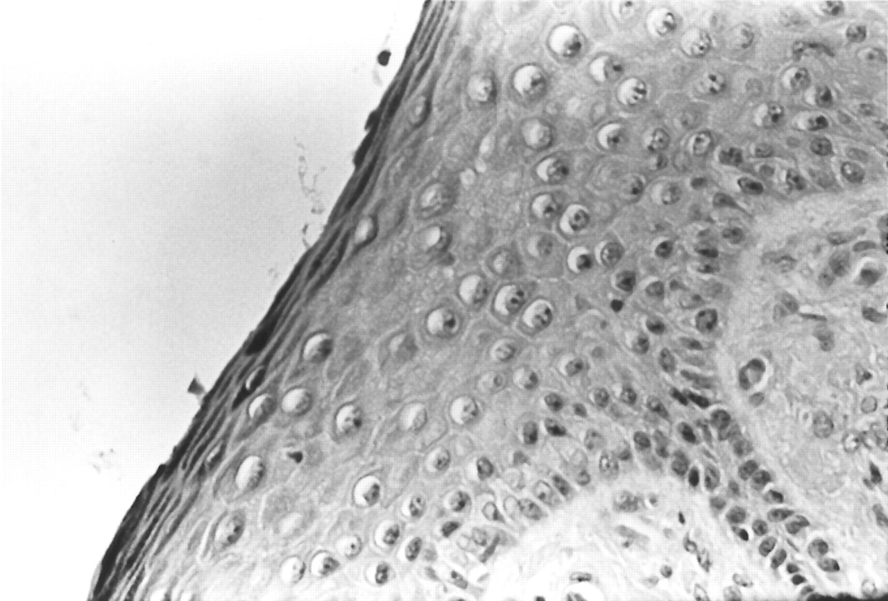
Microscopic analysis of normal anatomy revealed a thick layer of epidermal cells (
Fig. 1). The basal cell layer and rete ridges were readily identified. The cells were normochromatic.
Figure 1.
Normal skin (H&E, ×40). Rete ridges are seen and the basal layer is visible and covered by a thick epidermal layer.
Figure 1.
Normal skin (H&E, ×40). Rete ridges are seen and the basal layer is visible and covered by a thick epidermal layer.
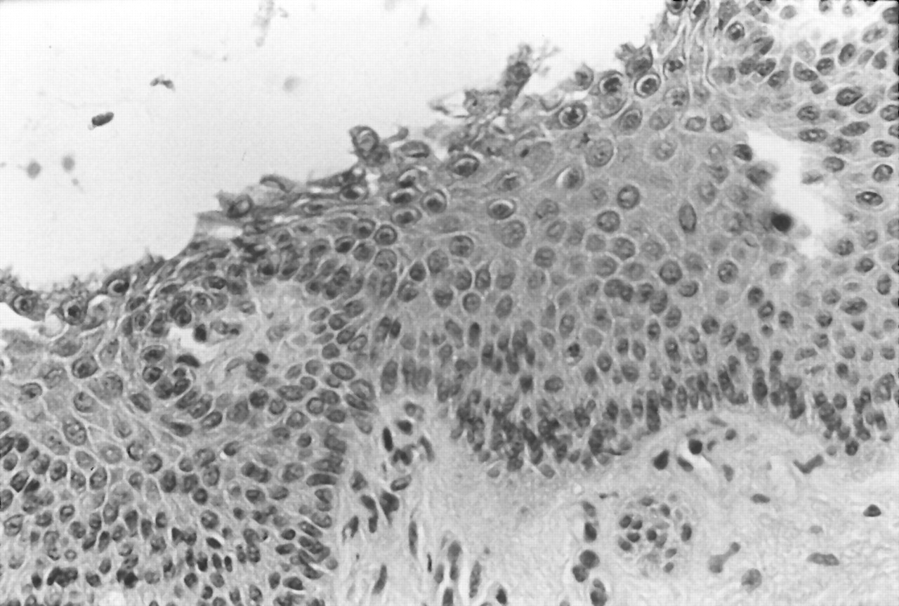
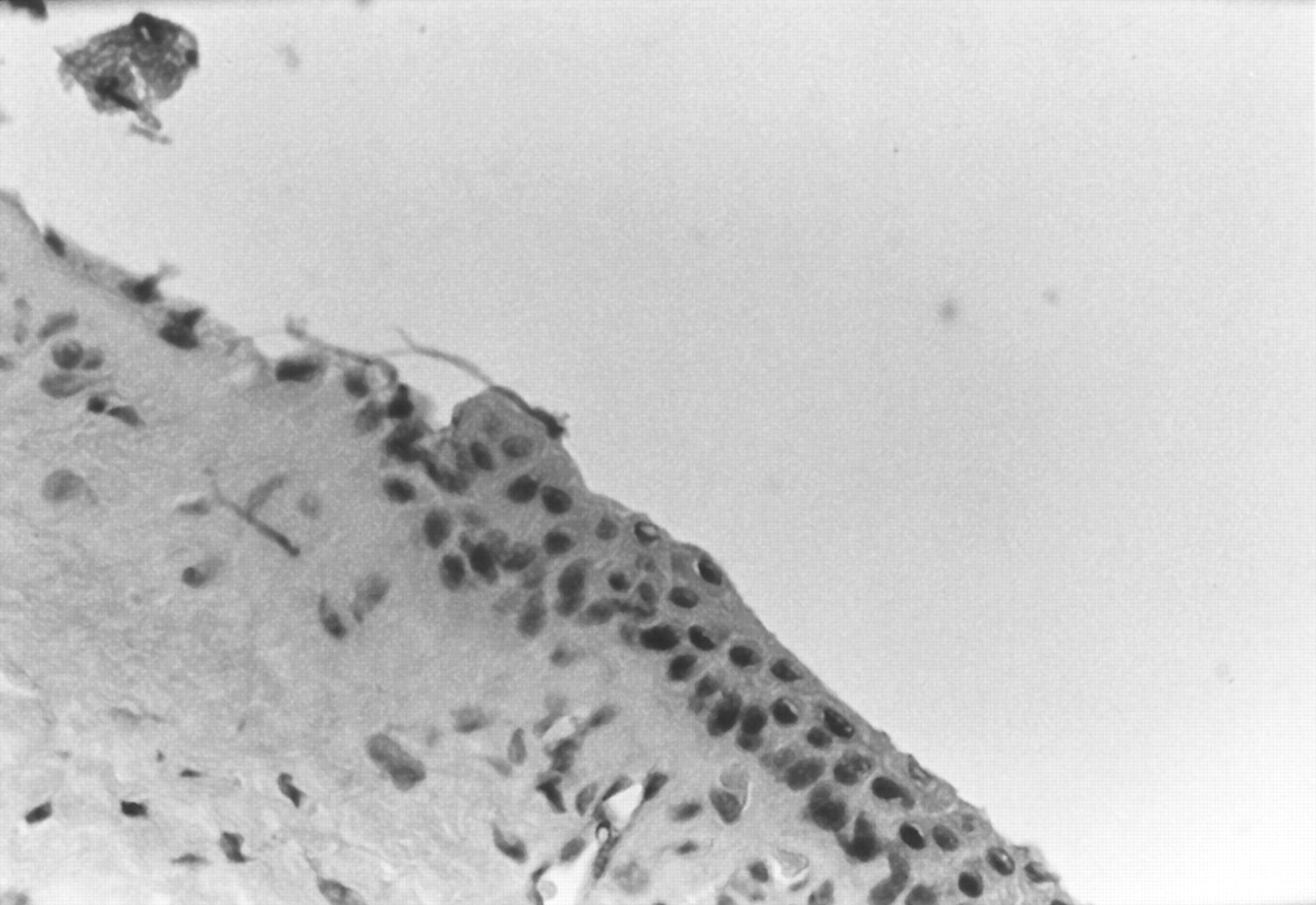
After a 30-sec application of 89% phenol solution, only superficial damage and mild coagulation of the squamous cells were noted (
Fig. 2). Some hypochromatic cells were noted. The basal layer was primarily intact, and the potential for regrowth was high.
Figure 2.
Minimal damage of the epidermal layer only after 30-sec application of 89% phenol solution (H&E, ×40).
Figure 2.
Minimal damage of the epidermal layer only after 30-sec application of 89% phenol solution (H&E, ×40).
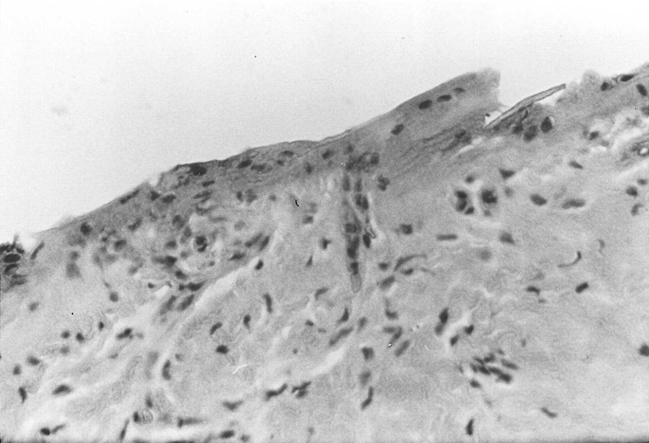
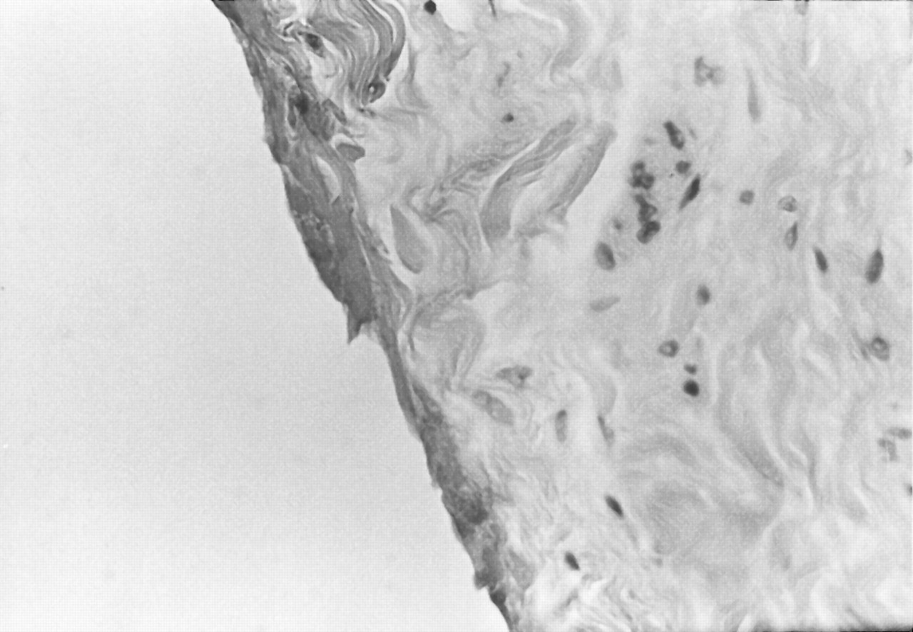
Following a 1-min application of 89% phenol solution, full-thickness necrosis of the epithelium was noted (
Fig. 3). Some hyperchromatic nuclei were present in the dermis, and the dermal cells were shrunken. The basal layer was completely destroyed.
Figure 3.
Full-thickness epidermal necrosis with destruction of the basal layer after 1-min application of 89% phenol solution (H&E, ×40).
Figure 3.
Full-thickness epidermal necrosis with destruction of the basal layer after 1-min application of 89% phenol solution (H&E, ×40).
The effects of 90-sec and 2-min applications of the 89% phenol solution were similar (
Figs. 4 and
5). There was complete destruction of the basal layer with a more intense necrosis of the underlying dermis without full-thickness loss of the dermis.
Figure 4.
Full-thickness necrosis of the epidermal layer after 90-sec application of 89% phenol solution (H&E, ×40).
Figure 4.
Full-thickness necrosis of the epidermal layer after 90-sec application of 89% phenol solution (H&E, ×40).
Figure 5.
Full-thickness necrosis similar to that shown in
Figure 4 after 2-min application of 89% phenol solution (H&E, ×40).
Figure 5.
Full-thickness necrosis similar to that shown in
Figure 4 after 2-min application of 89% phenol solution (H&E, ×40).
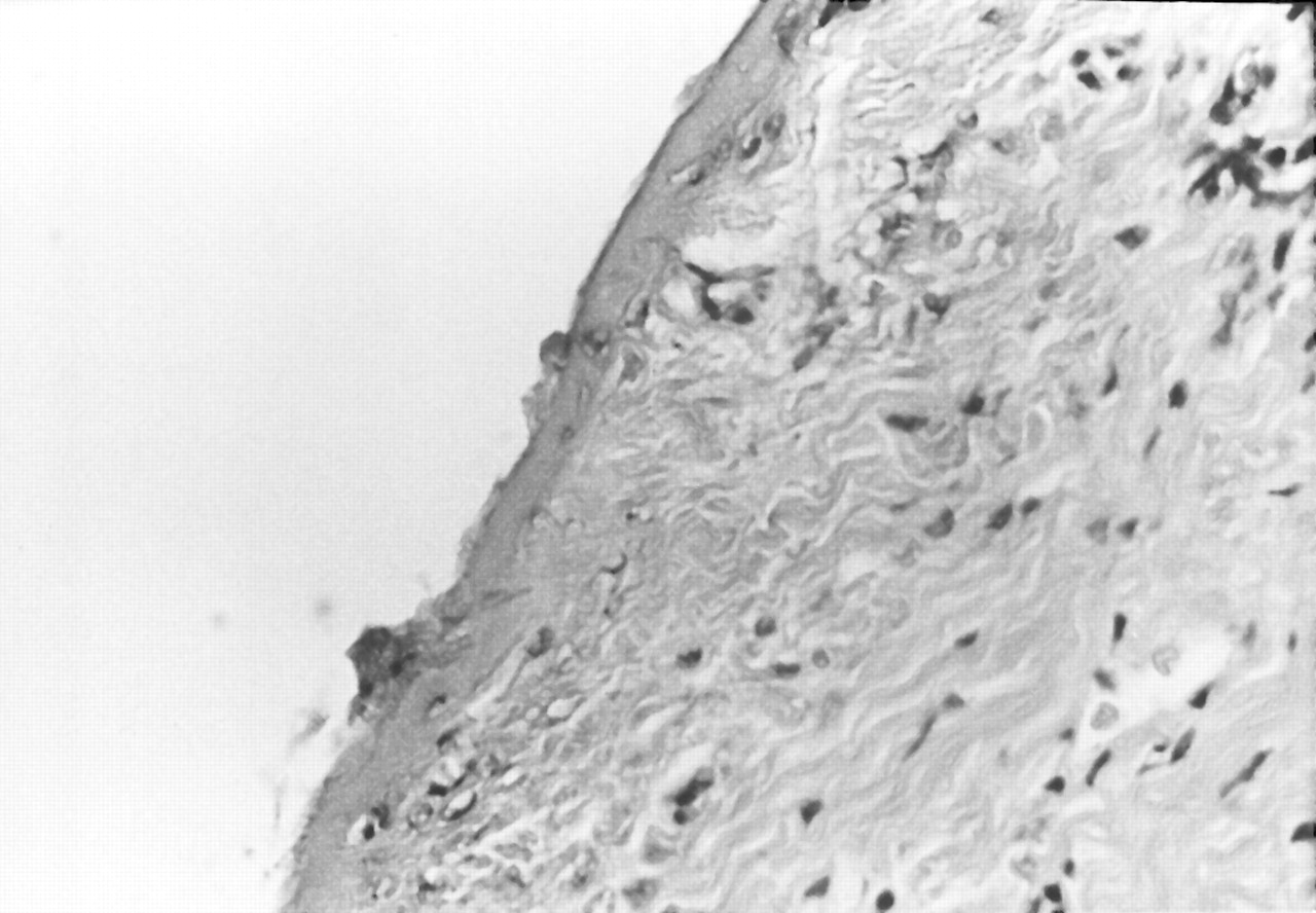
Finally, a 45% phenol solution was applied for 1 min (
Fig. 6). Little destruction of the basal layer occurred, indicating likely regrowth of the nail.
Figure 6.
Mild coagulation of the superficial layer after 1-min application of 45% phenol solution; the basal layer is partially intact (H&E, ×40).
Figure 6.
Mild coagulation of the superficial layer after 1-min application of 45% phenol solution; the basal layer is partially intact (H&E, ×40).
Discussion
The first medical use of phenol was to decontaminate compound fractures. Today, phenol is used clinically primarily to create chemical burns of the skin or skin appendages. Examples include chemical facial peels and nail matrix cauterization. Phenol is a weak acid that does not produce full-thickness dermal loss. Topical application of phenol causes denaturation and precipitation of the skin’s proteins. This unique action has allowed dermatologists and plastic surgeons to use it as a skin resurfacing technique. For these procedures, a 45% solution of phenol is typically applied directly to the face and immediately occluded for 24 to 48 hours.
Brown et al[
26] postulated that the precipitated proteins formed a coagulated protein barrier to prevent further penetration of the phenol. The higher the concentration, the more complete the barrier. They suggested that lowering the concentration would decrease the coagulum, leading to increased phenol penetration.
In a review of the literature, the authors found only one comparative study analyzing the histologic effects of phenol when applied to the skin. In a study by Spira et al,[
27] phenol was applied to the inner thigh of six volunteers. Four different concentrations were used: 25%, 50%, 75%, and 100%. One-half of these phenol applications were occluded with waterproof tape for 48 hours. Punch biopsies were then taken from these phenol-induced wounds at intervals ranging from immediately following phenol application to 3 months later. These histologic specimens were studied and showed that the reaction sequence began with an initial keratocoagulation and epidermolysis, with a deeper zone of cellular destruction involving only the papillary dermis. No full-thickness destruction of the dermis was noted. Epidermal regeneration was noted as early as the second day and was complete by 1 week, while total epidermal healing was complete by 2 weeks. However, the dermis continued to heal at 3 months. Occlusion of the test sites resulted in a broader zone of destruction and an increase in the inflammatory response with each increase in phenol concentration.
The study found that phenol concentrations of 50% or more had similar destructive effects on the skin; a 25% phenol solution was only slightly less destructive. The study concluded that the destructive effects of phenol depend in part on its concentration but that there is not a linear relationship between skin necrosis and phenol concentration. Furthermore, occlusion of the phenol-induced wound caused an increase in inflammation and delayed the wound healing without providing any further penetration. The epidermis completely regenerated in all cases, in spite of its complete destruction by the phenol. This partly explains nail regrowth despite applications of phenol.
The present study found the amount of destruction to be concentration-dependent. A 45% phenol solution applied for 1 min resulted in significantly less destruction than an 89% solution applied for the same length of time. In all instances, a full-thickness loss never occurred. There was a significant increase in the amount of tissue destruction between 30 sec and 1 min, but little change from 1 min to 2 min.
Conclusion
Prolonged application of phenol increases the inflammatory response, leading to prolonged pain, drainage, and increased risk of infection. Optimizing the duration of application should reduce the potential complications and lead to reduced healing times. This study showed that the application of 89% phenol solution for 1 min is the minimum time and concentration required for complete destruction of the germinal nail matrix.