The plantar fascia extends from the calcaneus to the deep tissues of the forefoot and affects the overall function and structure of the foot. The central band of the plantar fascia extends to the sesamoid apparatus, the base of the hallux, and the lesser digits, causing a biomechanical change in these joints.[
1,
2]
Several studies have been performed to calculate the relative change in arch height after the plantar fascia has been sectioned.[
3-
9] These studies have proven that the overall structure of the foot is altered with sectioning of the plantar fascia; however, there are no studies describing the effect on the range of motion at the first metatarsophalangeal joint.
The purpose of this study was to investigate whether sectioning the plantar fascia would affect the range of motion at the first metatarsophalangeal joint. The authors were particularly interested in investigating the average change in first metatarsophalangeal joint dorsiflexion obtained with fascia release because of the possible benefit of sectioning the plantar fascia during hallux limitus repair.
Anatomical Review of the Plantar Fascia
The plantar fascia, or plantar aponeurosis, is a thick band of strong connective tissues located on the plantar aspect of the foot. Its anatomy has been well described by a number of authors.[
2,
10-
12] The plantar fascia is located between the subcutaneous tissues and the plantar musculature. It originates from the medial tubercle of the calcaneus and divides into a complex, superficial, and deep network distally.[
2] The fascia is divided into three major well-defined bands: the medial, central, and lateral bands.
The central band plays a major biomechanical role in the anatomical distribution of the plantar fascia complex. It originates from the medial aspect of the calcaneus and extends distally, where it divides into numerous sections. The fascia is narrow and thick at its origin, measuring 1 to 2 cm in width, but it widens and becomes thinner as it extends distally.[
2,
11] According to Bøjsen-Möller and Flagstad,[
12] the central band divides into superficial and deep layers at the metatarsal diaphysis. The deeper layer splits into two distinct septa that encompass the flexor tendons, separating them from surrounding structures. The septa terminate by blending with the deep transverse metatarsal ligament, the fibrous sheaths, the flexor plate of the metatarsophalangeal joint, and the periosteum at the base of the proximal phalanges. Medially, the septa located plantar to the first metatarsal have fascial investment into the tibial and fibular sesamoids together with the fascia overlying the flexor hallucis brevis muscle.[
2,
11,
13] The plantar fascia inserts mostly into the medial forefoot, and decreasingly firm attachments are seen laterally.[
14]
The superficial layer is divided into five bands. The most lateral band attaches proximal to the base of the fifth toe; the central three bands attach between the fourth and fifth toes, at the base of the third toe, and between the first and second toes.[
11] The fifth band attaches proximal to the base of the hallux. All five bands course distally and vertically to attach to the skin and subcutaneous tissues.[
2]
The medial component of the plantar fascia is thinner than the central band. It encompasses the abductor hallucis muscle, which partially originates from a deeper and more posterior portion of this fascia. The medial band thickens distally, where it wraps medially to connect to the deep fascia of the dorsum of the foot.[
11]
The lateral component of the plantar fascia is thicker than the medial band and is well defined. This structure originates from the plantar medial tubercle of the calcaneus and extends distally, where it splits into two parts: a medial part that attaches to the base of the fifth metatarsal, and a lateral part that connects to the muscle fascia of the abductor digiti quinti minimi.[
11,
14] A comprehensive study by Loth[
15] found that this structure is absent in some people.
Biomechanical Function
The plantar fascia is a biomechanically active structure that does not work independently. It acts passively when the digits are dorsiflexed and the metatarsal is plantarflexed. Hicks[
16] first described this phenomenon, comparing it to a “windlass action” that occurs when the digits, particularly the hallux, are dorsiflexed. He noticed that supination of the foot and an increase in the arch height occurred when the digits were extended. This phenomenon occurred when the digits were dorsiflexed, pulling the fascia over the metatarsal heads and causing them to become plantarflexed. This function appeared to be lost in fresh cadaver specimens when the fascia was transected.
The fascia works differently in stance than it does during gait. Plantarflexion of the metatarsal does not readily occur because of ground reaction forces. However, when tension is placed on the plantar aponeurosis, the base and the head of the metatarsal move proximally, shortening and raising the medial arch.[
8] As tension is placed on the plantar medial tubercle of the calcaneus, the rearfoot inverts, supinating the subtalar joint and the oblique axis of the midtarsal joint. This chain reaction affects the whole foot, including the more distal joints.[
13,
14]
The windlass mechanism functions during the propulsive phase of the gait cycle. As heel-off begins, the toes are dorsiflexed at the metatarsal owing to the ground reaction forces. Prior to toe-off, maximal tension is placed on the plantar fascia, as the proximal phalanges of the digits are maximally dorsiflexed at the metatarsals. This mechanism is partially responsible for locking the foot and changing it from a mobile adapter to a rigid lever in an effort to provide an effective propulsive mechanism.[
7,
11] During this phase, the heel cord contracts, rotating the talus and calcaneus in the sagittal plane. The fascia acts as an important restraint to prevent collapse of the medial arch.[
6]
Methods
Patients selected for this study were healthy, with no evidence of either subjective or objective pathology of the first metatarsophalangeal joint, who had undergone surgery for recalcitrant plantar fasciitis. Radiographs were reviewed and patients who exhibited severe spurring, joint destruction, or fusion of the first metatarsophalangeal joint were excluded.
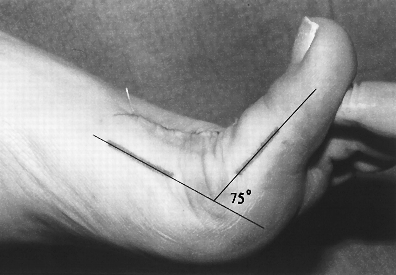
Preoperatively, attention was directed to the medial aspect of the first metatarsal, which was palpated and bisected proximally and distally. A mark was placed on the skin at each bisection. These two marks were then connected with a straight line to form the medial longitudinal bisection of the first metatarsal. The bisection of the proximal phalanx was obtained in a similar manner. All measurements were taken by the same clinician (F.M.H.) throughout the study.
The first metatarsophalangeal joint was considered at 0° or neutral position when the two lines previously drawn were parallel. The joint was then dorsiflexed to maximal resistance and measurements were obtained with a tractograph and recorded as preoperative dorsiflexion (
Fig. 1). The first metatarsophalangeal joint was then plantarflexed to maximal resistance and measurements were obtained using the previously marked area. The amount of plantarflexion obtained was recorded as preoperative plantarflexion. These measurements were taken while the patient was sedated and anesthetized to reduce variability due to muscle spasm.
An instep fasciotomy as described by Fishco et al[
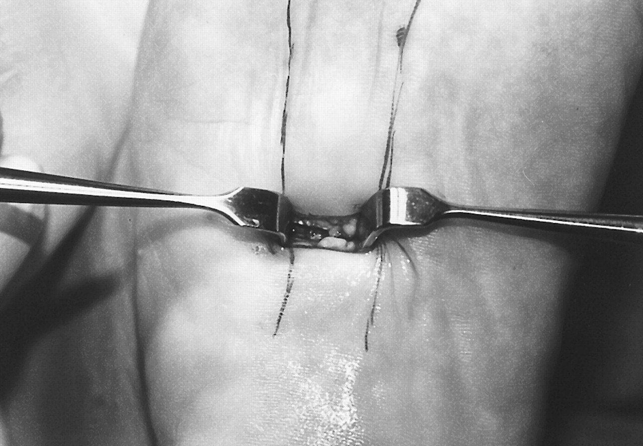
17] was performed. The medial and lateral borders of the central band of the plantar fascia were marked. This band can be palpated by dorsiflexing the first metatarsophalangeal joint. The plantar aspect of the foot was divided into thirds and a line was created over the central band of the plantar fascia at the junction between the proximal and central thirds. The area was then injected with local anesthetic and an incision was created parallel to the skin lines. This incision was deepened to the level of the central band of the plantar fascia. Once the fascia was well identified, it was transected with a surgical knife (
Fig. 2). The hallux was dorsiflexed and the central band of the plantar fascia was simultaneously palpated for any remaining fibers that required transection. Once the central band had been totally sectioned, the surgical site was irrigated and vertical mattress sutures were applied.
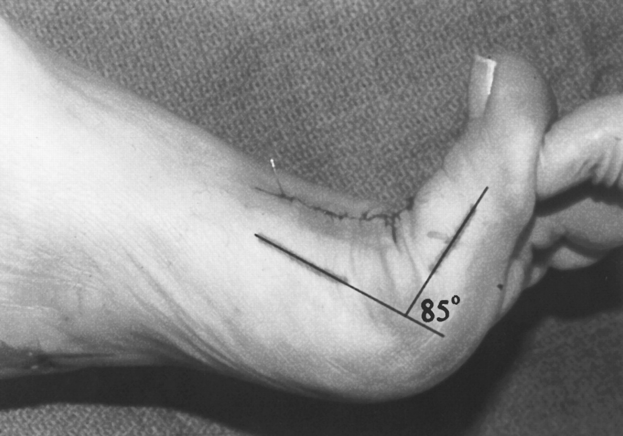
After the instep fasciotomy procedure, postoperative dorsiflexion and plantarflexion measurements were obtained and recorded in the same manner as the preoperative measurements (
Fig. 3). To ensure consistency with the preoperative method, the postoperative measurements were obtained immediately after transecting the plantar fascia while the patient was still sedated.
Figure 1.
Preoperative measurements of first metatarsophalangeal joint range of motion after the bisection lines have been drawn.
Figure 1.
Preoperative measurements of first metatarsophalangeal joint range of motion after the bisection lines have been drawn.
Figure 2.
Instep plantar fasciotomy.
Figure 2.
Instep plantar fasciotomy.
Figure 3.
Postoperative measurements of first metatarsophalangeal joint range of motion.
Figure 3.
Postoperative measurements of first metatarsophalangeal joint range of motion.
Long-term measurements were not taken for two reasons. First, the majority of the studies evaluating the effect of sectioning the fascia on the arch were performed on cadavers, and measurements were recorded immediately following the procedure; the authors of the current study wanted to maintain consistency with the previous studies. Second, adaptation of the soft tissues or osseous structures could affect the results. The purpose of this study was to evaluate the immediate results of plantar fascia transection on the motion of the first metatarsophalangeal joint.
Results
Eighteen patients with no first metatarsophalangeal joint pathology who had undergone an instep plantar fasciotomy for recalcitrant plantar fasciitis participated in this study on the effect of sectioning the plantar fascia on first metatarsophalangeal joint range of motion. The average preoperative dorsiflexion and plantarflexion measurements were 69.9° and 14.5°, respectively. The average postoperative dorsiflexion and plantarflexion measurements were 79.7° and 14.7°, respectively. The average increases in dorsiflexion, plantarflexion, and total range of motion of the first metatarsophalangeal joint after release of the plantar fascia were 9.8°, 0.2°, and 10.0°, respectively. A paired t-test performed on the data revealed a statistically significant increase in dorsiflexion and total range of motion of the first metatarsophalangeal joint to a level of P < .001. Changes in plantarflexion were not statistically significant.
Discussion
Many studies have shown that sectioning of the plantar fascia has an effect on the biomechanical function of the foot. Previous research has focused on how sectioning the plantar fascia affects arch height, length, and stiffness.[
3-
9] Kitaoka et al[
8] examined how sectioning the plantar fascia affects midfoot and rearfoot joints. However, no research has been conducted on more distal joints, including one of the most important functional joints in the foot, the first metatarsophalangeal joint.
Hicks[
16] was the first to investigate the effect of the plantar fascia on the major joints of the foot when he described the windlass mechanism. He noted that toe dorsiflexion and metatarsal plantarflexion affected all of the joints in the foot as arch height increases. Thordarson et al[
5] observed that sequential sectioning of the plantar fascia with the toes dorsiflexed at 30° and at maximal dorsiflexion consistently decreased the arch-supporting function. The most significant loss of the windlass mechanism was found after complete sectioning of the plantar fascia. Sectioning the fascia had a combined effect on the height and length of the arch, which contributed to a 25% decrease in arch stiffness. Elongation of the arch was found to be more consistent with sequential sectioning than a decrease in arch height. In 1997, Arangio et al[
3] demonstrated a 17% increase in vertical displacement and a 15% increase in horizontal elongation after releasing the link between the metatarsal and the calcaneus. In 1998, Arangio et al[
4] showed that cutting the plantar fascia altered the mechanical response of the arch significantly more in the high-arch foot as compared with the low-arch foot. The authors concluded that cutting the fascia in a low-arch foot had less impact on the arch height.[
3,
4]
Kitaoka et al[
8] quantified the increased amount of movement that occurs at the rearfoot and ankle area after resection of the plantar aponeurosis. Pontious et al[
14] documented a case in which development of hammer toes was seen following resection of a portion of the plantar aponeurosis for a fibroma, concluding that when placed under tension, the plantar fascia stabilizes the digits against the ground.
Some authors have indicated that the plantar fascia may have a significant effect on the function of the first metatarsophalangeal joint, including limiting its range of motion. Durrant and Siepert[
1] theorized that for a structure to limit dorsiflexion of the first metatarsophalangeal joint,
that structure must: 1) cross the transverse axis of motion of the first metatarsophalangeal joint; 2) lie plantar to this axis of motion; 3) attach to the distal hinge (proximal phalanx); 4) exert a force parallel to the longitudinal axis of the first ray; and 5) be equally present on both sides of the longitudinal axis of the first ray.
The medial attachment of the central band of the plantar fascia meets all of these criteria. If the plantar fascia is congenitally, iatrogenically, or idiopathically altered, this may have a direct effect on the first metatarsophalangeal joint. A tight plantar fascia may contribute to the development of hallux limitus.
Using the windlass model, Fuller[
13] described how tension from the fascia may prevent hallux dorsiflexion in gait, thus creating a functional hallux limitus. The long-term sequela is a repetitive retrograde force applied to the metatarsal head. Compression of the phalanx on the metatarsal head will cause reactive bone formation in the form of osteophytes leading to structural hallux limitus.
In 1927, Cochrane[
18] first described the use of a plantar incision to transect the soft-tissue structures that attach the base of the proximal phalanx to the hallux to increase range of motion at the first metatarsophalangeal joint. He used this procedure for decreased hallux dorsiflexion in the absence of osteoarthritis. Chang[
19] presented a stepwise approach to the surgical treatment of hallux limitus. His sequential approach to hallux limitus repair included cheilectomy, metatarsal osteotomy, and soft-tissue release. This soft-tissue release was performed with a small medial stab incision to release the tight fascial band. Although the release can also be performed proximally, it was carried out distally to reduce the possible effect on rearfoot stability.
In the current study, the release was performed proximally, anterior to the plantar calcaneal tubercle. The fascia was released solely for treatment of plantar fasciitis and not to increase the first metatarsophalangeal joint range of motion. However, in each case, increased motion was noticed when the hallux was dorsiflexed at the metatarsal after the fascia was released. The results of the authors’ study show that the average change in motion at the first metatarsophalangeal joint was 10.0° with complete sectioning of the plantar fascia. The greatest contribution to the change in range of motion was through dorsiflexion, with an average increase of 9.8°. There was an average increase of 0.2° in plantarflexion postoperatively.
A limitation of this study is that the measurements were taken manually. Although the same person took the measurements throughout the study, some degree of uncertainty probably exists. Although more data would be useful, the number of patients participating in this study demonstrated reliable and statistically significant results.
Although long-term results for this procedure would be of interest, this procedure can now be used in conjunction with other procedures to increase the range of motion in cases of hallux limitus. Other long-term studies of this procedure, along with the osseous procedure for hallux limitus repair, would yield more data to show the benefit of this procedure for hallux limitus.
Conclusion
The results of this study indicate that an increase in range of motion at the first metatarsophalangeal joint occurs immediately after the release of the plantar fascia. The data extracted from this study quantify the increased range of motion at the first metatarsophalangeal joint following plantar fasciotomy. Dorsiflexion increased by an average of 9.8°; however, no significant change in plantarflexion was observed immediately following plantar fascia release.