Onychomycosis is one of the most common conditions affecting the feet of the geriatric population. The exact incidence of onychomycosis is unknown, but it is reported to range between 2% to 13% in the general population. Onychomycosis is reported to affect 15% to 20% of the population from 40 to 60 years of age, 32% of the population from 60 to 70 years of age, and more than 48% of the population older than 70 years of age.[
1] In subgroups such as miners, military personnel, and athletes, the incidence has been reported to rise to 20% due to the use of communal showers and changing rooms.[
2] With immunocompromised patients, including acquired immunodeficiency syndrome (AIDS) patients, the incidence rises 11% to 67%.[
1] Although onychomycosis can be caused by dermatophyte, saprophyte, or yeast infections, most studies attribute the diagnosis of onychomycosis to dermatophytes in the general population. Currently, there have been no large-scale studies of onychomycosis that have been limited to the geriatric population. In an effort to determine the diagnosis of onychomycosis in a geriatric population, the authors sent toenail specimens from 450 South Florida patients with suspected onychomycosis to an independent mycology laboratory. The laboratory performed a fluorescent potassium hydroxide (KOH) preparation and microscopic fungal culture examination to determine the specific organism for each patient.
Clinical Studies
Onychomycosis is often thought of as a cosmetic problem; however, it has a great impact on the overall health of a patient, and can decrease mobility and diminish peripheral circulation.[
3] In addition, pressure necrosis of the nail bed, along with minor ulcerations on adjacent toes caused by sharp, brittle, infected toenails, can occur in neuropathic diabetic patients.[
4] Toenails also can serve as a reservoir for organisms that can spread to other body areas or even other individuals. Onychomycosis also can interfere with daily living and diminish a patient’s quality of life. Psychological and social consequences resulting from this visible disease can lead to feelings of embarrassment, self-consciousness, loss of confidence and self-esteem, lack of intimate contact, and feelings of anxiety or depression.[
3,
5]
Onychomycosis, by definition, is a fungal infection that can lead to thickening, splitting, roughening, and discoloration of the nail.[
6] The causative organisms are collectively termed “fungi” and encompass the diverse morphological forms of dermatophytes, saprophytes, and yeasts. The dermatophytes that can cause onychomycosis,
Trichophyton, Epidermophyton, and
Microsporum genera, have the ability to colonize keratinized tissues of the human body, such as skin, hair, and nails. These organisms produce keratinases that allow them to break down keratin in the stratum corneum for penetration. The host immune system can affect dermatophyte pathogenicity and when there are defects in the immune system, locally invasive dermatophyte disease may ensue.[
7] Of the dermatophytes,
Trichophyton rubrum and
Trichophyton mentagrophytes are the leading causes of onychomycosis, accounting for a reported 90% of toenail infections.[
8] Saprophytes are a ubiquitous filamentous fungus that can grow on dead organic matter and are commonly referred to as nondermatophytic molds. These organisms, unlike dermatophytes, are nonkeratolytic and only affect patients who have altered keratin.[
7] Saprophyte-induced onychomycosis usually is seen in the elderly, patients with other skin diseases, and immunocompromised individuals. It is more common in toes than in fingers, because of shoe confinement and associated pressures.[
7,
9 ]
Aspergillus, Scopulariopsis, and
Scytalidium are the main saprophytes diagnosed in onychomycosis. Yeast is a general term denoting true fungi of the family
Saccharomycetaceae. These organisms, like the saprophytes, are pathogenic when the host’s immune status is diminished.
Candida albicans is the most common yeast diagnosed in onychomycosis and it has been reported in more than 70% of cases.[
9] Yeast can reproduce through budding when a daughter cell completely separates from the parent cell. This form of reproduction is in contrast to the dermatophytes and saprophytes, which both reproduce by long chains of cells called hyphae.[
7] Other nail pathology can be mistaken for fungal infections, such as bacterial infection, psoriasis, lichen planus, and trauma.[
10] Therefore, the diagnosis of onychomycosis can be difficult when multiple etiologies could be the causative agent in nail pathology.
Dermatophyte test medium, fluorescent KOH preparation, and microscopic examination of a fungal culture are the current techniques used to diagnose onychomycosis. Dermatophyte test medium was not used in the present study because a recent study by Scherer and Kinmon[
10] demonstrated that only 50% of positive dermatophyte test medium cultures correlated with a positive microscopic fungal culture for dermatophytes in a geriatric population. The authors believe the most reliable methods used to diagnose fungal nail infections are fluorescent KOH preparation and microscopic examination of a fungal culture by a certified mycology laboratory.
When nail specimens of suspected onychomycosis are submitted to a mycology laboratory, identification of the organism begins with direct microscopic examination, including the KOH preparation. Most sophisticated laboratories enhance the traditional KOH preparation method by adding the fluorescent dye Calcofluor white and observing the specimen under fluorescent microscope. The KOH preparation dissolves the bonds holding keratinized cells together and exposes the fungal elements that may be present. The fungus absorbs the fluorescent dye, which enhances its visibility with a bright, apple-green fluorescence and increases the accuracy of diagnosis.[
11]
Microscopic examination of a fungal culture performed by a mycology laboratory is the definitive technique to identify the pathogenic genus and species of suspected onychomycosis. The specimens are grown on two different types of media for 3 to 6 weeks, then examined macroscopically for gross colony morphology and microscopically for exact species identification. A culture is performed using nail samples to inoculate two media. The first is Sabouraud’s dextrose agar+chloramphenicol, which allows for a broad range of fungi growth. The second medium is the same as the first with the addition of cycloheximide to inhibit the overgrowth of dermatophytes by molds. With enough culture growth, presumptive macroscopic identification can be made. In order to identify the pathologic genus and species, a wet-mount microscopic examination is usually performed.[
10]
As mentioned, onychomycosis can be caused by dermatophytes, saprophytes, and yeast infections. In a study by Elewski,[
12] dermatophytes accounted for 90% of onychomycosis of the toenails. In a study of more than 3,000 nails, Summerbell et al [
13] found that 91% of fungal infections were caused by dermatophytes, 6% by
Candida species, and 3% by nondermatophyte molds. In another study, Elewski [
14] reported similar results, finding that 90% of fungal infections were caused by dermatophytes, 7% by
Candida species, and 3% by nondermatophytic molds. Clayton [
15] performed a study of 699 specimens in the UK and reported that 81% of nail infections were caused by dermatophytes, 17% by yeasts, and 2% by nondermatophyte molds.
Historically, dermatophytes have been implicated as the most common causative agent in diagnosed cases of onychomycosis. However, in a study from Poland, the authors reported only 51.7% of the fungal infections over a 9-year period in 25,737 patients were caused by dermatophytes.[
16] In this same study, nondermatophyte infections accounted for 48.3%, and it was determined that the only fungi to have a direct correlation to age were the
Aspergillus species. In Pakistan, dermatophytes are the second most frequent pathogen in onychomycosis. Bokhari et al [
17] attributed the increase of nondermatophyte pathogens to the hot and humid climate of Pakistan. In addition, this study reported that saprophytes could primarily infect nails to produce disease. This study conflicts with some authors who consider saprophytes nonpathogenic contaminants and unable to infect human nail.[
18] The differences in causative organisms have been attributed to geographical and temporal variances. In tropical countries, such as Nigeria, Thailand, and Jamaica, 10% to 50% of fungal infections are caused by a saprophyte, the
Scytalidium species. Other reports indicate that yeasts, such as
Candida albicans, are a common pathogenic agent of toenail infections in Egypt and Saudi Arabia, where bathing the feet is a ritualistic religious practice.[
14]
It has been reported that nondermatophyte onychomycosis accounts for 1.5% to 6% of all cases of onychomycosis, but there is debate on the significance of the nondermatophyte molds and yeasts when they are isolated along with dermatophytes.[
2] These “mixed infections” are increasing in frequency and the true causative organism is still unknown. The controversy is based on whether these organisms are true causative pathogenic isolates or simply contaminates from a nonsterile environment.[
18] Summerbell [
19] attributes misinterpretation of the collected sample as a contributing factor in the debate. The author provides several examples: 1) 20% of all samples with dermatophytes present have “dead filaments” included, which can lead to false-negative results when there is still an active dermatophyte colony; 2) when dead filaments of dermatophytes are seen under microscopy in the presence of spores, this could be misinterpreted as a nondermatophyte infection; 3) if dead dermatophyte filaments are interposed with living elements of nondermatophytes, the incorrect diagnosis of a pure nondermatophytic infection could be made; and 4) a true nondermatophyte infection can be missed when nondermatophyte filaments are interpreted as dead dermatophyte filaments and the nondermatophytes are mislabeled as contamination, suggesting that growth of a nondermatophyte on a culture following a positive microscopy is not enough to diagnose a nondermatophyte infection. Current medical literature implies that nondermatophytes are simply opportunists that invade an already dermatophyte-degraded nail.[
2] A recent study found that nondermatophytic mold could invade healthy nails and that local factors were not important for the occurrence of this type of onychomycosis.[
20] Other authors have reported that even if the mixed infection is the cause of onychomycosis, the treatment for the patient still will not change. Ellis et al [
18] reported that the treatment for dermatophytes successfully cleared the nails of infection in all cases. Tosti et al [
20] reported that all of the patients in their study with
Aspergillus-induced onychomycosis were cured after systemic or topical treatment. Furthermore, the eradication of the mold produced a complete cure of nail abnormalities in all of the patients who responded to treatment. These conflicting reports fuel the ongoing debate on the interactions of dermatophytes and nondermatophytes in the diagnosis of onychomycosis.
Materials and Methods
Four hundred fifty subjects, 65 years of age and older, with clinical signs and symptoms of onychomycosis, were selected at random from a private practice office and two nursing homes, the National Healthcare Center (Nursing Home 1) and the Palm Court Nursing and Rehabilitation Center (Nursing Home 2), both located in Fort Lauderdale, Florida. Specimens were not submitted from patients who currently were on any type of antifungal therapy. Photographs were taken of the patient’s nails before a culture was taken. Samples were taken from the right or left hallux and were chosen based on the most clinically infected-looking toenail. The patient’s affected toenail and surrounding skin were prepared using a sterile alcohol pad. The infected toenail and subungual debris were debrided and the proximal margin of the affected nail was placed in an individual sealed bag to prevent contamination; the distal nail clippings were discarded. The bags were then sent to Podiatric Pathology Laboratories (Baltimore, Maryland) for fluorescent KOH preparation and microscopic fungal culture examination within 24 hours of collection.
The mycology laboratory prepared and microscopically read a KOH preparation slide enhanced with a fluorescent dye, Calcofluor white. The readings were based on the presence or absence of the fungal elements and presented in the following format: no fungal elements, rare to few hyphae, moderate hyphae, many hyphae, hyphae and arthrospores, rare to few yeast cells, moderate yeasts cells, many yeast cells, and yeast cells and pseudohyphae observed. Fungal cultures then were performed to determine the genus and species of each organism. The mycology culture tube media used included Mycosel agar and Potato flake agar, which were screened weekly for the presence of fungal growth. The molds were identified from a Lactofuscin scotch tape prep slide and the yeasts were identified from the microscan yeast identification panel. After 4 to 6 weeks of incubation, all of the positive and negative cultures for fungal growth were finalized.
The data were compiled into a computer database with the associated photographs for further analysis. The database included the patient’s laboratory identification number, age, sex, clinic setting, right or left hallux, KOH preparation results, number of organisms cultured, genus and species of each organism, and type of organism. A photograph of each subject’s affected toenail also was included in the database. The complete database and photographs of all 450 subjects are available on the Internet at
http://www.onychomycosis.com.
Results
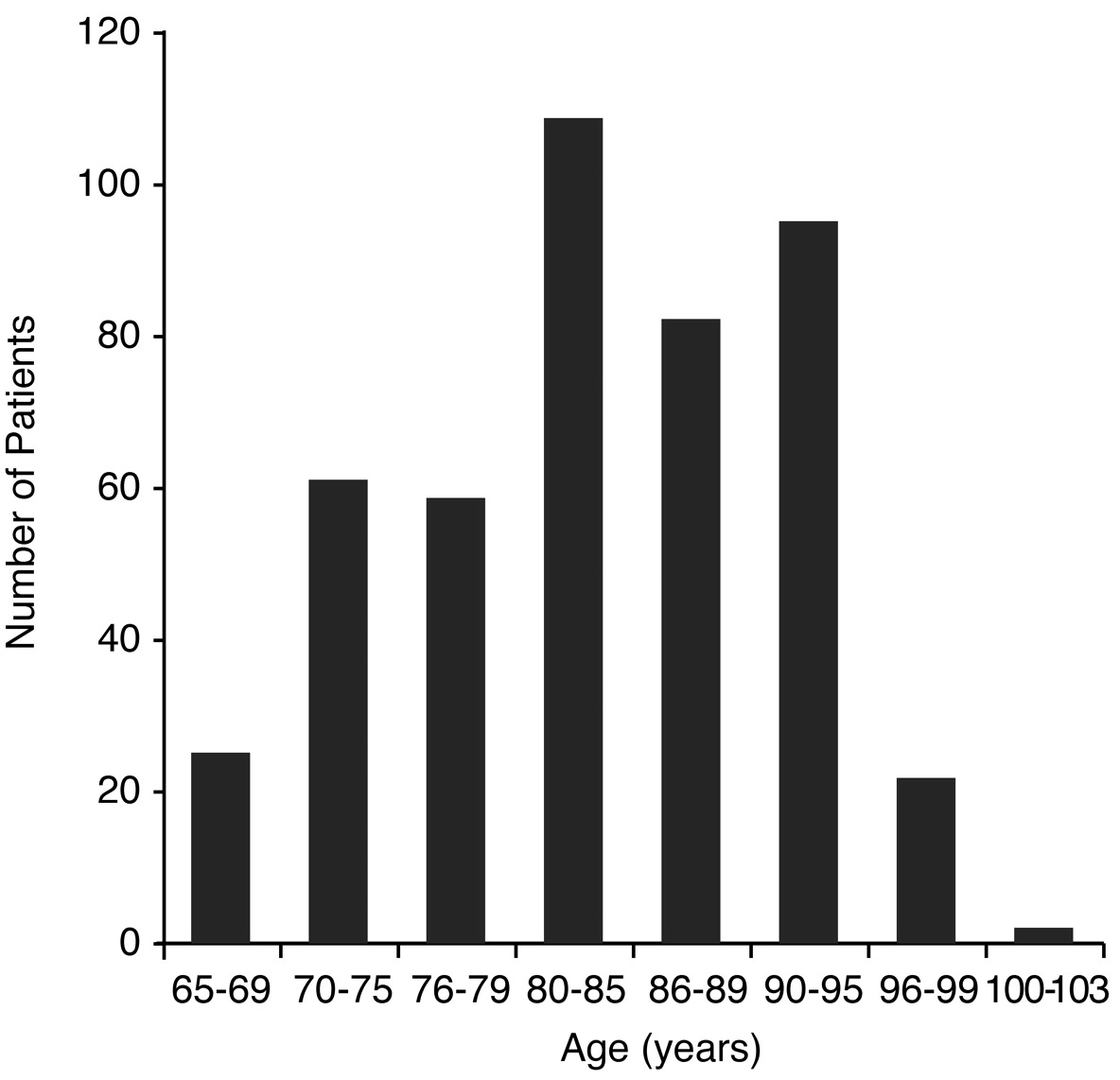
A total of 450 subjects were used in this study, 326 women and 124 men. The median age and mode for the patient population were both 84 years. The mean age was 83.5 years old, with a range from 65 to 103 years (
Fig. 1).
Figure 1.
Age distribution of patient population.
Figure 1.
Age distribution of patient population.
Patient sampling was taken from three clinical locations consisting of a private practice office and two nursing homes. The private practice office setting used 130 subjects, Nursing Home 1 had 203 subjects, and Nursing Home 2 had 117 subjects. Nail clippings were performed on the hallux, with 224 taken from the right hallux and 226 from the left hallux.
Of the 450 subjects sampled, 293 (65.1%) had positive fluorescent KOH preparation readings and 157 (34.9%) had negative results. On microscopic examination, 51 (11.3%) had many hyphae, 92 (20.4%) had moderate hyphae, 86 (19.1%) had rare to few hyphae, 56 (12.4%) had hyphae and arthrospores, 4 (0.9%) had many yeast cells, 2 (0.4%) had moderate yeast cells, 1 (0.2%) had rare to few yeast cells, 1 (0.2%) had yeast cells and pseudohyphae, and 157 (34.9%) had no fungal elements observed (
Table 1).
Table 1.
KOH Preparation Results of 450 Patients with Suspected Onychomycosis
Table 1.
KOH Preparation Results of 450 Patients with Suspected Onychomycosis
A total of 526 organisms were cultured from the total patient population. A total of 346 (76.9%) subjects had positive fungal cultures and 104 (23.1%) had no growth. Of the 450 subjects, 209 (46.4%) subjects had a single organism and 137 (30.4%) had mixed (two or more) organisms grown. One hundred two (22.7%) had two organisms, 27 (6%) had three organisms, and 8 (1.8%) subjects had four organisms cultured. An average of 1.5 organisms were identified per positive fungal culture (
Table 2).
Table 2.
Fungal Culture Results of 450 Patients with Suspected Onychomycosis
Table 2.
Fungal Culture Results of 450 Patients with Suspected Onychomycosis
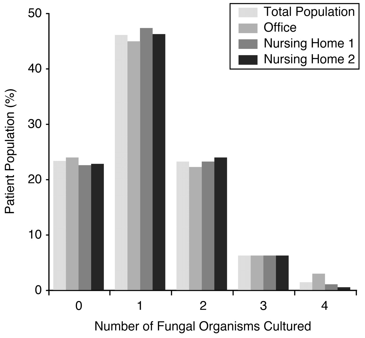
Of the 130 subjects from the private practice office, 99 (76.2%) had positive fungal cultures and 31 (23.8%) had no growth. Fifty-nine (45.4%) had a single organism and 40 (30.8%) had mixed organisms grown. Twenty-eight (21.5%) of the subjects had two organisms, 8 (6.2%) had three organisms, and 4 (3.1%) had four organisms present. An average of 1.6 organisms were identified per positive fungal culture.
Of the 203 subjects from Nursing Home 1, 157 (77.3%) had positive fungal cultures and 46 (22.7%) had no growth. Ninety-six (47.3%) had a single organism and 61 (30%) had mixed organisms grown. Forty-six (22.7%) of the subjects had two organisms, 12 (5.9%) had three organisms, and 3 (1.5%) had four organisms present. An average of 1.5 organisms were identified per positive fungal culture.
Of the 117 subjects from Nursing Home 2, 90 (76.9%) had positive fungal cultures and 27 (23.1%) had no growth. Fifty-four (46.2%) had a single organism and 36 (30.8%) had mixed organisms grown. Twenty-eight (23.9%) of the subjects had two organisms, 7 (6%) had three organisms, and 1 (0.9%) had four organisms present. An average of 1.5 organisms were identified per positive fungal culture.
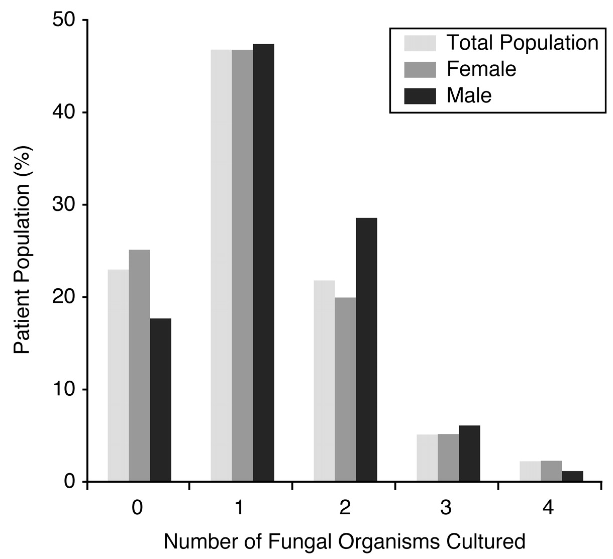
Of the 326 female subjects, 243 (74.5%) had positive fungal cultures and 83 (25.5%) had no growth. One hundred fifty-one (46.3%) had a single organism and 67 (20.6%) had mixed organisms grown. Ninety-two (28.2%) of the subjects had two organisms, 19 (5.8%) had three organisms, and 6 (1.8%) had four organisms present. An average of 1.5 organisms were identified per positive fungal culture.
Of the 124 male subjects, 103 (83.1%) had positive fungal cultures and 21 (16.9%) had no growth. Fifty-eight (46.8%) had a single organism and 45 (36.3%) had mixed organisms grown. Thirty-five (28.22%) of the subjects had two organisms, 8 (6.5%) had three organisms, and 2 (1.6%) had four organisms present. An average of 1.6 organisms were identified per positive fungal culture.
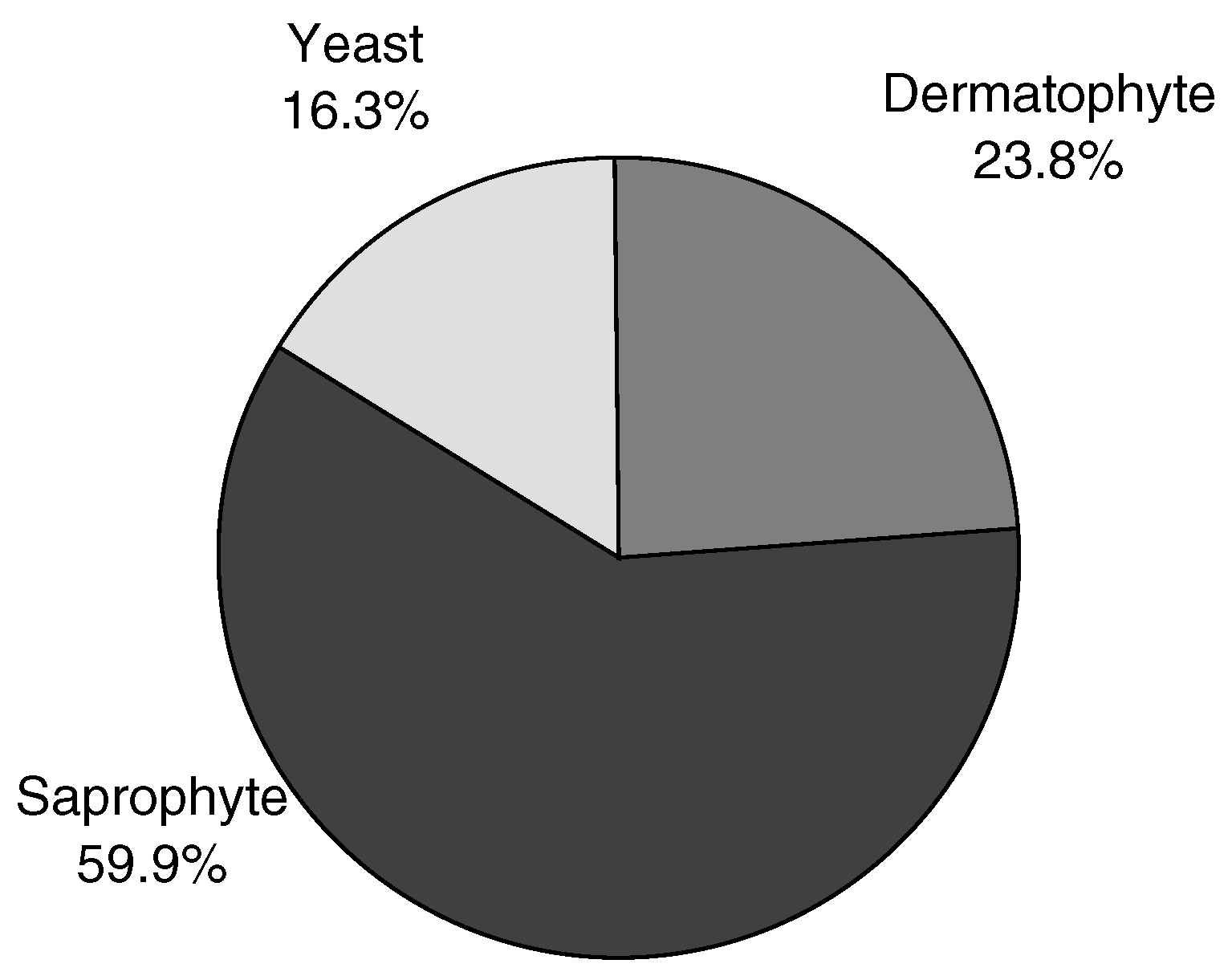
From a total of 526 fungal organisms cultured, 125 (23.8%) were dermatophytes, 315 (59.9%) were saprophytes, and 86 (16.3%) were yeasts (
Fig. 2). Among the 209 subjects with a single fungal organism cultured, 54 (25.8%) had dermatophytes only, 124 (59.3%) had saprophytes only, and 31 (14.8%) had yeasts only (
Table 2).
Figure 2.
Types of fungal organisms from 526 total organisms.
Figure 2.
Types of fungal organisms from 526 total organisms.
Of the 102 subjects with two organisms cultured, 38 (37.3%) had dermatophyte+saprophyte combinations, 32 (31.4%) had saprophyte+saprophyte combinations, 19 (18.6%) had saprophyte+yeast combinations, and 13 (12.7%) had dermatophyte+yeast combinations.
Of the 27 subjects with three organisms cultured, 10 (37%) had saprophyte+saprophyte+yeast combinations, 7 (25.9%) had dermatophyte+saprophyte+saprophyte combinations, 5 (18.5%) had dermatophyte+saprophyte+yeast combinations, 4 (14.8%) had saprophyte+saprophyte+saprophyte combinations, and 1 (3.7%) had a dermatophyte+yeast+yeast combination.
Of the 8 subjects with four organisms cultured, 6 (75%) had saprophyte+saprophyte+saprophyte+saprophyte combinations, 1 (12.5%) had a dermatophyte+saprophyte+saprophyte+saprophyte combination, and 1 (12.5%) had a dermatophyte+saprophyte+saprophyte+yeast combination.
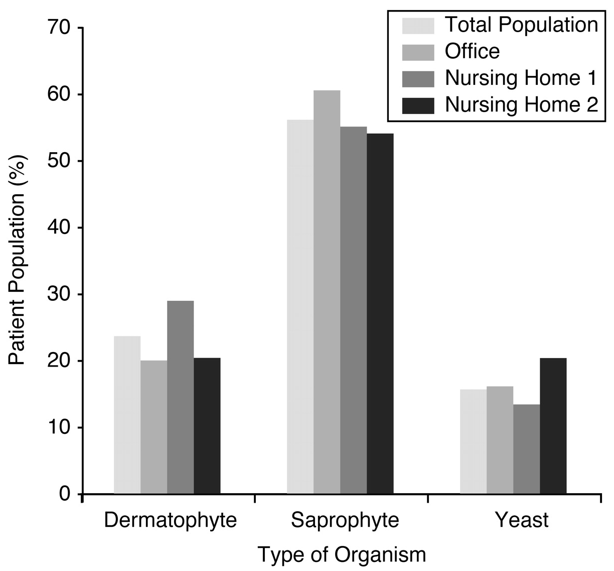
Of the 155 organisms cultured from the office location, 31 (20%) were dermatophytes, 98 (63.2%) were saprophytes, and 26 (16.8%) were yeasts. Of the 236 organisms cultured from Nursing Home 1, 66 (28%) were dermatophytes, 138 (58.5%) were saprophytes, and 32 (13.6%) were yeasts. Of the 135 organisms cultured from Nursing Home 2, 28 (20.7%) were dermatophytes, 79 (58.5%) were saprophytes, and 28 (20.7%) were yeasts.
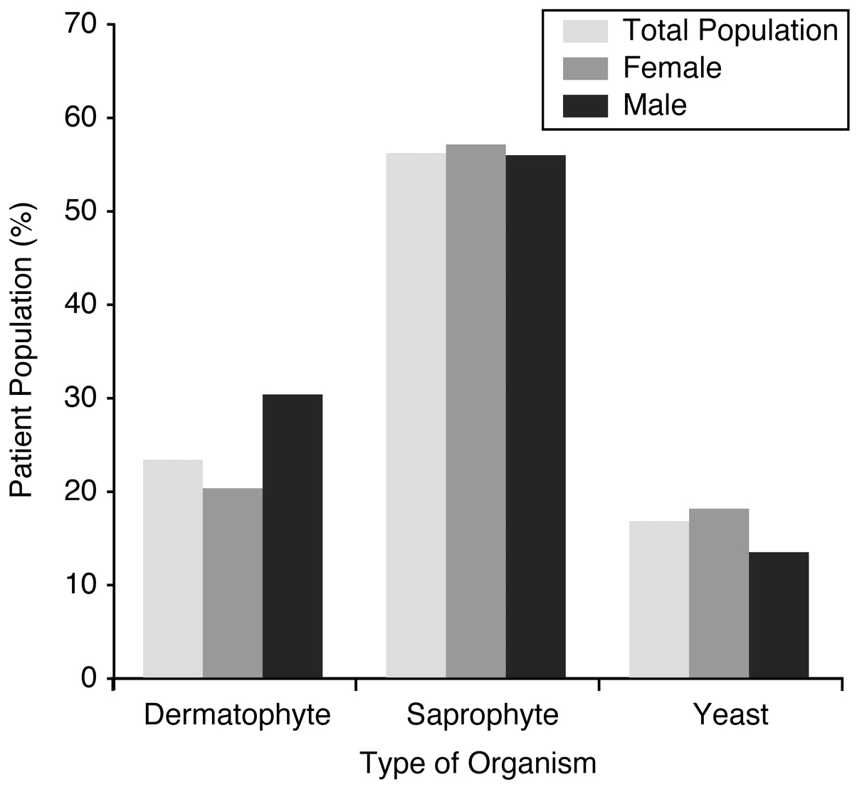
Of the 366 organisms cultured from the female population, 26 (7.1%) were dermatophytes, 203 (55.5%) were saprophytes, and 67 (18.3%) were yeasts. Of the 160 organisms cultured from the male population, 49 (30.6%) were dermatophytes, 92 (57.5%) were saprophytes, and 19 (11.9%) were yeasts.
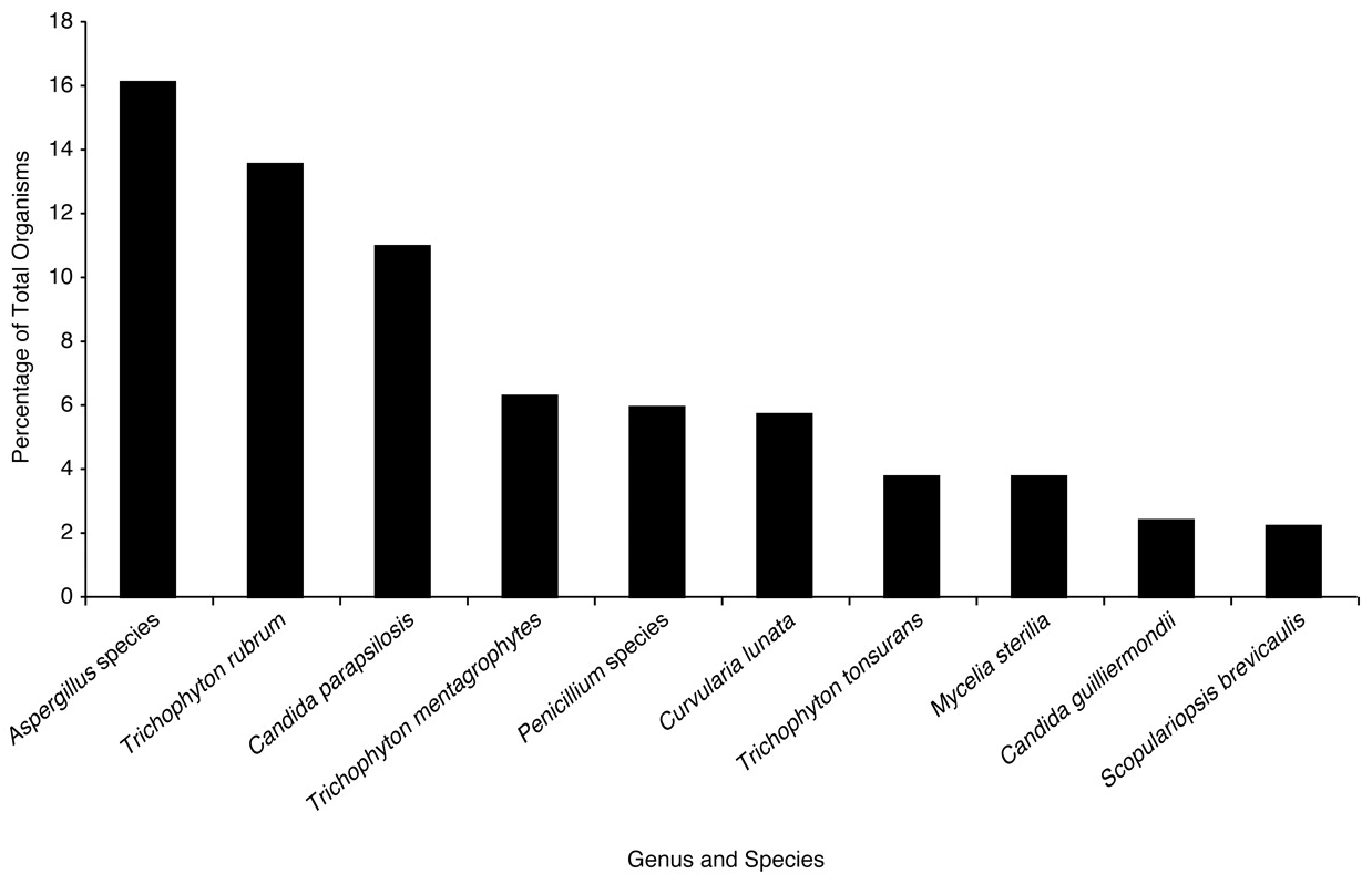
Of the population of 450 patients, 526 total organisms were cultured and 63 types of organisms were identified by genus and species (
Table 3). Among the highest number of organisms identified, 85 (16.2%) belonged to unspecified
Aspergillus species, 71 (13.5%) were
Trichophyton rubrum, 58 (11.0%) were
Candida parapsilosis, 33 (6.3%) were
Trichophyton mentagrophytes, 32 (6.1%) were of unspecified
Penicillium species, 30 (5.7%) were
Curvularia lunata, 19 (3.6%) were
Trichophyton tonsurans, 19 (3.6%) were
Mycelia sterilia, 13 (2.5%) were
Candida guilliermondii, and 12 (2.3%) were
Scopulariopsis brevicaulis (
Fig. 3).
Table 3.
The 526 Organisms Identified from 346 Positive Cultures
Table 3.
The 526 Organisms Identified from 346 Positive Cultures
Figure 3.
Top 10 fungal culture organisms from 526 total organisms.
Figure 3.
Top 10 fungal culture organisms from 526 total organisms.
Discussion
The average age of the total patient population in the study was 83.5 years, demonstrating a geriatric population. Of the 450 subjects in the study, 65.1% had positive fluorescent KOH preparation and 34.9% were negative. In the study population, 76.9% had positive microscopic fungal cultures and 23.1% had no fungal growth. Of the entire population, 46.4% of the subjects had a single organism and 30.4% had mixed (two or more) organisms grown. In the isolated organism types, 12% of the population had dermatophyte-only involvement, 27.5% had saprophyte-only involvement, and 6.9% had yeast-only involvement (
Table 3). Of the 526 organisms isolated, 23.8% had dermatophyte involvement, 59.9% had saprophyte involvement, and 16.3% had yeast involvement.
When the results of the number of fungal organisms cultured for the total patient population were compared to the three individual patient locations, no major differences were observed by location (
Fig. 4). When the results of the type of organism cultured for the total patient population were compared to the three individual patient locations, no major differences were observed by location (
Fig. 5). When the female patient population and male patient population were compared to each other and to the total patient population, no major differences were observed for the number of organisms cultured (
Fig. 6). When the type of organism cultured among the male and female subjects was compared to each other and to the total patient population, there was a slight increase of dermatophytes in male subjects (30.6%) to female subjects (20.8%). In addition, there was a slight increase in yeasts among female subjects (18.3%) as compared to male subjects (11.9%) (
Fig. 7).
Figure 4.
Comparison of the number of fungal organisms cultured.
Figure 4.
Comparison of the number of fungal organisms cultured.
Figure 5.
Comparison of the type of fungal organisms cultured.
Figure 5.
Comparison of the type of fungal organisms cultured.
Figure 6.
Comparison of the number of fungal organisms cultured by gender.
Figure 6.
Comparison of the number of fungal organisms cultured by gender.
Figure 7.
Comparison of the type of fungal organisms cultured by gender.
Figure 7.
Comparison of the type of fungal organisms cultured by gender.
Conclusion
The results of this study of 450 cases of onychomycosis in a South Florida geriatric population demonstrate that 46.4% of the patients had a single fungal organism cultured, 30.4% had mixed fungal infections cultured, and 23.1% had no fungal growth, according to the mycology laboratory reports. Of the 137 patients with a mixed fungal culture, 74.4% had two organisms present, 19.7% had three organisms present, and only 5.8% had four organisms present. A total of 526 organisms were identified, with an average of 1.5 organisms per positive fungal culture. Saprophytes were found in more than half of the 526 total fungal organisms cultured and more than twice as many saprophytes (59.9%) were observed than dermatophytes (23.8%). There was at least one saprophyte in 89.8% of the subjects with mixed infections while only 48.2% subjects had a dermatophyte present. In addition, 35.8% of the subjects had yeast present in a mixed infection. Among the 209 subjects with a single fungal organism cultured, 25.8% had dermatophytes only, 59.3% had saprophytes only, and 14.8% had yeasts only. The most common organism cultured out of 526 fungi identified was the Aspergillus species, a saprophyte that was found in 16.2% of the results. Of the two most common dermatophytes, Trichophyton rubrum was found in 13.5% of the results and Trichophyton mentagrophytes was found in 6.3% of the results. The most frequent yeast identified was Candida parapsilosis, which was found in 11% of the results.
This study’s findings demonstrate an increase of mixed fungal infections and saprophytes in a South Florida geriatric population with onychomycosis. The authors believe that age, circulation, health, environment, and geography all may play an equal role in the pathogenesis of onychomycosis, but these findings are beyond the scope of this investigation. The study’s results are contradictory to most reports in the medical literature that state onychomycosis is mainly due to dermatophyte infection. However, the present study is the first large-scale investigation of the diagnosis of onychomycosis in a population limited to geriatrics; most current medical studies do not restrict age as a parameter. The results of this investigation demonstrate that there may be a shift from isolated dermatophyte infection to mixed saprophyte infections in a geriatric population with onychomycosis.