Lower-extremity injuries frequently have a multifactorial etiology. Abnormal rearfoot motion has been implicated as a contributing factor in a number of pathologies, including metatarsal stress fractures, [
1] shin splints, [
2] and plantar fasciitis. [
3] One of the basic premises of clinical podiatric biomechanics is that static foot posture is associated with foot motion during gait. Thus, clinical diagnosis of abnormal foot function frequently involves measuring static foot posture and using the results in the prescription of foot orthoses. [
4] It is important, therefore, that the techniques used to evaluate static foot posture are both reliable and valid. [
5]
A number of techniques for the clinical assessment of foot posture have been described in the literature, including the valgus index, [
6] footprint indexes, [
7] arch height, [
8] and frontal plane measurements of the rearfoot. [
9] Although many of these techniques are widely used in clinical practice, their reliability and validity have not been fully established. In particular, the commonly performed frontal plane rearfoot measurements have been reported to exhibit poor to moderate reliability [
10-
12] and may not accurately represent the dynamic motion of the rearfoot during walking. [
13,
14]
Recent studies using roentgen stereophotogrammetry indicate that the talonavicular joint is a significant contributor to pronation and supination motion of the foot. [
15,
16] Unfortunately, this process is highly invasive and requires special equipment, and is therefore inappropriate for routine clinical assessment of foot posture. There is thus a need for simple clinical techniques to assess talonavicular joint function. One way to clinically determine the position and motion of the talonavicular joint may be to assess the position and motion of the navicular, as this bone is an easily accessible surface landmark. [
17]
Brody [
17] initially reported the measurement of “navicular drop” in 1980 to detect hyperpronation in runners, and this measurement is thought to represent the sagittal plane displacement of the navicular from a neutral position to a relaxed standing position. Excessive navicular drop has been reported in subjects with a history of anterior cruciate ligament rupture, [
18,
19] and it is also thought to predispose to shin splints. [
20] Furthermore, Cornwall and McPoil [
21] suggest that as the navicular undergoes greater displacement than the calcaneus, measurement of navicular position and motion may provide more useful information about the function of the foot in normal walking than frontal plane rearfoot measurements. Therefore, this measurement may be of benefit in the clinical assessment of patients with overuse symptoms of the lower limb.
The reliability of navicular drop measurement has been evaluated by Sell et al, [
22] who reported fair intertester reliability and good intratester reliability when two testers measured 30 healthy volunteers. Weiner-Ogilvie and Rome [
23] compared three methods of measuring foot position and found that navicular height measurement exhibited the least intratester and intertester variability. In contrast to these two studies, Picciano et al [
24] reported poor intertester reliability and poor to moderate intratester reliability of navicular drop measurement; however, this study used two inexperienced testers to perform the measurements.
Despite the potential benefits of the navicular drop measurement, it has a major limitation: it is capable of measuring displacement only in the sagittal plane, whereas motion of the navicular actually takes place in all three planes simultaneously. [
21,
25] Although the frontal plane motion of the navicular is small, the transverse plane component is larger and may therefore be clinically significant. To address this, Menz [
26] proposed the measurement of navicular drift to provide an indicator of the change in medial prominence of the talonavicular joint when the foot moves from a neutral to a resting position. However, the reliability of navicular drift measurement has not been evaluated.
The aim of this study was to evaluate the reliability of clinical measurement of navicular drop and drift as a way of establishing the usefulness of these techniques in podiatric clinical practice.
Methods
Subjects
The participants in this study consisted of 7 males and 13 females (mean [±SD] age of 20 ±2 years). Participants were excluded from the study if they had any medical or orthopedic conditions affecting the lower limb, were currently in foot pain, or were receiving treatment for any lower-limb condition. The clinicians performing the measurements consisted of five podiatric physicians with a minimum of 3 years’ postgraduate experience (mean, 4.8 years; range, 3–7 years).
Protocol
The clinicians attended three 1-hour training sessions in which they were instructed on how to perform the measurement techniques. They practiced the techniques to make sure that they were taking the measurements correctly. Data collection consisted of two sessions, 1 week apart, in which 20 participants were measured with both techniques by the five clinicians. Both feet of each participant were measured three times so that the reliability of the average of three measurements could be compared with the reliability of a single measurement. To obtain intertester data, each participant was measured by each clinician with the use of each method. The repeat of the process 1 week later allowed for intratester comparisons.
Measurement of Navicular Drop
In the current study, a modified version of the navicular drop described by Brody [
17] was used to determine the sagittal plane displacement of the navicular between resting and neutral stance positions. The participant was seated with both feet flat on the ground and knees flexed at 90°. The most medial aspect of the navicular was marked. The participant was then asked to stand. The neutral stance position was determined by palpation of talar head congruency, as described by Wernick and Langer. [
27] When the neutral position was located, the participant was asked to stand on one leg by flexing the contralateral knee. The single-limb stance position was selected because recent work by McPoil and Cornwall [
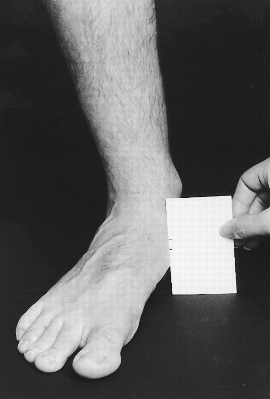
28] has shown that measurements taken from this position more accurately represent the position of the foot during the midstance phase of gait. The position of the talar head was then checked and, if there was any change, was adjusted back into neutral. A blank card was held at right angles to the foot against the navicular marking with the base of the card flat on the supporting surface. The height of the navicular was marked on the card. Next, the participant was asked to stand on both feet in a relaxed position, then to stand only on the foot being measured. Again, the height of the navicular was marked on the card (
Fig. 1). The difference between the height of the navicular in neutral and resting positions was recorded as navicular drop.
Figure 1.
Measurement of navicular drop.
Figure 1.
Measurement of navicular drop.
Measurement of Navicular Drift
The navicular drift measurement is the projection onto the transverse plane of the navicular displacement that occurs with an alteration in rearfoot position. [
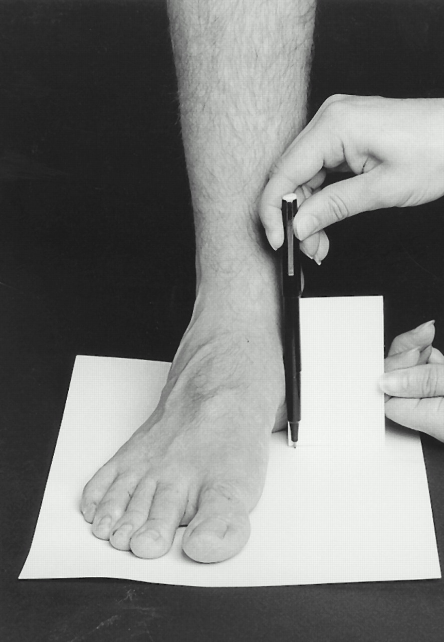
26] Before either measurement was taken, the participant was asked to stand in a relaxed position and lift each foot so that the clinician could place a sheet of paper under it. The participant was instructed to look straight ahead while doing this to avoid any subconscious alteration in foot placement that might occur from, for example, trying to place the foot squarely on the piece of paper. Neutral stance and single-limb stance were determined in the manner described above. The position of the navicular, as indicated by the bottom corner of the business card, was marked on the paper (
Fig. 2). The relaxed foot position was obtained as for measurement of navicular drop; the new position of the navicular was then recorded on the paper. The difference between the location of the navicular in neutral and resting positions was recorded as navicular drift.
Figure 2.
Measurement of navicular drift.
Figure 2.
Measurement of navicular drift.
The pieces of paper and cards were given to one of the authors (A.V.), who measured the distance between the two points on the paper or card. As the clinicians did not actually measure the displacement, they were blinded to the results. Each measurement was performed three times.
Statistical Analysis
To determine the difference in reliability between taking a single measurement and taking an average of three measurements, all statistical analyses were performed twice: for the first of three repeated measurements and for an average of the three repeated measurements. First, the Pearson’s
r correlation coefficient was calculated for intertester and intratester comparisons. Pearson’s
r describes the association between two variables measured on an interval or ratio scale. To determine the correlations for the averaged measurements, a Fisher’s
r to
z transformation was first employed; this involves converting
r scores to
z scores, averaging the
z scores, and then converting the result back into an
r value. [
29]
A limitation of the Pearson’s r statistic as a measure of reliability is that it represents only an association between scores, not the level of agreement. Consequently, although the r statistic can detect random variation in scores (ie, random error), it cannot identify a consistent difference between scores (ie, systematic error). It is therefore also necessary to determine whether a consistent difference exists between the measurement variables under investigation. A series of paired t-tests were used for this purpose.
As intraclass correlation coefficients (ICCs) are more widely used in the literature than Pearson’s
r and the two correlation coefficients are calculated differently, ICCs were also calculated to make the results more comparable to other studies. For intertester data, the ICC(2,1) model was used to determine the reliability of the first of the three measurements, and the ICC(2,3) model was used to assess reliability when the average of three measurements was taken. For intratester data, the ICC(3,1) and (3,3) models were used, as suggested by Shrout and Fleiss. [
30] The ICC is a commonly used reliability statistic that offers a measure of correspondence between scores based on an analysis of variance model. The ICC differs from the Pearson’s
r statistic in that it describes overall agreement (ie, random and systematic error combined). While this overcomes the aforementioned limitation of the Pearson’s
r statistic, the disadvantage of the ICC is that if a low score (indicating poor reliability) is obtained, the statistic cannot delineate the source of the low agreement (ie, random or systematic error). [
29]
Finally, for the results to be more clinically applicable, the standard error of measurement was calculated. The standard error of measurement has an advantage over other reliability statistics in that it presents the degree of measurement error in the units in which the measurement is taken. This allows the study findings to be more directly applicable to the clinical situation. For each standard error of measurement, 95% confidence intervals were also calculated.
Results
The mean, SD, and range of scores obtained for the measurement of navicular drop and navicular drift are presented in
Table 1. The navicular drop values are similar to those that have been reported previously. [
17,
22,
24,
25]
Table 1.
Descriptive Data for Navicular Measurements
Table 1.
Descriptive Data for Navicular Measurements
Averaged correlation coefficients demonstrated a consistency of measurement of navicular drift, ranging from .44 to .77 for intratester data and from .32 to .53 for intertester reliability. Correlation coefficients of navicular drop measurements varied between .44 and .91 for intratester data and between .56 and .78 for intertester data. The ICCs were generally very similar to Pearson’s r values.
The paired t-tests for intratester reliability showed that only in four situations did scores vary significantly when compared with an alpha level of .05. Overall, little significant difference was noted in the mean of the scores obtained by each clinician during the two sessions. Clinically, this means that error associated with measurements taken by the same clinician was generally random and not due to systematic differences from one trial to the next. Conversely, paired t-tests of intertester data found that a significant difference existed in almost half of the scores obtained. In these cases, the correlations reported show that the measurements taken by different clinicians were consistently different (ie, one clinician consistently recorded a larger or smaller value than another).
The standard error of measurement, expressed in 95% confidence intervals, ranged from ±2.82 mm to ±4.39 mm for navicular drift and ±1.47 mm to ±3.66 mm for navicular drop for intratester comparisons. Intertester confidence intervals varied from ±4.02 mm to ±4.84 mm for navicular drift and ±2.29 mm to ±3.23 mm for navicular drop.
A summary of the averaged correlation coefficients (Pearson’s
r and ICC) and standard error of measurement is shown in
Tables 2 through
5.
Table 2.
Intertester Reliability of Navicular Drop
Table 2.
Intertester Reliability of Navicular Drop
Table 3.
Intertester Reliability of Navicular Drift
Table 3.
Intertester Reliability of Navicular Drift
Table 4.
Intratester Reliability of Navicular Drop
Table 4.
Intratester Reliability of Navicular Drop
Table 5.
Intratester Reliability of Navicular Drift
Table 5.
Intratester Reliability of Navicular Drift
Discussion
Interpretation of Results
For measurement of navicular drop, reliability coefficients ranged from poor to good; however, the 95% confidence interval revealed a high level of variability over the two measurement sessions, both between clinicians and within clinicians. The results suggest that clinicians can be 95% certain that the true value for navicular drop lies between ±1.5 mm and ±3.5 mm from one session to the next and between ±2.5 mm and ±3.5 mm from their measurement when compared with that of other clinicians. Given that the range of values recorded for navicular drop in this investigation was 12.5 mm, it is clear that the potential source of error is a large component of this total range (12% to 28%). Thus, the information on foot posture provided by measurement of navicular drop is only moderately reliable between clinicians. However, the intratester reliability was slightly better.
For the measurement of navicular drift, reliability coefficients ranged from poor to moderate; again, however, the 95% confidence interval revealed a high level of variability, both between clinicians and within clinicians over the two measurement sessions. The results suggest that clinicians can be 95% certain that the true value for navicular drift lies between ±3 mm and ±4.5 mm from one session to the next, and ±4 mm to ±5 mm from their measurement when compared to other clinicians. The range of values recorded for navicular drift in this investigation was 9 mm, which suggests that it is also questionable as to whether measurement of navicular drift provides useful clinical information regarding foot posture (33% to 55% of the measurement range is potential error).
When deciding whether a measurement technique is reliable enough for clinical use, one must consider the purpose of the measurement. If, for example, the measurement is to be used to guide surgical intervention, a high degree of accuracy is imperative. On the other hand, if the measurement is to be used as part of a complete biomechanical assessment, to aid diagnosis and possible orthotic prescription, a slightly larger error range is perhaps more acceptable. Even in the latter context, however, it is important to be aware of the magnitude of the error associated with the technique and to take this into account when interpreting the results.
For both techniques, the intratester reliability was higher than intertester reliability. Intratester reliability may appear to be more clinically relevant, as it is unlikely that more than one practitioner will be required to take the same measurements from the same patient. However, poor intertester reliability may present a problem when a measurement is compared with normative data.
This study’s conclusions regarding navicular drop reliability differ from those of Sell et al, [
22] who reported high reliability for this measurement and suggested that navicular drop is an acceptable technique for clinical assessment of foot posture. Sell et al based much of their conclusion on the high ICC values they obtained, which ranged from .73 to .83. However, Portney and Watkins [
29] suggest that for clinical measurements to be useful, an ICC value of at least 0.90 is desirable. Furthermore, the 95% confidence intervals and range of measurements reported by Sell et al were similar to the findings of this study. These statistics have an advantage over correlation coefficients in that they present the degree of measurement error in the units in which the measurement was taken, thus making the results more clinically applicable. In this context, the results of this study are actually similar to those of Sell et al, despite their higher ICCs.
Potential Sources of Error
The large measurement error in the assessment of navicular displacement could arise from a number of sources. First, the clinicians reported difficulty in the location of the navicular tuberosity owing to anatomical variations among individuals. In some cases, the medial prominence of the navicular was easily palpated and marked; in other cases, the morphology of this bone made reference point location difficult. Secondly, the reliability of navicular drop and drift is dependent on the reliability of locating the neutral reference position. Previous authors have suggested that the reliability of locating this position is moderate to poor. [
31] To investigate this issue further, the navicular drop measurements were broken down into resting and neutral navicular height and re-analyzed. Neutral navicular height represented the distance between the bottom of the card and the neutral marking, whereas resting navicular height was from the bottom of the card to the resting stance position marking. As navicular drift did not have a consistent reference point, this breakdown was not possible with navicular drift. Interestingly, analysis of the resting and neutral heights revealed little difference in reliability between the two measurements. This finding suggests that the location of the neutral position may not be the major source of error in these measurements, as the error involved in measuring the height of the navicular in a resting position was similar. It is possible that the single-limb standing position may have contributed to the unreliability of these two techniques, as the subjects intermittently altered their standing posture to maintain balance.
Study Limitations
The population measured in the present investigation consisted of healthy adults with feet that were, by definition, considered normal. Such factors as pain-induced restricted range of motion and ankle edema may affect the reliability of the measurement techniques. It could therefore be anticipated that the reliability of the two techniques might change when applied in a clinical setting. Therefore, the results of this study can be extrapolated only to a nonpathological population.
Another limitation of the study is that the effect of clinical experience on reliability of measurements was not fully investigated. Clinicians who carried out the measurements had an average of only 4.8 years of postgraduate clinical experience. Previous studies indicate that the reliability of foot and ankle measurements increases slightly with experience. [
31] It appears feasible that the same pattern would occur with the techniques under investigation; however, this hypothesis could not be tested in the current study, as navicular drop and drift measurements are relatively new techniques that are not widely used in clinical practice. Nevertheless, comparisons of intratester reliability did not reveal that the most experienced clinicians produced the most reliable measurements. This suggests that experience in performing foot and ankle measurements in general does not necessarily improve the reliability of measurements of navicular drop and drift.
Finally, this study was designed to simulate a clinical situation as closely as possible. This was not achieved with respect to the repetitive nature of the data collection. The large number of measurements required of each clinician may have led to fatigue, which may in turn have affected the consistency of the results.
Indications for Further Research
Quantification of foot posture is a necessary, yet problematic, area of clinical biomechanics. For clinical evaluation of foot posture to be scientifically credible, reliable and valid techniques need to be developed. Measurements taken from the navicular bone have face validity, as they may indicate the effect of altered foot position on the talonavicular joint, the major contributor to pronation and supination movements of the foot. [
15,
16] However, the results of this study show that the measurement of navicular drop and drift is only moderately reliable, owing to the large error ranges associated with both measurements. Nonetheless, these techniques are more reliable than the more traditional methods of rearfoot evaluation and may provide more useful information on foot motion during gait. [
21] Further research therefore needs to be undertaken to address and minimize the sources of error inherent in these clinical measurements. As some clinicians performed the measurements more reliably than others, determining the factors that improve reliability of an individual’s measurements would increase the reliability of the techniques.
Conclusion
Navicular drop and navicular drift measurements, as described in this study, are only moderately reliable and are thus of somewhat limited value in the clinical evaluation of foot posture. Physicians using these techniques in clinical practice should interpret their measurements with caution and make informed clinical decisions in light of the error associated with each technique. Additional research is warranted to further refine these techniques and improve the scientific credibility of foot posture evaluation.