Weightbearing radiographs are thought to provide an accurate reflection of foot structure [
1,
2] and are frequently used by podiatric and orthopedic surgeons in the preoperative assessment of foot deformities [
3,
4]. Metatarsus primus elevatus has been cited in the literature as a cause of hallux limitus [
5-
8]. Such an assumption, however, has been based on clinical experience rather than scientific evidence. Camasta [
9] maintained that the dorsal cortices of the central areas of the first and second metatarsals in normal feet are parallel, while in hallux limitus the first metatarsal is elevated, forming a measurable angular divergence between the two metatarsal shafts.
Other authors, however, have disputed the relationship between metatarsus primus elevatus and hallux limitus. Meyer et al [
10] found no differences in first metatarsal elevation between normal, hallux valgus, and hallux limitus feet, and Rzonca et al [
11] found only one example of metatarsus primus elevatus in 35 cases of hallux limitus studied. In a recent study, Bryant et al [
12] found no statistical correlation between first metatarsal declination angle and hallux limitus in 30 control and 30 hallux limitus subjects. Unfortunately, a common measurement technique was not used in each study, which may have affected the results and interpretation of the findings.
This study, based on the descriptive work of Camasta [
9], attempted to determine if an association could be demonstrated between metatarsus primus elevatus and hallux limitus by measuring the sagittal plane divergence of the dorsal cortices of the first and second metatarsals from weightbearing foot radiographs. A new podiatric radiographic measurement term—lateral intermetatarsal angle—is introduced to describe the sagittal plane angular relationship between the first and second metatarsals.
Methods
This study was part of a larger research project investigating the effects of surgery on foot function of patients with hallux valgus and hallux limitus. The Human Research and Ethics Committee of Curtin University of Technology (Shenton Park, Western Australia) approved x-raying the feet of 30 control subjects. The nature and purpose of the research was explained to all subjects and written consent was obtained from them.
Subjects
Thirty control subjects were randomly selected and 30 subjects with hallux limitus were recruited from a private podiatry practice. Control subjects were excluded from the study if they had clinical symptoms or radiographic signs of hallux limitus, obvious musculoskeletal abnormality of the lower limb, had undergone osseous foot surgery, or suffered a significant injury to the foot or leg in the previous 12 months.
All of the hallux limitus subjects had clinical and radiographic evidence of hallux limitus without concurrent signs of hallux valgus. They also had painful restriction of first metatarsophalangeal joint dorsiflexion with passive joint motion and radiographic stage I or II hallux limitus according to the classification system proposed by Reginauld [
13]. Subjects were excluded from the study if they had a history of previous osseous foot surgery, inflammatory joint disease, or any other syndrome that could predispose to hallux limitus. Subject demographics are listed in
Table 1.
Table 1.
Subject Demographics.
Table 1.
Subject Demographics.
Radiographic Measurement Technique
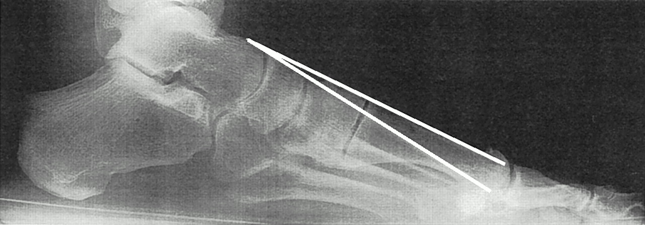
With subjects in a relaxed stance position, lateral radiographs were taken in a standardized manner with the central beam targeted on the cuboid. On each weightbearing lateral radiograph, the central region of the dorsal cortex of the first and second metatarsal shafts were marked with a fine x-ray pencil, and the angular deviation thus formed was measured with a tractograph (
Fig. 1). Six radiographs were selected at random for intrarater measurement-reliability testing. The lateral intermetatarsal angle between the first and second metatarsals was measured for each selected radiograph and wiped clean of any marking. The measurement was performed a total of three times, at 1-week intervals. An intraclass correlation coefficient was then computed. Angular measurements of all control and test subjects were entered into a spreadsheet for statistical analysis by means of SPSS-X (SPSS, Inc; Chicago, Illinois), where descriptive statistics were generated and an independent two-tailed
t-test was performed.
Figure 1.
Lateral radiograph of a representative hallux limitus subject showing lateral intermetatarsal angle.
Figure 1.
Lateral radiograph of a representative hallux limitus subject showing lateral intermetatarsal angle.
Results
The lateral intermetatarsal angle between the dorsal surfaces of the first and second metatarsal shafts was compared in 30 controls and 30 hallux limitus subjects.
The intraclass correlation coefficient for the intrarater reliability test of the lateral intermetatarsal angle was .97, indicating acceptable reliability of the measurement [
14].
Descriptive statistics of the measured sagittal plane angular divergence for both groups are summarized in
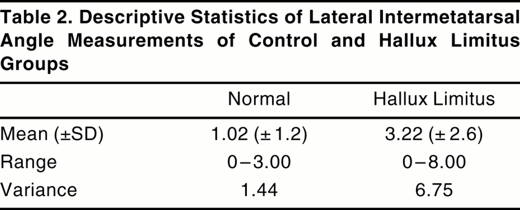
Table 2. The mean (±SD) lateral intermetatarsal angle for the control group was 1.02° ± 1.2°, while the mean lateral intermetatarsal angle for the hallux limitus group was slightly higher, 3.22° ± 2.6°. An independent two-tailed test was calculated under the assumption of unequal variances; it indicated the existence of a significant difference (
P < .05) between the first and second intermetatarsal sagittal plane angular divergence in the control and hallux limitus groups.
Table 2.
Descriptive Statistics of Lateral Intermetatarsal Angle Measurements of Control and Hallux Limitus Groups.
Table 2.
Descriptive Statistics of Lateral Intermetatarsal Angle Measurements of Control and Hallux Limitus Groups.
Discussion
Most radiographic measurements of the foot have been found to have reasonably good interrater and intrarater reliability [
15,
16], with intraobserver measurement reliability seen to improve with experience [
17]. With respect to radiographic hallux valgus measures, linear measurements have been reported to yield more accurate results than angular measurements [
18]. Interestingly, given the small angular values measured in the study, the results of the intrarater reliability study suggest that the lateral intermetatarsal angle formed between the dorsal cortices of the first and second metatarsals may be measured with acceptable reliability.
While an association between metatarsus primus elevatus and hallux limitus has been suggested in the literature [
6,
7,
19], some authors have found no such relationship to exist [
10,
12]. Meyer et al [
10] measured first metatarsal elevation as a linear measurement (in millimeters) between the height of the first and second metatarsal shafts. By comparison, Bryant et al [
12] measured the first metatarsal declination angle by longitudinal bisection of the metatarsal shaft. Both authors reported no significant differences in their measurements between subjects with normal feet and those with hallux limitus or hallux valgus.
The present study identified a significant difference between the angular divergence of the dorsal cortices of the first and second metatarsal shafts between normal and hallux limitus feet. Therefore, the measurement technique used in the present study, as opposed to the previous reported techniques [
10-
12], possibly may be a more reliable measure of the actual structural deformity of metatarsus primus elevatus and potentially useful in both clinical practice and podiatric research.
With respect to possible limitations of the study, the homogeneity of control and hallux limitus groups was considered reasonable, with a similar gender ratio and body weight in each group. However, the hallux limitus group had a mean age approximately 10 years older than the control group, which may have adversely influenced the results. A further potential limitation to the study was that while all of the radiographs were taken in a standardized manner, not all radiographs were taken by the same radiographer or with the same radiographic equipment.
Conclusion
The lateral intermetatarsal angle between the first and second metatarsals derived from weightbearing foot radiographs may be measured with good reliability and, although relatively small, was found to be significantly higher in a sample of 30 feet with hallux limitus compared with a sample of 30 normal feet. The authors suggest the lateral intermetatarsal angle may, with practice, be measured with confidence. However, given the relatively small angles involved and the variance of measurement seen, particularly with respect to hallux limitus subjects, the authors believe further research with a larger number of subjects is desirable to compare various radiographic measuring techniques for metatarsus primus elevatus.
Given the contemporary surgical practice of plantarflexing the first metatarsal at operation, the radiographic signs of metatarsus primus elevatus should be considered, along with clinical examination of the foot, in the preoperative assessment of patients presenting with hallux limitus deformity. The most meaningful objective radiographic measurement of metatarsus primus elevatus, however, is yet to be determined.