Onychomycosis is the most common foot condition diagnosed and treated by podiatric physicians in the United States. According to the American Podiatric Medical Association (APMA), in 1998 approximately 25% of all Medicare reimbursements made to podiatric physicians were for the treatment of onychomycosis (APMA Department of Educational Services, personal communication, 2000). In an effort to control costs, several managed-care organizations have established diagnostic criteria that must be met before a treatment plan for onychomycosis will be approved. A specific example is the need to obtain a positive in-office dermatophyte test medium (DTM) culture result prior to initiating oral antifungal therapy for the treatment of onychomycosis. In an effort to determine the validity of in-office DTM culture results, the authors have compared those results for 100 cases of suspected onychomycosis in a geriatric population with the results of an independent mycology laboratory. The mycology laboratory performed a fluorescent potassium hydroxide (KOH) preparation and a microscopic examination of a fungal culture to determine the specific organism for each patient.
Clinical Studies
Onychomycosis, or tinea unguium, is generically thought of as a fungal infection of the nails, characterized by thickening, splitting, roughening, and discoloration. [
1] Onychomycosis can be caused by dermatophytes, saprophytes (nondermatophytic molds), and yeasts. Fungal infections can also be mimicked by other nail conditions, such as bacterial infection, psoriasis, lichen planus, and trauma. Of the three groups of fungi infecting toenails, dermatophytes have traditionally been documented as the most common organisms causing onychomycosis.
In a study by Elewski, [
2] 90% of fungal nail infections were caused by dermatophytes, 7% by
Candida species, and 3% by nondermatophytic molds. Another study of 3,000 nails had similar results, finding dermatophytes responsible in 91% of the cases,
Candida species in 6%, and nondermatophytic molds in 3%. [
3] A study performed in the United Kingdom had comparable results, with 81% of nail infections caused by dermatophytes, 17% by yeasts, and 2% by nondermatophytic molds. [
4] Current literature identifies
Trichophyton rubrum and
Trichophyton mentagrophytes as the two most common dermatophytes infecting nails, but acknowledges that other non-pathogenic fungi (contaminants) and
Candida may also infect the nail. [
5,
6]
There are significant geographic variations in the causative organisms in onychomycosis. [
2] In some tropical countries such as Nigeria, Thailand, and Jamaica, between 10% and 50% of fungal nail infections are caused by
Scytalidium species. [
3] In contrast,
Candida albicans is reported to be a common causative agent in toenail infections in Egypt and Saudi Arabia. [
4] Treatment of onychomycosis is often unsuccessful, for a number of reasons. One of the major factors in failure of therapy is incorrect identification of the infecting organism. [
7] Several methods are available for diagnosing onychomycosis, including DTM cultures, KOH preparations, and microscopic examination of fungal cultures.
Dermatophyte test medium cultures were introduced in 1969 to provide medics in Vietnam with a simple and rapid method of differentiating between dermatophyte and ringworm infection in soldiers. [
8] Dermatophyte test medium is a selective medium intended for use in the detection of dermatophytes from cutaneous sources (skin, hair, and nails), and is based on the following features of dermatophytes: 1) they are keratinophilic, 2) they produce and tolerate an alkaline pH in culture media, and 3) they are resistant to 500 μg/mL or less of cycloheximide, 100 μg/mL or less of gentamicin, and 100 μg/mL or less of chlortetracycline. [
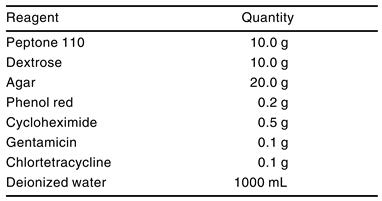
9] Dermatophyte test medium consists of peptone 110 (papaic digest of soy protein) and dextrose-impregnated agar, which are conducive to the growth of dermatophytes. Cycloheximide, gentamicin, and chlortetracycline are three antibiotics that are added to DTM in order to inhibit the growth of most bacteria and many nonpathogenic (saprophytic) contaminating fungi. Phenol red indicator is added to allow easy interpretation of the results. Dermatophytes release alkaline metabolites into the medium, which results in an increase in pH within 10 to 14 days, at which time the color of the medium will change from yellow to red, theoretically indicating growth of a dermatophyte. [
7]
Many disadvantages of DTM cultures for the confirmation of onychomycosis have been reported. Several studies have demonstrated that growth of some nondermatophytic fungi and bacteria has not been inhibited by the concentrations of the antibiotics used in DTM cultures and that such growth can also activate the pH indicator system, resulting in the color change from yellow to red and producing a false-positive result. [
9,
10,
11,
12] In addition, typical colony morphology is not manifested by dermatophytes grown on DTM, and the color change makes it difficult to identify specific organisms. For these reasons, several mycologists discourage the routine use of DTM cultures in cases of suspected onychomycosis. [
10]
Several authors believe that the most important methods used to diagnose fungal nail infection are KOH preparation and microscopic examination of a fungal culture by a mycology laboratory. [
2] When nail specimens are submitted to a mycology laboratory, identification of the organism begins with direct microscopic examination, including the KOH preparation. Most sophisticated laboratories enhance the traditional KOH method with the addition of the fluorescent dye Calcofluor white and observe the specimens under a fluorescent microscope. The KOH preparations dissolve the bonds holding keratinized cells together and expose the fungal elements that may be present. The fungus absorbs the fluorescent dye and enhances their visibility. The fluorescent dye technique increases the accuracy of diagnosis because fungi that are present will exhibit a bright apple-green fluorescence. However, a negative KOH preparation, with or without fluorescent enhancement, does not necessarily indicate the absence of fungal infection. In addition, KOH preparation is incapable of determining the specific genus and species of a fungus or its pathogenicity. [
13]
Specific identification of fungal pathogens is accomplished by fungal culture prepared in a mycology laboratory. The specimen is grown on two different types of media for 3 to 6 weeks and then examined macroscopically for gross colony morphology, and then microscopically for exact species identification. The first medium is Sabouraud dextrose agar plus chloramphenicol, which allows a wide range of fungi to grow. Because molds frequently overgrow a dermatophyte, a second, more selective medium containing the first two ingredients plus cycloheximide is used. The cycloheximide inhibits the growth of the molds to allow dermatophytes to flourish. [
13] Some fungi have a colony morphology that is distinctive enough to allow presumptive identification based solely on macroscopic culture appearance. However, in order to make a definitive identification of fungal genus and species, the resultant culture should be examined microscopically as a wet mount.
Davies[
14] examined 3,955 nail specimens by means of KOH preparation and fungal cultures from patients infected with dermatophytes. He concluded that the fungus would have been missed 15% of the time if fungal cultures had been omitted and 53% of the time if direct microscopy (KOH preparation) had been omitted.
There is a false-negative rate of approximately 30% for fungal cultures and KOH preparation studies, and that number is probably higher among less-experienced laboratory analysts and physicians. [
15] Most authors agree that the reason for this discrepancy is the lack of viable hyphae in the distal portion of the nail, where most samples are obtained.
Additional techniques that may be used for diagnosing onychomycosis include histopathologic examination, immunohistochemistry, flow cytometry, and dermatophyte identification medium. [
11,
13] These techniques have been reported to be beneficial adjunct methods when combined with the traditional techniques, but have yet to gain widespread acceptance and were not included in this study.
Materials and Methods
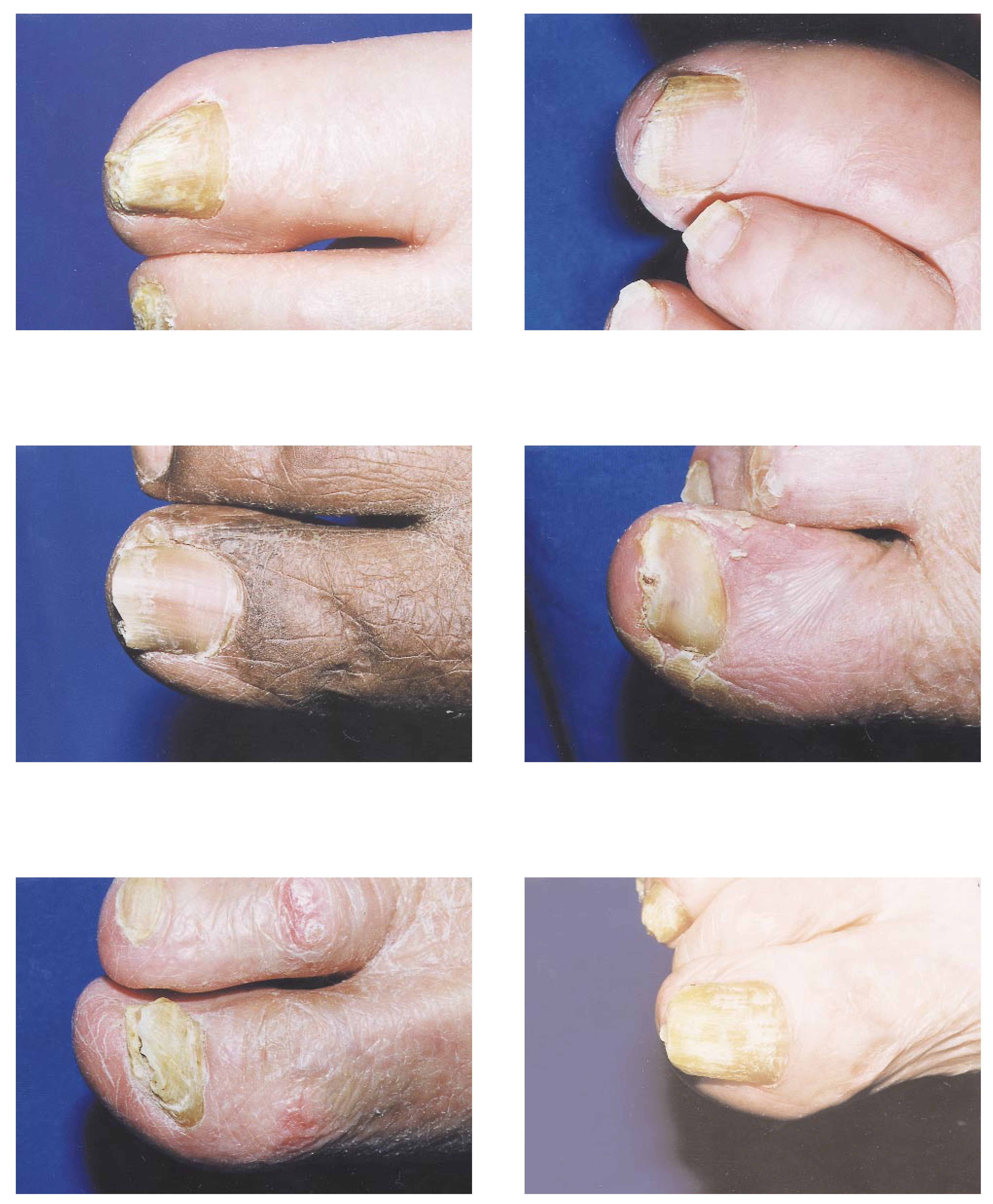
The study population consisted of 100 patients over the age of 65 with clinical signs and symptoms of onychomycosis who were selected at random from a nursing-home population in south Florida. Specimens were not submitted from patients who were currently receiving any type of antifungal therapy. A clinical photograph was taken of each patient’s most-affected hallux prior to specimen collection (
Figure 1). Specimens were obtained by first cleansing the affected toenail and surrounding skin with alcohol and then aggressively clipping the toenail as proximally as possible without causing excessive discomfort to the patient. The nail specimen and subungual debris were obtained from the proximal end of the growing edge of the suspected infection; the distal nail clippings were discarded. The specimens were placed in small individual plastic bags, labeled with the patient’s name, the date of collection, and location of the specimen source (right or left hallux), and transported to the office of the senior author (W.P.S.).
All 100 individual specimens were divided in half. Half of each specimen was left in the plastic bag and sent to the Mycology Division of the Laboratory of Podiatric Pathology in Philadelphia for fluorescent KOH preparation and microscopic fungal culture examination. The other half was inoculated in an ACU-DTM (Acuderm, Inc, Ft Lauderdale, Florida) culture in the manner described by the manufacturer. The specimens were placed on the surface of the medium with aseptic forceps, breaking the surface of the medium without becoming completely embedded. The cap was loosely replaced to ensure the presence of an aerobic environment, and an orange biohazard label was applied to the top of each vial cap. The DTM vials were labeled with the patient’s name, date of inoculation, and location of the specimen source (right or left hallux) and incubated at room temperature (68° to 86° F).
ACU-DTM is the preferred medium for the isolation and early identification of members of the
Microsporum, Trichophyton, and
Epidermophyton genera by means of a distinct color change from yellow to red effected by a pH indicator (
Table 1). [
16] The DTM cultures were examined daily for colony growth or color changes from yellow to red. All DTM culture color changes from yellow to red that occurred on or before 14 days of inoculation were interpreted as a positive result. After 15 days, the cultures were discarded.
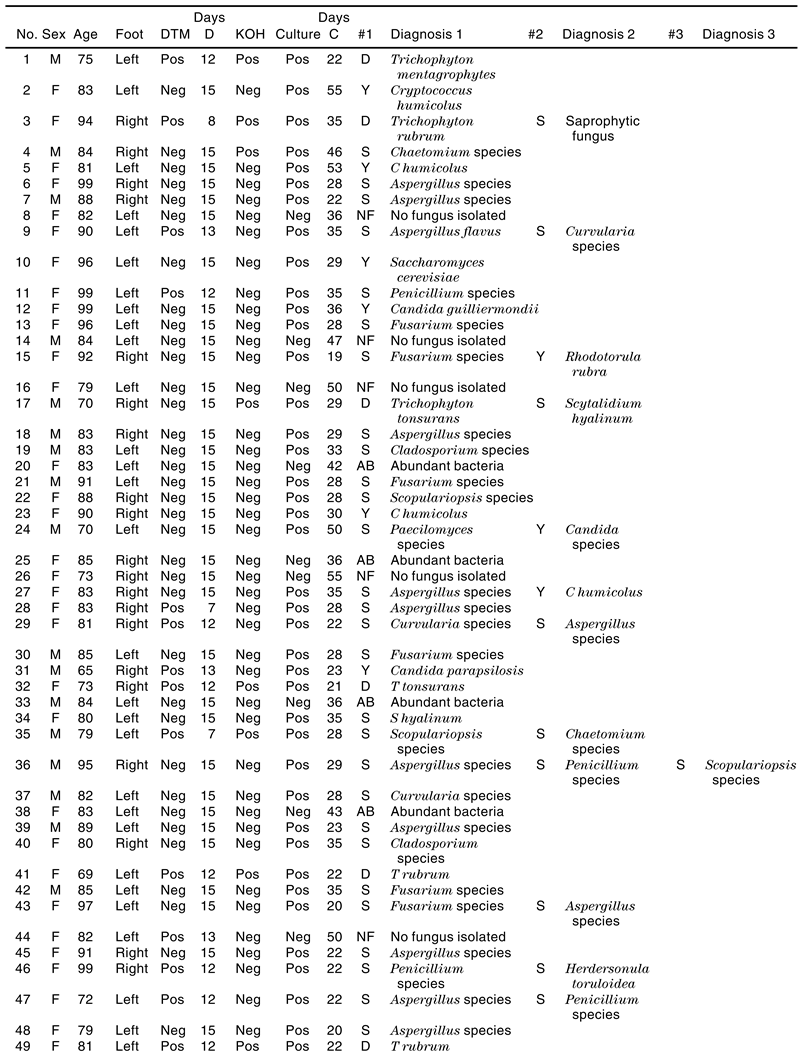
A computer database was created with each patient’s identification number, sex, age, foot (left or right), DTM culture result, number of days for DTM culture result, fluorescent KOH preparation result, microscopic fungal culture examination result, number of days for fungal culture examination report, identification of the organism genus and species, and the type of organism (dermatophyte, yeast, or saprophyte) for positive cultures. Additional columns were created for specimens from which multiple organisms were isolated (
Table 2).
Results
The study group consisted of 72 women and 28 men. The ages of the patients ranged from 65 to 99 years, with a mean age of 84.4 years. There were 52 specimens collected from the right hallux and 48 specimens collected from the left hallux. The total number of days required for a positive DTM result ranged from 7 to 14 days, with a mean of 11.2 days. Negative DTM cultures were identified 15 days after inoculation when there was no color change from yellow to red on the medium. The total number of days required for fluorescent KOH preparation and microscopic fungal culture examination results to be reported ranged from 16 to 55 days, with a mean of 32.1 days.
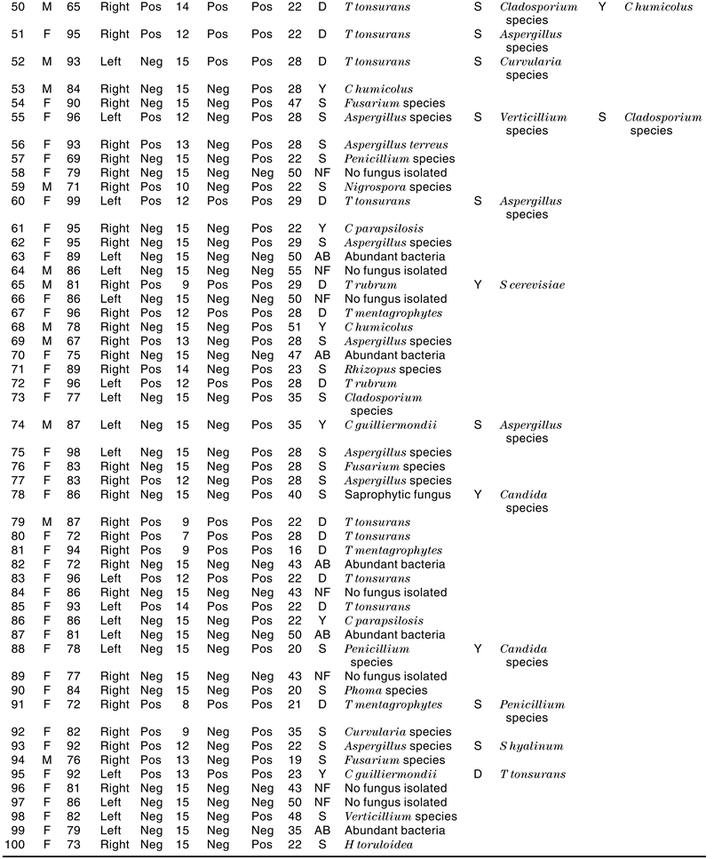
Of the 100 subjects with suspected onychomycosis, 36 had positive DTM culture results and 64 had negative results; 22 had positive fluorescent KOH preparation results and 78 had negative results; 79 had microscopic fungal cultures that were positive for fungal growth, 12 had cultures with no fungus isolated, and 9 had cultures that grew abundant bacteria. Of the 100 microscopic fungal cultures, 11 were dermatophyte only, 34 were saprophyte only, and 10 were yeast only. Of the 79 positive microscopic fungal culture results, 55 (69.6%) were from a single organism and 24 (30.4%) demonstrated mixed fungal growth. Of the 24 mixed fungal growth cultures, 21 (26.6%) of the positive microscopic fungal cultures grew two different organisms and 3 (3.8%) grew three different organisms (
Table 3).
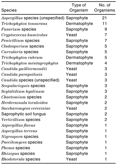
A total of 106 organisms were identified, with 26 different types of organisms observed, from the 79 positive microscopic fungal cultures. An average of 1.3 different organisms were identified per positive fungal culture. Of the 106 total organisms identified, 20 (18.9%) were dermatophytes, 67 (63.2%) were saprophytes, and 19 (17.9%) were yeasts.
Of the 20 dermatophytes isolated, 4 were
T mentagrophytes, 5 were
T rubrum, and 11 were
Trichophyton tonsurans. Of the 67 saprophytes isolated, 1 was
Aspergillus flavus, 1 was
Aspergillus terreus, 21 were unspecified
Aspergillus species, 2 were
Chaetomium species, 5 were
Cladosporium species, 5 were
Curvularia species, 9 were
Fusarium species, 2 were
Herdersonula toruloidea, 1 was
Nigrospora species, 1 was
Paecilomyces species, 7 were
Penicillium species, 1 was
Phoma species, 1 was
Rhizopus species, 2 were unspecified saprophytic soil fungus, 3 were
Scopulariopsis species, 3 were
Scytalidium hyalinum, and 2 were
Verticillium species. Of the 19 yeasts isolated, 3 were
Candida guilliermondii, 3 were
Candida parapsilosis, 3 were unspecified
Candida species, 7 were
Cryptococcus humicolus, 1 was
Rhodotorula species, and 2 were
Saccharomyces cerevisiae (
Table 4).
Of the 24 patients with mixed organism growth on culture, 9 (37.5%) had dermatophyte involvement and 15 (62.5%) had saprophyte/saprophyte or saprophyte/ yeast combinations.
Discussion
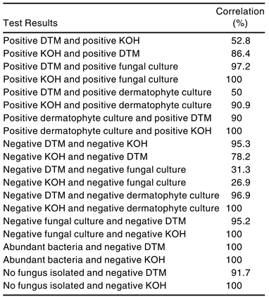
When positive DTM culture results were compared with the corresponding fluorescent KOH preparation results for the same patients, a low correlation was observed. Of the 36 patients with positive DTM culture results, 19 (52.8%) had positive fluorescent KOH preparation results, while 17 (47.2%) had negative results (
Table 5). However, when positive fluorescent KOH preparation results were compared with the corresponding DTM culture results for the same patients, a high correlation was observed. Of the 22 patients with positive fluorescent KOH preparation results, 19 (86.4%) had positive DTM culture results and 3 (13.6%) had negative results. A positive fluorescent KOH preparation result indicates the visible presence of fungi in the specimen. A negative fluorescent KOH preparation result does not preclude the presence of fungal infection; it only indicates that fungal hyphae were not observed in the sampled tissue, and fungi may still be grown and identified on microscopic fungal culture examination.
When positive DTM culture results were compared with the corresponding microscopic fungal culture examination findings for the same patients, a very high correlation was observed. Of the 36 patients with positive DTM culture results, 35 (97.2%) had positive culture results for some type of fungal growth, with only 1 (2.8%) negative result. However, when the comparison was limited to include positive DTM culture results and the corresponding microscopic fungal culture examination results for the same patients that were positive only for dermatophyte growth, a much lower correlation was found. Only 18 (50%) of the 36 positive DTM cultures corresponded with positive microscopic fungal culture examination results for dermatophytes.
When positive fluorescent KOH preparation results were compared with the corresponding microscopic fungal culture examination results for the same patients, a perfect correlation was observed. Of the 22 patients with positive fluorescent KOH preparation results, 22 (100%) had positive culture results for some type of fungal growth. When the comparison was limited to include positive fluorescent KOH preparation results and the corresponding microscopic fungal culture examination results for the same patients that were positive only for dermatophyte growth, a very high correlation was found. Of the 22 patients with positive fluorescent KOH preparation results, 20 (90.9%) had positive culture results for dermatophyte growth.
Only 20 patients had positive microscopic fungal culture examination results for dermatophytes. A comparison of these results with the corresponding DTM culture results for the same patients revealed a very high correlation. Of the 20 patients with positive microscopic fungal culture examination results for dermatophytes, 18 (90%) had positive DTM culture results and 2 (10%) had negative results. A comparison with the corresponding fluorescent KOH preparation results for the same patients revealed a perfect correlation. All 20 fluorescent KOH preparations were positive. A 100% correlation existed between microscopic fungal culture examination results and fluorescent KOH preparation results in this geriatric population.
When negative DTM culture results were compared with the corresponding fluorescent KOH preparation results for the same patients, a very high correlation was observed. Of the 64 patients with negative DTM culture results, 61 (95.3%) had negative fluorescent KOH preparation results and 3 (4.7%) had positive results. When negative fluorescent KOH preparation results were compared with the corresponding DTM culture results for the same patients, 61 (78.2%) had negative DTM culture results.
When negative DTM culture results were compared with the corresponding microscopic fungal culture examination results for the same patients, the following correlations were observed. Of the 64 patients with negative DTM culture results, 20 (31.3%) had negative culture results for fungal growth of any type and 44 (68.8%) had positive results for some type of fungal growth. However, the positive fungal culture growth results could have been attributed to fungi other than dermatophytes. Of the 64 negative DTM culture results, 62 (96.9%) of the corresponding samples demonstrated no dermatophyte growth on microscopic fungal culture examination and 2 (3.1%) demonstrated dermatophyte growth.
When negative fluorescent KOH preparation results were compared with the corresponding microscopic fungal culture examination results for the same patients, the following correlations were observed. Of the 78 patients with negative fluorescent KOH preparations, 21 (26.9%) had fungal culture examinations that demonstrated no fungal growth of any type. Of the 78 negative fluorescent KOH preparations, none of the corresponding cultures demonstrated dermatophyte growth.
There were 21 negative microscopic fungal culture examinations that demonstrated no fungus isolated or abundant bacterial growth. A comparison with the corresponding fluorescent KOH preparation results and DTM culture results for the same patients revealed an extremely high correlation. There were 21 (100%) negative fluorescent KOH preparations: 20 (95.2%) with negative DTM culture results and only 1 (4.8%) with a positive DTM culture result.
When microscopic fungal culture examinations demonstrating abundant bacterial growth or no fungus isolated were compared with the corresponding fluorescent KOH preparation and DTM culture results for the same patients, an extremely high correlation was observed. Nine of the 100 microscopic fungal culture examinations demonstrated abundant bacterial growth, which most likely was a contaminant growing that may have inhibited the growth of fungus. Of the nine microscopic fungal culture examinations that demonstrated abundant bacterial growth, all had negative fluorescent KOH preparation results and negative DTM culture results. Of the 12 microscopic fungal culture examinations that demonstrated no fungus isolated, there were 12 (100%) negative fluorescent KOH preparation results: 11 (91.7%) with negative DTM culture results and 1 (8.3%) with a positive DTM culture result.
In this study of 100 patients with suspected onychomycosis, saprophytes accounted for the majority of the organisms isolated, occurring in 56 of the patients and 56 (70.9%) of the 79 positive fungal cultures.
Aspergillus was the most common organism, accounting for 23 (21.7%) of the 106 organisms identified. This is contradictory to the majority of current and past literature on onychomycosis. Dermatophytes, which have been demonstrated in several other studies to account for over 90% of onychomycosis infections, occurred in only 20 of the patients and 20 (25.3%) of the 79 positive fungal cultures.
Trichophyton rubrum and
T mentagrophytes have traditionally been thought of as the most common dermatophytes responsible for onychomycosis.[
4,
5] In this study, only 5 (4.7%) cases of
T rubrum, 4 (3.8%) cases of
T mentagrophytes, and 11 (10.4%) cases of
T tonsurans were observed out of the 106 organisms identified. Yeast was found in 19 of the patients and 19 (24.1%) of the 79 positive fungal cultures.
Candida was the most common organism, accounting for 9 (8.5%) of the 106 organisms identified.
Conclusion
The results of this study of 100 cases of suspected onychomycosis in a geriatric population demonstrated that 20% had dermatophyte involvement, 56% had saprophyte involvement, 19% had yeast involvement, 24% had mixed fungal infections, 9% had abundant bacteria, and 12% had no fungus isolated according to the mycology laboratory report.
For the entire study group, 36% of the subjects had a positive in-office DTM culture result; however, the DTM culture results had a 52.8% correlation with a positive fluorescent KOH preparation and only a 50% correlation with a positive microscopic fungal culture for dermatophytes. In comparing in-office DTM cultures with fluorescent KOH preparations, a positive fluorescent KOH preparation had a 90.9% correlation with a positive microscopic fungal culture for dermatophytes and a negative fluorescent KOH preparation had a 100% correlation with a negative microscopic fungal culture for dermatophytes. In comparing abundant growth and no fungus isolated results from microscopic fungal culture with in-office DTM cultures and fluorescent KOH preparations, both diagnostic techniques yielded excellent results.
It is questionable whether in-office DTM cultures should be routinely used in geriatric patients for the diagnosis of onychomycosis, for several reasons. With only 20% of the study group demonstrating dermatophyte growth, DTM cultures would not diagnose the approximately 59% of the patients with saprophyte or yeast involvement. There were too many false-positive results with in-office DTM cultures for it to be used as a definitive diagnostic test for onychomycosis; only 50% of positive DTM cultures correlated with positive microscopic fungal cultures for dermatophytes. The only case in which DTM culture results had a high correlation with mycology laboratory testing results was when a DTM culture was negative for dermatophytes; this had a 95.3% correlation with a negative fluorescent KOH preparation and a 96.9% correlation with a negative microscopic fungal culture for dermatophytes.
These findings should influence the current diagnosis and treatment methods of physicians treating onychomycosis. The authors believe mycology laboratory testing with fluorescent KOH preparations and microscopic fungal cultures to be superior to in-office DTM cultures for the diagnosis of suspected onychomycosis in geriatric patients.