Chondromyxoid fibroma is a rare benign tumor of cartilaginous origin that typically presents in the second or third decade of life. [
1,
2,
3,
4,
5,
6,
7] Although many authors have described a preponderance of males affected by this condition, some studies have shown no predilection for either sex. [
3,
8]
The most common location for the formation of a chondromyxoid fibroma is the proximal one-third of the tibia,1, 4 with 95% of tibial cases occurring at the metaphysis.6 Although chondromyxoid fibroma accounts for less than 1% of all bony neoplasms, [
1,
6,
8] the foot is affected in about 20% of all cases.6 The most common locations in the foot are the calcaneus, the metatarsals, and the phalanges. Rahimi et al1 reported on 56 cases involving the foot; 21 of these occurred in the metatarsals. There was no discussion of the technique used for the resection of these neoplasms in the metatarsals.
The typical radiographic presentation is a well-circumscribed, eccentrically located, lobulated, roundto- ovoid, metaphyseal, lytic lesion. [
1,
3,
4,
5,
6,
8] Chondromyxoid fibroma frequently has a border of sclerotic reactive bone that varies in thickness from 1 mm to 1.5 cm. [
3,
4,
6,
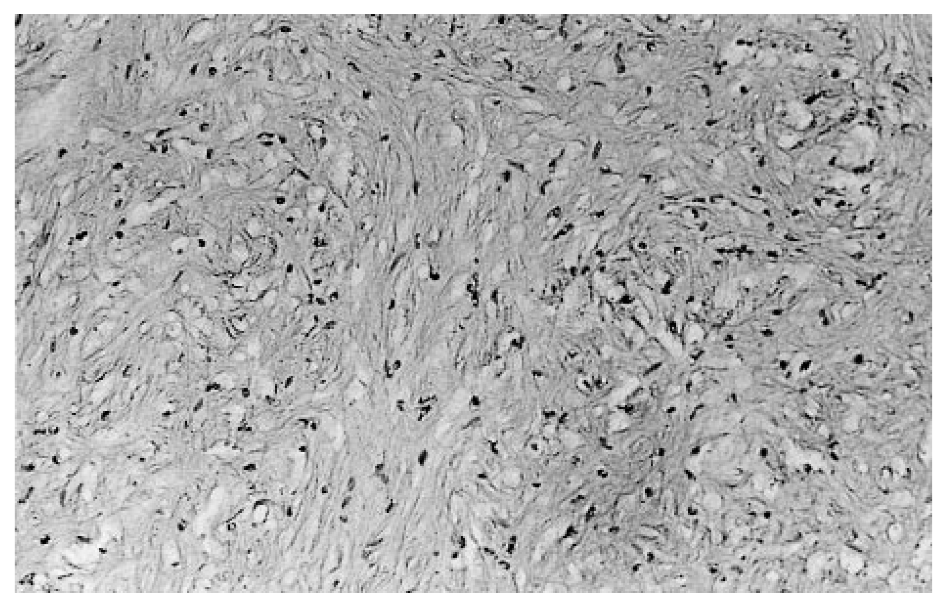
8] Histologically, chondromyxoid fibroma appears as an unusual mixture of fibroid, chondroid, and myxoid tissues. Most lesions show peripheral condensation of fibroid cells around chondroid lobules. The lesion may be quite cellular and contain stellate cells. Mitotic figures are rare and always typical. [
6]
The typical clinical presentation of chondromyxoid fibroma is pain associated with a palpable mass or area of swelling that is extremely tender on palpation. The periosteum usually remains intact except in rare cases of pathologic fractures. A hemispheric “bite-out-of-bone” appearance from the cortical margins without periosteal activity has been described. [
9]
Typically, chondromyxoid fibroma in the foot progresses slowly and is expansile in nature. Most reported cases have responded well to simple operative measures, such as local resection or curettage with bone grafting. Although recurrences have been reported with some frequency, they are usually due to incomplete curettage or incomplete resection. [
1,
2,
8,
10] When the tumor does recur, it usually does so within 1 to 3 years following resection.10
Radiation therapy is contraindicated in the foot because of the benign nature of the lesion, which is composed of tissue unlikely to be radiosensitive. In addition, radiation therapy may increase the risk of malignant degeneration.2 Although this tumor is usually benign and innocuous, the physician should be aware of its potential for local malignancy. [
10,
11,
12] There have been rare cases of conversion to sarcoma and, more specifically, chondrosarcoma. [
3,
10,
13,
14] Some authors have suggested that chondromyxoid fibroma may not invariably be benign, but may be a slowgrowing tumor of low-grade malignancy from the outset. [
15]
When an expansile bony neoplasm occurs in the metatarsal head region, it is likely to result in metatarsalgia, nerve impingement, and muscle dysfunction. Some bony neoplasms in the foot are often associated with pain due to cortical fractures that develop as a result of their thin cortical margins (eg, aneurysmal bone cysts, giant cell tumors). This rarely occurs with a chondromyxoid fibroma because of its thick cortical rim of bone. Chondromyxoid fibroma is therefore often asymptomatic until it grows larger. It then invades the surrounding soft tissues and neurovascular structures, resulting in secondary symptoms.
Case Report
A 13-year-old girl presented to the surgical clinic at the California College of Podiatric Medicine in San Francisco complaining of pain at the plantar aspect of her left forefoot. The pain had begun 2 years previously and had become progressively worse. The first noticeable change was the inability to spread the toes on the left foot to the same degree as the toes on the right foot. The patient then developed an aching and throbbing pain with occasional tingling in the left forefoot.
The pain increased with ambulation, particularly in tight-fitting shoes. Cold weather also significantly increased the patient’s level of forefoot pain. Previous treatment had included nonsteroidal anti-inflammatory drugs, which resulted in an approximately 50% improvement in her symptoms, and shoe modifications, which also provided some relief.
Physical examination demonstrated mild edema of the left forefoot. There was moderate pain on palpation at the plantar aspect of the fourth metatarsal and mild pain on palpation dorsally. There was a palpable, expansile mass extending plantarly into the soft tissues. There was moderate pain with lateral compression of the forefoot. The patient had moderate pain with end range of motion at the metatarsophalangeal joint. There was also pain with impaction of the fourth metatarsophalangeal joint. The neurologic and vascular examinations were unremarkable. Gait evaluation demonstrated a compensatory antalgic gait favoring the left foot.
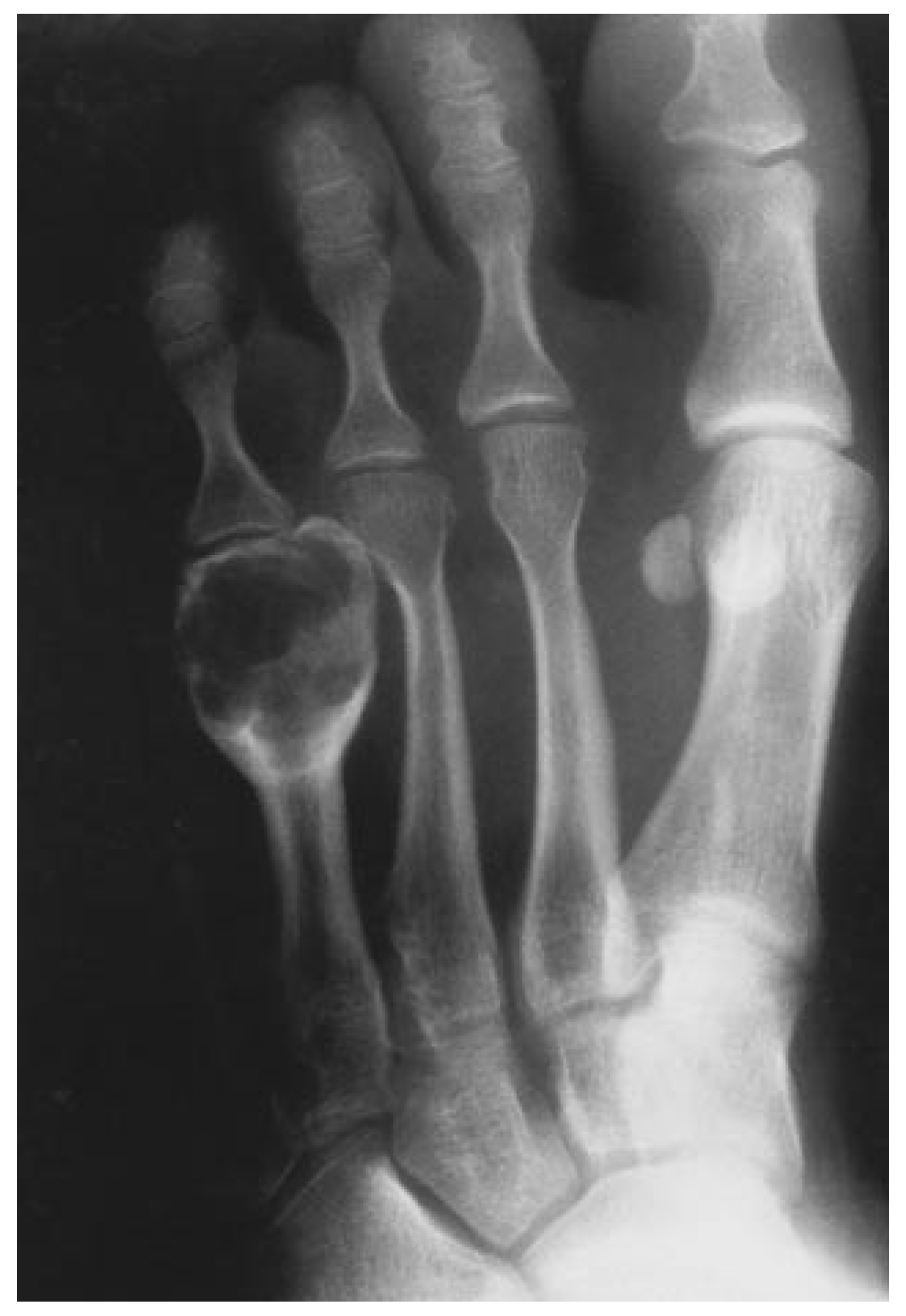
Plain radiographic evaluation revealed an expansile bone tumor of the left fourth metatarsal head and neck region (
Fig. 1 and
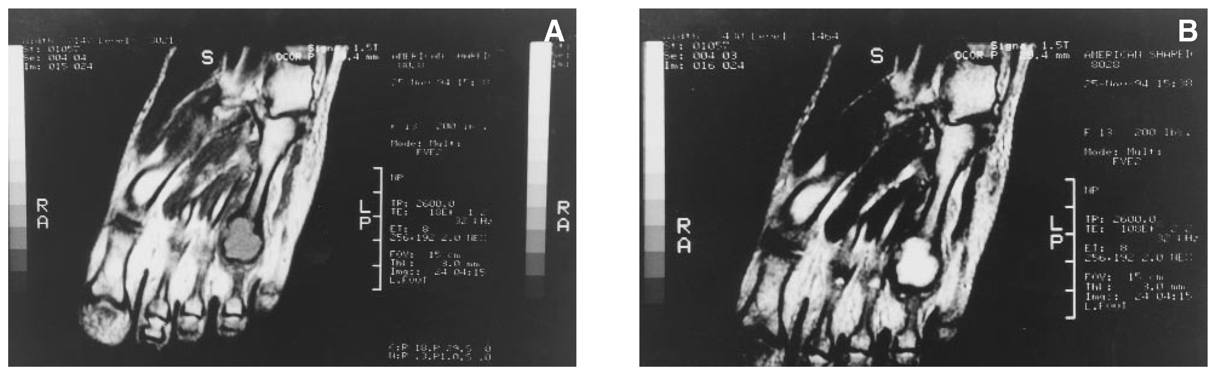
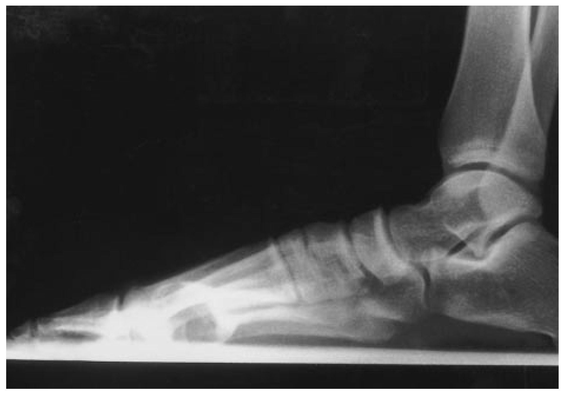
Fig. 2). At its widest point, the lesion measured 1.9 cm on the anteroposterior projection and approximately 3 cm along the length of the metatarsal. The lesion encompassed the entire fourth metatarsal head, with an apparently normal joint space. There were no obvious signs of cortical fracture. On the lateral view, the lesion was expanded plantarly. The lesion had a well-defined cortical margin with thick sclerotic bone. Magnetic resonance imaging (MRI) was undertaken to further evaluate the lesion prior to surgery and to better determine the extent of the lesion’s invasion into the soft tissues. The MRI scans demonstrated increased T1 and T2 signal intensity of the fourth metatarsal head and neck region, with no evidence of invasion into the adjacent bones (
Fig. 3).
A pneumatic thigh tourniquet was used for hemostasis. Attention was initially directed to the dorsal aspect of the fourth metatarsal of the left foot. A linear incision was made beginning distal to the metatarsophalangeal joint and extending proximally to the base of the fourth metatarsal. Dissection was carried down to the dorsum of the fourth metatarsal. The metatarsal demonstrated a large bony prominence that included the distal one-third of the metatarsal and the entire head. The tumor was resected
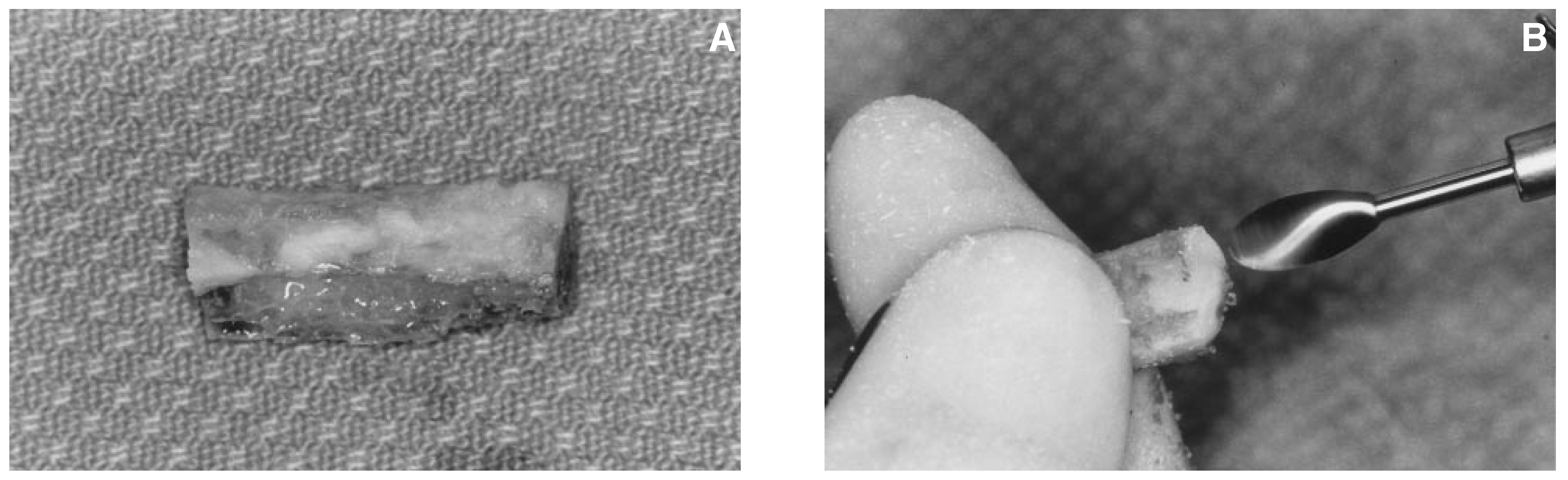
in toto at the proximal margin. It was noted to be well encapsulated with a 1-cm intact cortical margin. The tumor measured 3 cm in length and 5 cm in width. This measurement helped to determine the size of the bone graft needed from the fibular donor site in order to reconstruct the fourth metatarsal. The tumor was then sent for pathologic analysis (
Fig. 4).
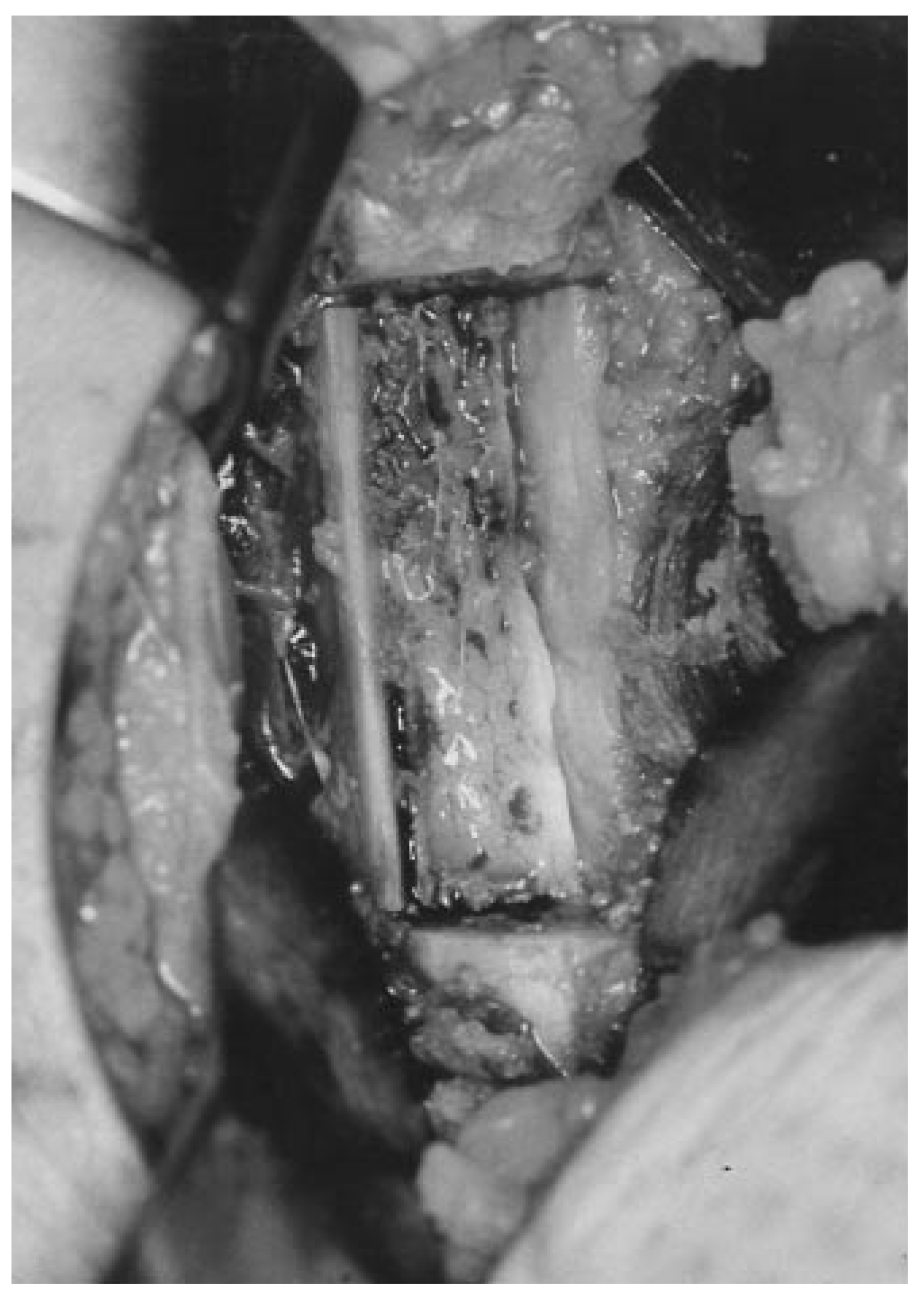
Attention was then directed to the lateral aspect of the left fibula. An incision was made over the distal one-third of the fibula. Dissection was carried down to the lateral aspect of the peroneal tendons. The anterior aspect of the muscle belly was dissected and retracted posteriorly, thus exposing the posterior aspect of the fibula. The bone graft was then taken from a site 6 cm proximal to the distal tip of the fibula. The graft measured 3 cm in length and 2 cm in width. The medial cortex of the fibula was left intact with some cancellous bone remaining (
Fig. 5). The periosteum was carefully dissected off of the fibula before resection and was reapproximated at the time of closure. The muscle belly and skin were then reapproximated over the bone graft site.
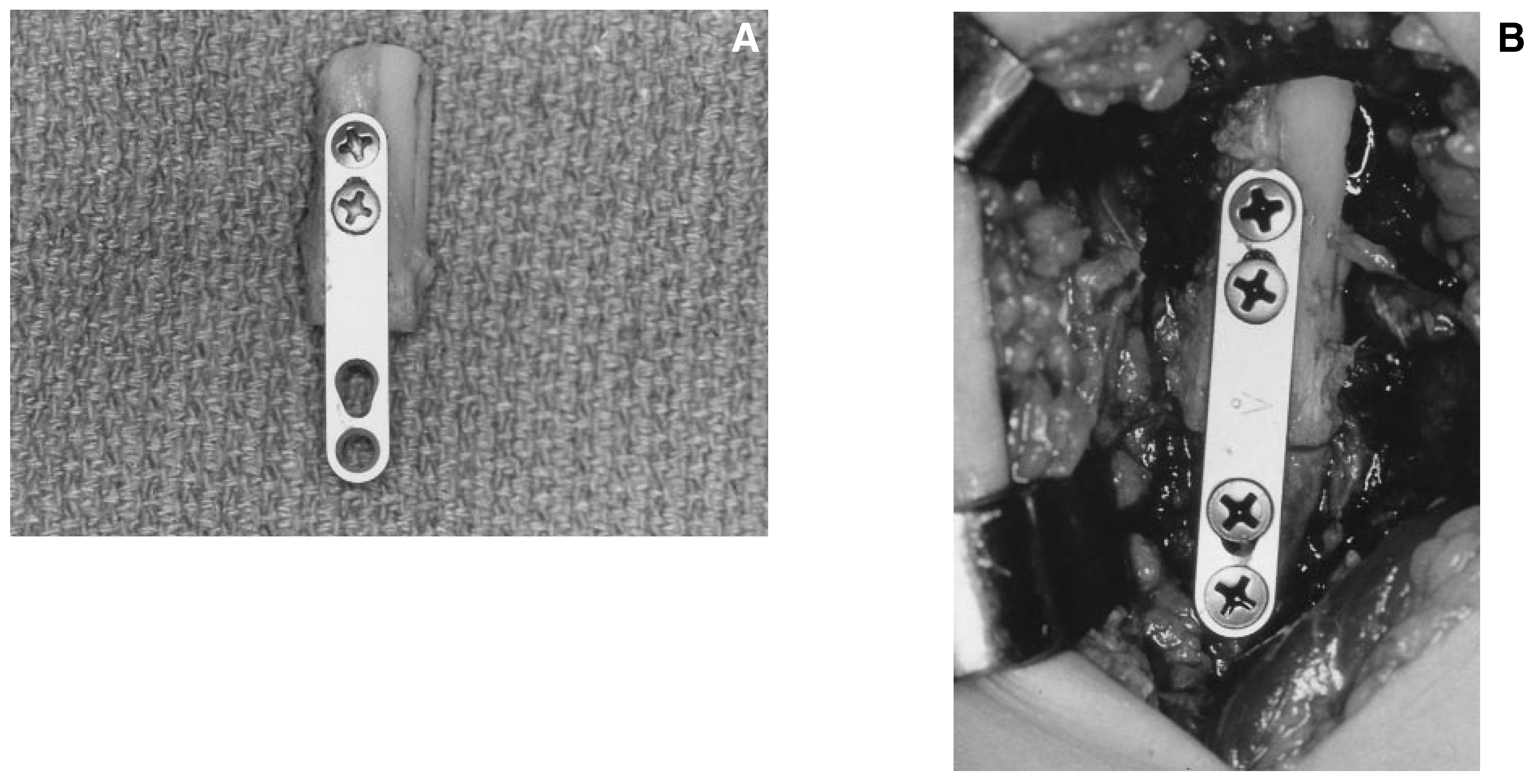
Attention was then redirected to the fourth metatarsal. The surgical site was irrigated with 20 mL of 3% hydrogen peroxide to help remove any remaining neoplastic cells, and then immediately irrigated with lactated Ringer’s solution. The bone graft was then remodeled into the shape of a metatarsal. The distal aspect of the graft was rounded to simulate the head of a metatarsal (
Fig. 6). The bone graft was then fixated on the back table with a 4-hole Luhr plate (How Medica, Rutherford, New Jersey) with two 2-mm screws in the bone graft. The graft was then placed in the foot, and two screws were inserted through the plate onto the distal aspect of the metatarsal shaft (
Fig. 7). The screws were slightly angled toward the center of the plate and graft site to allow for some compression at the recipient site. The bone graft was stable in all planes and well apposed to the metatarsal shaft. The patient was placed in a Jones compressive dressing with a fiberglass shell and kept nonweightbearing with crutch-assisted ambulation.
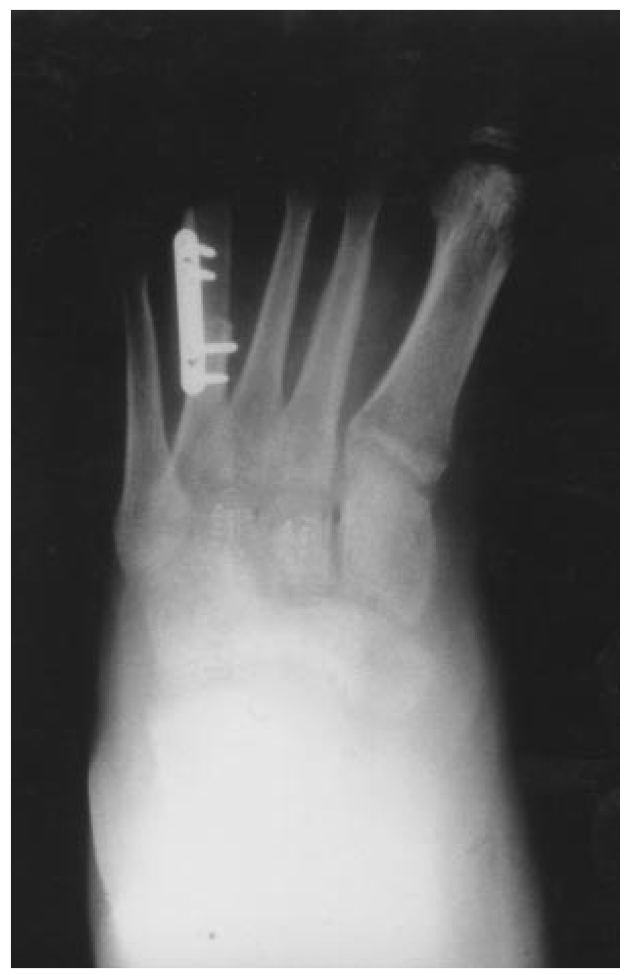
Immediately postoperatively, the patient began using an EBI Bone Healing System (EBI Medical Systems, Parsippany, New Jersey) to facilitate bone healing. The patient remained nonweightbearing for 3 months in a below-the-knee fiberglass cast. She then wore a below-the-knee walker for 1 month before resuming wearing shoes. At 1 month postoperatively, the patient demonstrated a fracture through the medial cortex of her fibular bone graft site. Despite the fracture, the bone graft site filled in with bone and the patient was asymptomatic during the recovery period. At 3 months postoperatively, the bone graft was completely incorporated and the patient returned to wearing shoes (
Fig. 8). At 2 years postoperatively, the patient was walking without pain and with no signs of recurrence in either the foot or the ankle.
Discussion
Determining the most appropriate bone graft for a patient with a neoplasm of the foot is always difficult, particularly in a child. The authors prefer to use autogenous bone because of its more rapid incorporation by means of its osteoinductive and osteoconductive properties. The autograft contains viable osteogenic cells, thereby allowing the graft not only to be incorporated, but also to help produce new bone. [
16,
17]
When a metatarsal bone is reconstructed with an autogenous bone graft, the two most common sites for graft harvesting are the iliac crest and the fibula. The main reason is because of the need for a corticocancellous graft, which has the benefits of cancellous incorporation properties and cortical structural support. In addition, the cortical bone will help support compressive fixation devices. Although only 25% of patients complained of hip pain in one large retrospective study on autogenous iliac crest grafts, [
18] the authors’ experience with hip grafts has shown a much higher incidence of pain. As a result, the authors prefer to avoid using iliac crest grafts in children. Iliac crest grafts are often tapered and larger than needed for metatarsal reconstruction, requiring more extensive remodeling as compared with fibular bone grafts.
The site from which a fibular graft is taken is very important because of the possibility of morbidity following resection of the fibula. Some studies have suggested that a valgus deformity of the ankle may develop after fibular resection. [
19,
20] In 1995, Babhulkar et al [
21] reported on 29 patients who had received distal fibular transplants. The average followup time was more than 2.5 years. The bone grafts were harvested from the distal half of the fibula. The results showed that six patients had ankle instability; in all six cases, the distal cut was less than 8 cm from the tip of the lateral malleolus. The remaining 23 patients had excellent results with no complaints. There was no correlation between the length of the fibula resected and ankle instability.
Studies have shown that the fibula moves distally during weightbearing to deepen the ankle mortise. [
22,
23] It has also been demonstrated that 10% to 16% of the total weightbearing load is transmitted through the fibula. [
24,
25] In this case study, the patient sustained a fracture of the medial cortex of the harvest site. This may have been due to premature weightbearing or to the fragility of the thin remaining cortex. Despite the fracture and mild posterior displacement of the distal remnant of the fibula, the fibula completely regenerated, and the patient remained asymptomatic at 2 years postoperatively. The patient will be monitored over the next few years for the possible development of ankle arthritis. One consideration for avoiding a possible harvest-site fracture may be to use an allogeneic fibular section to help stabilize the harvest site postoperatively. This could be inserted with a one-third tubular plate used in a buttressing technique. This may help prevent the development of ankle arthritis or instability in the long term.
When reconstructing the forefoot with a large bone graft, the surgeon is often concerned about the incorporation of the graft. This is particularly true when only one end of the graft is attached to the donor site, eg, when reconstructing a metatarsal head and shaft. The authors prefer to use electrical bone stimulation to facilitate incorporation of the graft with bone grafts longer than 3 cm. This technique has been performed in previous orthopedic studies. [
26,
27] When treating a patient with a bone tumor, the foot surgeon should use all available means of enhancing incorporation of the bone graft, thereby avoiding the need for further surgery.
Conclusion
The authors have described a case of chondromyxoid fibroma of the fourth metatarsal. The technique for reconstructing metatarsal bone with an autogenous fibular bone graft has been described. The principles illustrated here may apply to other, similar situations in which resection of tumors and bone grafting are needed.