To the Editor:
Traumatic etiology for the formation of neuromas is a well documented phenomenon in the literature [
1]. In fact, all neuromas have the common theme of traumatic origin and three scenarios in which they can form. The first type of neuroma occurs from sectioning of a nerve (eg, amputation or stump neuroma). The second type arises from repeated microtrauma of a nerve segment, and has been labeled as neuroma-in-continuity (eg, Morton’s neuroma). The final etiology occurs from blunt force trauma to a specific segment of nerve, and is also considered a type of neuroma-in-continuity [
1]. The blunt force trauma causes partial sectioning of the nerve that can allow for perineural structures and fibrous tissue to become interwoven between the sectioned nerve ends. An altered nerve signal is the result and leads to typical neuroma type dysesthesias [
1].
The innervation of the fifth toe is supplied by three nerves. Dorsally, the intermediate dorsal cutaneous nerve, a branch of the superficial peroneal nerve, supplies the medial half of the toe. The lateral half of the toe is supplied by the lateral dorsal cutaneous nerve, the terminal branch of the sural nerve.
Plantarly, the toe is supplied by branches of the lateral plantar nerve. The lateral plantar nerve splits into one deep and two superficial branches. The superficial branches are primarily responsible for the innervation of the fifth toe. The medial branch of the superficial division enters and serves the fourth intermetatarsal space. The lateral division of the superficial division innervates the intrinsic muscles of the fifth toe on its way to supply sensation to the lateral side of the toe as the proper digital nerve [
2,
3].
Case Report
A 28-year-old male presented with pain on the lateral aspect of his right foot 1 week after a 3,700-pound roll of paper fell on his foot. At the time of injury, radiographs were taken at a local emergency department and were negative for fracture. He was treated with immobilization and pain medication. He presented to the authors’ office 1 week after the initial injury. His medical history was unremarkable. The physical examination revealed moderate edema with ecchymosis around the fifth metatarsophalangeal joint with moderate hypesthesia on the dorsolateral aspect of the toe, presumably caused by edema. Additionally, he had a mild bunionette deformity.
A conservative approach was used, consisting of Unna boots for swelling, nonsteroidal anti-inflammatory drugs, and steroid injections sequentially over a period of 6 weeks without significant relief. Intense pain and altered sensation to the lateral aspect of the fifth toe persisted, so exploratory surgery of the dorsal digital nerve for possible excision of neuroma was scheduled.
At the time of surgery, the patient’s main symptoms were located on the dorsolateral aspect of the toe. A dorsal lateral incision was made on the enlarged dorsal nerve, which was resected and sent to pathology. The report was consistent with a digital neuroma. However, the patient’s symptoms did not improve; in fact, they became more severe and discriminant to the plantar lateral aspect of the fifth toe. Once again, a conservative approach was taken for approximately 4 weeks during the healing of the first incision.
Because of the excruciating pain the patient was experiencing and the impact on his life, it was decided that further surgery was needed for exploration of a concomitant plantar neuroma.
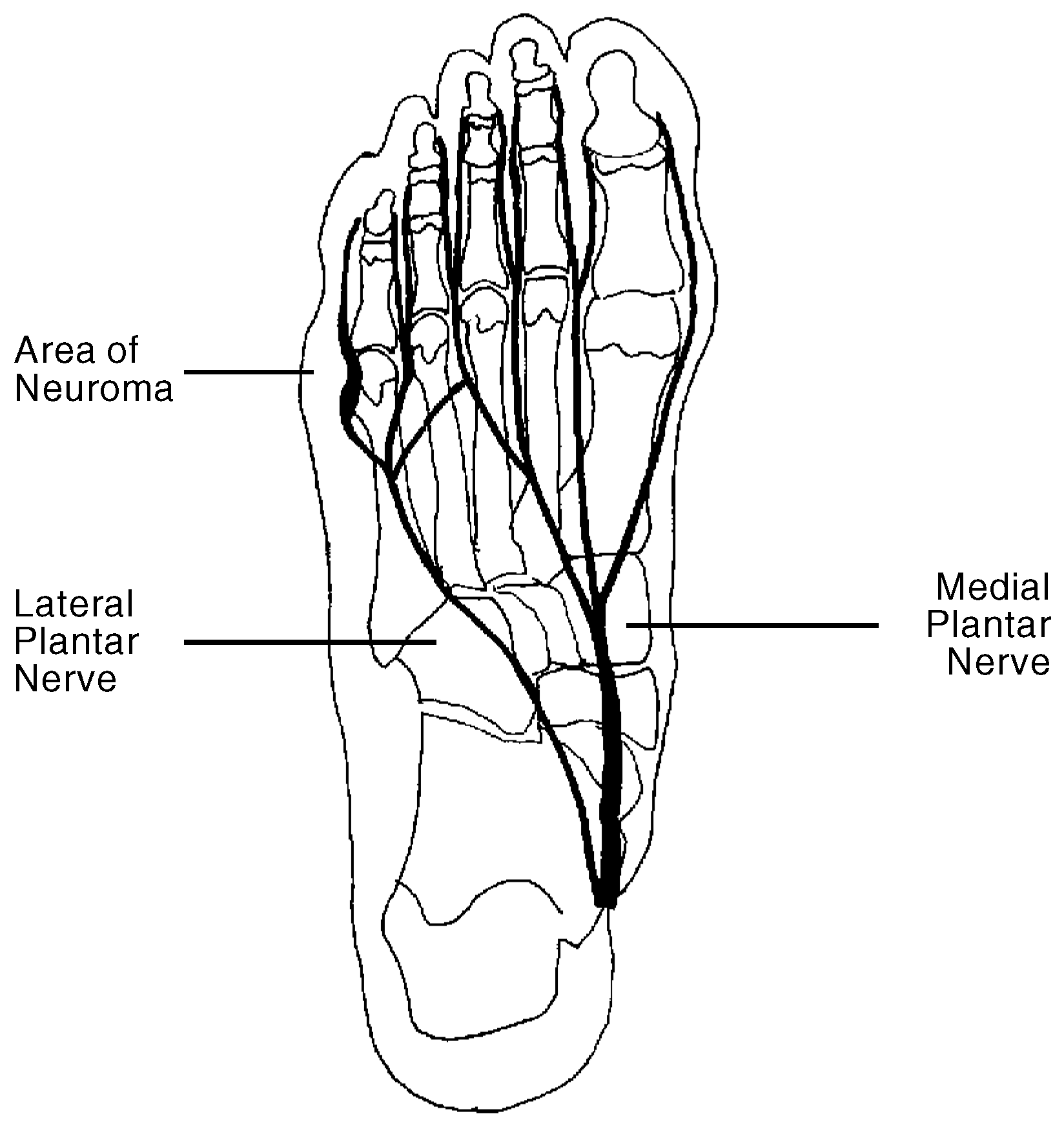
During surgery, the plantar proper digital nerve was extremely enlarged and somewhat convoluted along its course past the metatarsal neck and condyles. It was removed in toto and sent to pathology. The report revealed a large digital neuroma measuring 0.5 × 4.0 cm. The patient’s course was unremarkable at 10 weeks with obvious mild-to-moderate insensitivity of the toe.
Figure 1.
The main plantar branches of the tibial nerve with the enlarged division of the lateral plantar nerve outlined.
Figure 1.
The main plantar branches of the tibial nerve with the enlarged division of the lateral plantar nerve outlined.
Discussion
Neuroma of the proper digital nerve has been previously reported in the literature. However, in the previous report, the neuroma was found with preexisting bony pathology. That particular neuroma was encountered during the correction of a bunionette deformity.4 In the evaluation and treatment of this case, the authors reviewed the neuroanatomy of the fifth toe. Many anatomy texts and illustrations describe the proper digital nerve of the fifth toe as being normally enlarged and convoluted as it passes the neck and condyles of the fifth metatarsal (
Fig. 1) [
2,
3]. Given the course and size of the nerve and the proximity to the condyles of the fifth metatarsal, the possibility of neuroma of the proper digital nerve should be considered when dysesthesia of the fifth toe is associated with bunionette surgery or when a history of trauma is present.