The rheumatoid foot commonly shows deformities such as splay foot, rearfoot valgus, hammer toes, calluses, flatfoot, and lateral deviation of toes. The lower extremity joint involvement leads to greater dysfunction and pain than in the upper extremity often because of weightbearing structures [
1]. In descending order of frequency, rheumatoid arthritis characteristically affects the metatarsophalangeal, talonavicular, and ankle joints. Although rheumatoid arthritis affects the metatarsophalangeal joints in unison, frequently the earliest signs of destructive arthritis are seen at the fifth metatarsophalangeal joint on the radiography. It also shows the abnormalities of retrocalcaneal bursitis with posterosuperior calcaneal erosion, Achilles tendonitis, spurring at the posterior attachment of the Achilles tendon, well defined calcaneal spur, cysts, and ulcerations.
Rheumatoid nodules that appear in a quarter of the patients may or may not be treated unless symptomatic [
2]. Rheumatoid nodules usually develop with an insidious onset on areas of pressure or contact over tendons. Microscopically, there are central necrotic areas surrounded by an inner fibroblast layer and outer layer of inflammatory cells.2 Complications may include ulcerations and possible infection of soft tissue and bone. Also, these nodules may develop into large and painful deformities.
Treatment of a symptomatic rheumatoid arthritis nodule is excision. Surgically removing and debriding nodules can be challenging if neurovascular structures are nearby. Even after excision, recurrence is still a possibility.
Case Report
A 56-year-old female presented to Madigan Army Medical Center with bilateral foot deformities and draining, painful rheumatoid nodules on her heels. The nodules had been enlarging for 2 years with recent ulceration. Her pain was aggravated by weightbearing, ill-fitting shoes, and increased symptoms of the disease.
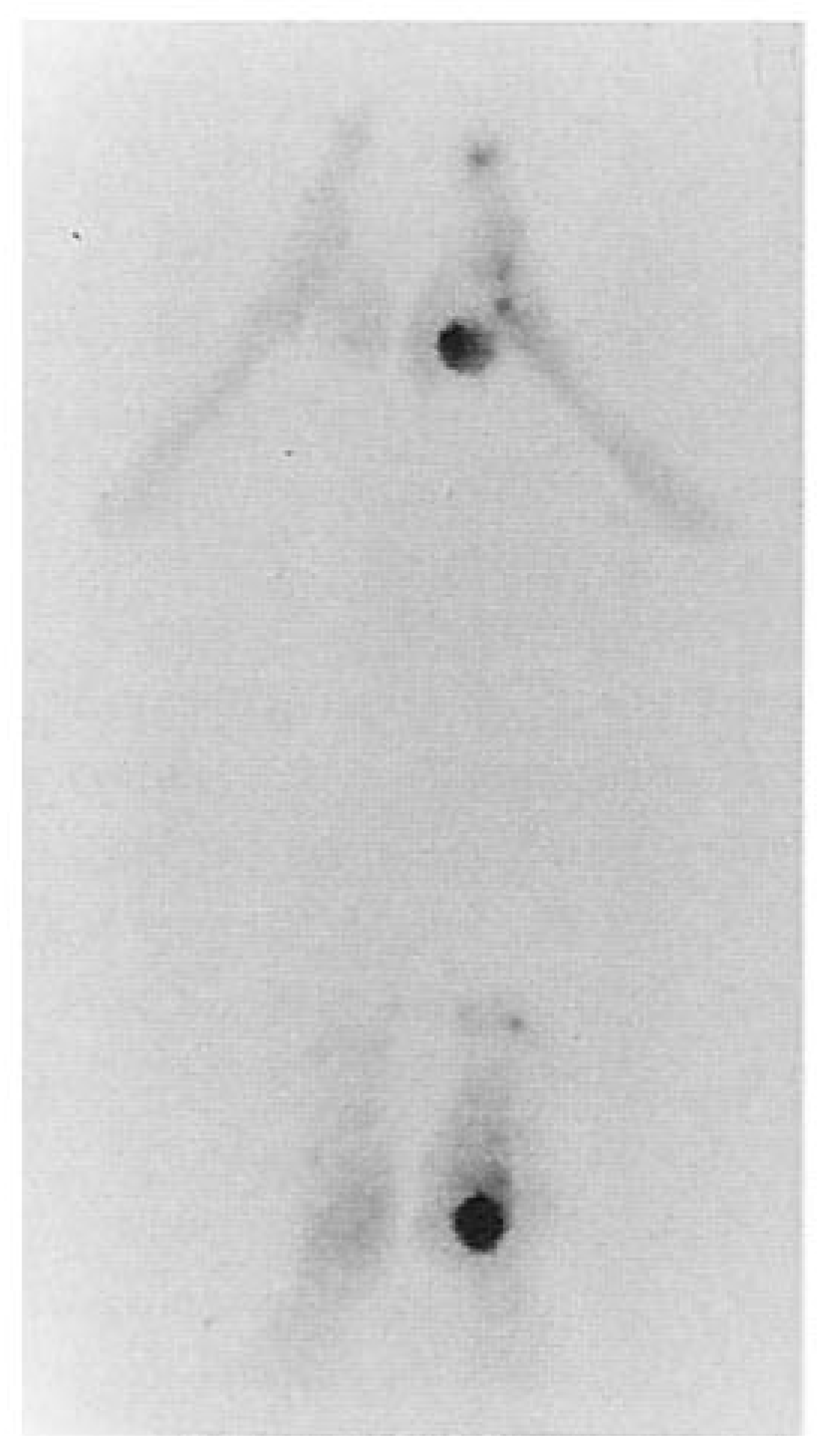
The patient had been placed on oral antibiotic therapy and was using a wheelchair to protect the sites. There was concern for osteomyelitis of her right calcaneus because of the chronic drainage. A triphasic technetium bone scan showed increased uptake involving the right calcaneus (
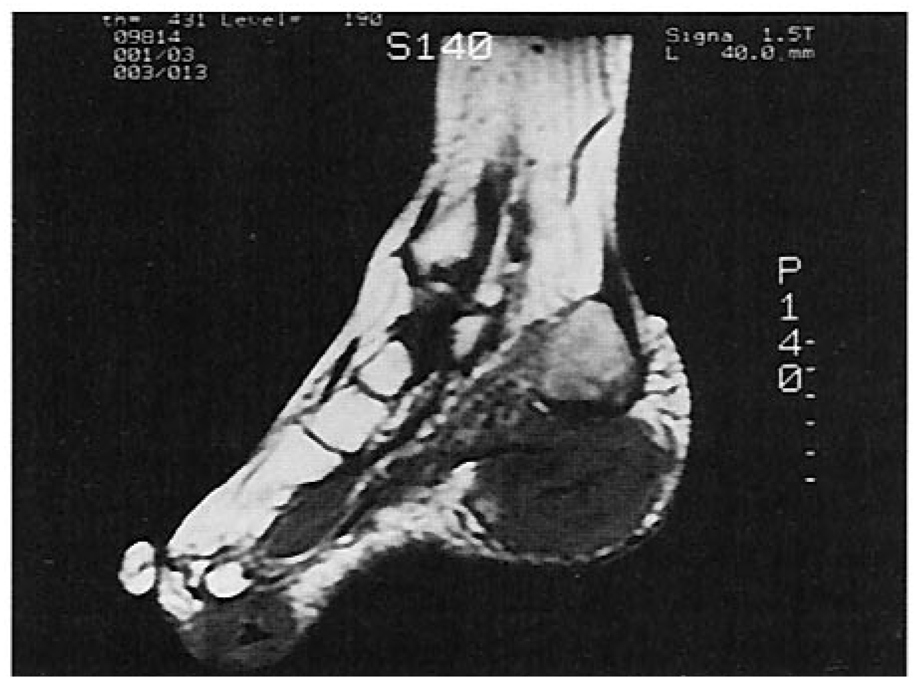
Fig. 1). A fine needle biopsy performed on the largest nodule of her right heel was inconclusive. A magnetic resonance image was also performed that showed the extent of soft tissue involvement (
Fig. 2).
Her medical history included rheumatoid arthritis for 12 years, hypertension, and obesity. Her medications included auranofin 3 mg twice a day, prednisone 5 mg per day, hydrochlorothiazide 50 mg per day, and piroxicam 20 mg as needed.
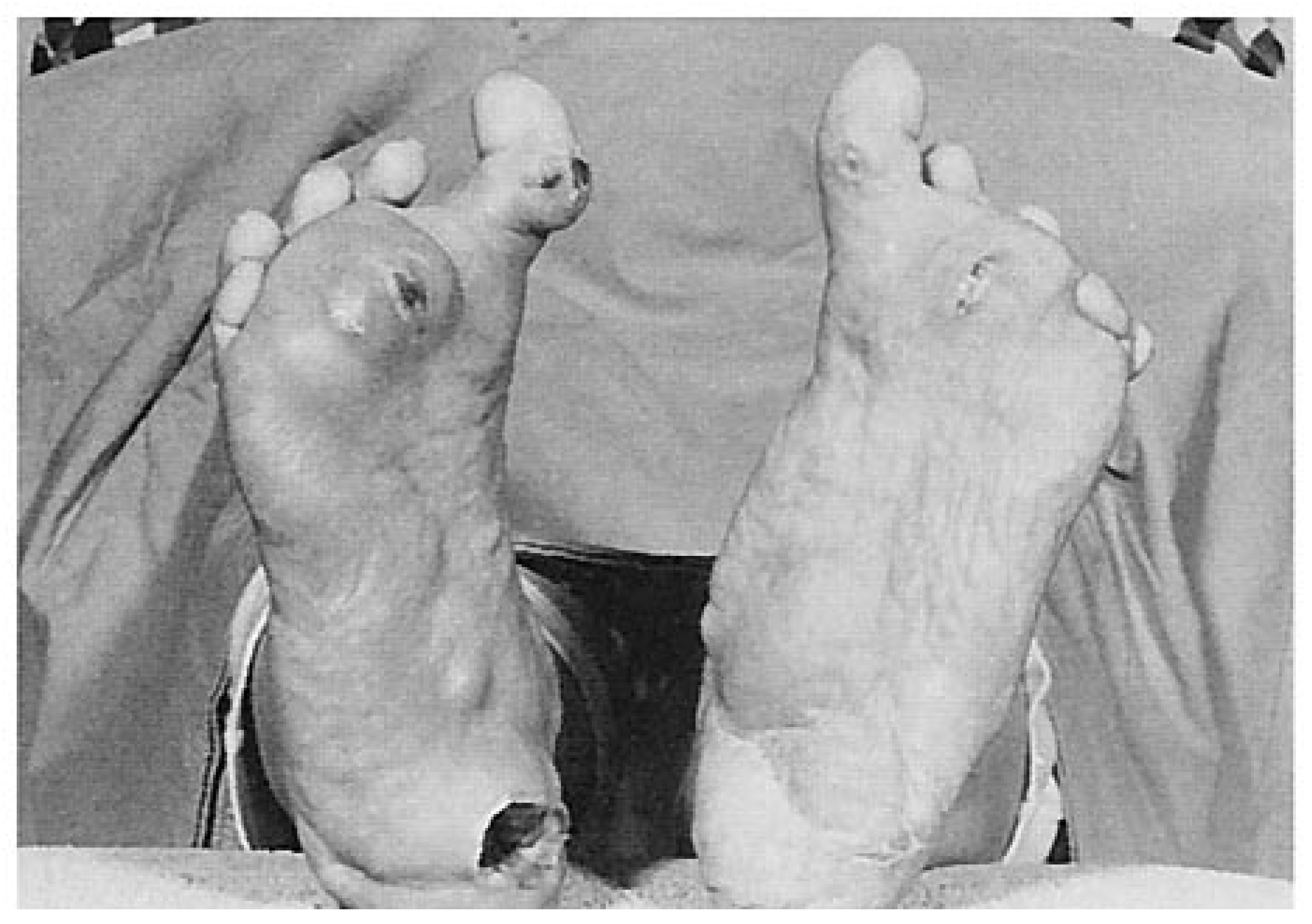
The physical examination revealed an obese female confined to a wheelchair because of painful draining lesions. The lower extremity vascular status revealed nonpalpable posterior tibial arteries secondary to grossly enlarged soft tissue masses of both heels. The dorsalis pedis pulses were palpable and increased. The neurologic examination was unremarkable. The dermatologic examination was significant for multiple rheumatoid arthritis nodules ranging in size from 4 cm at the heels to 2 cm at the plantar aspect of the third metatarsophalangeal joint and the plantar aspects of the hallux interphalangeal joints bilaterally. There were small sinus tracts with serosanguinous exudate without foul odor. Her heels were in valgus. A flexible pes planus deformity was present. The metatarsophalangeal joints were dorsally subluxed and the hallux in varus with plantarflexion bilaterally.
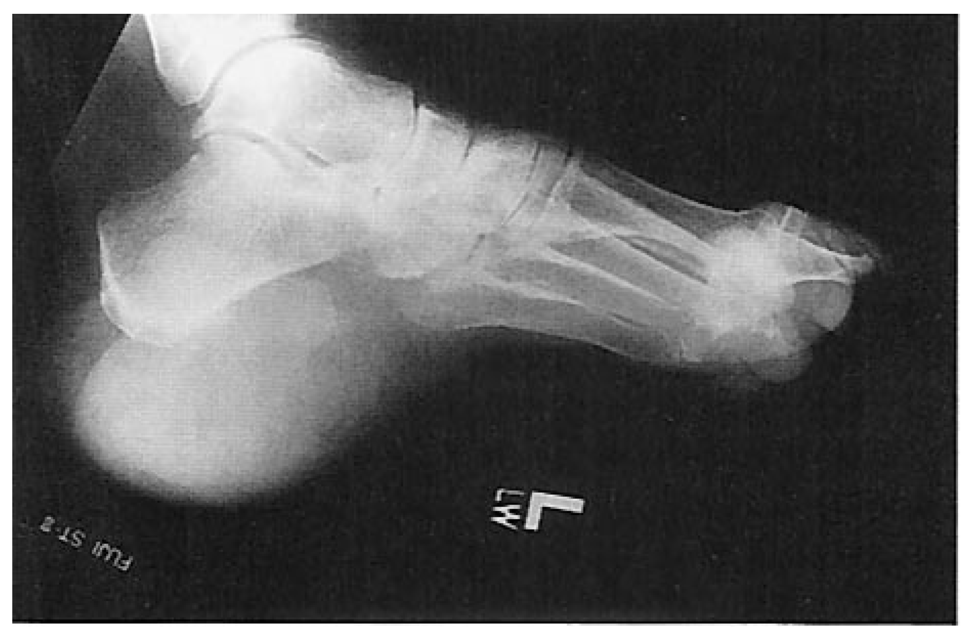
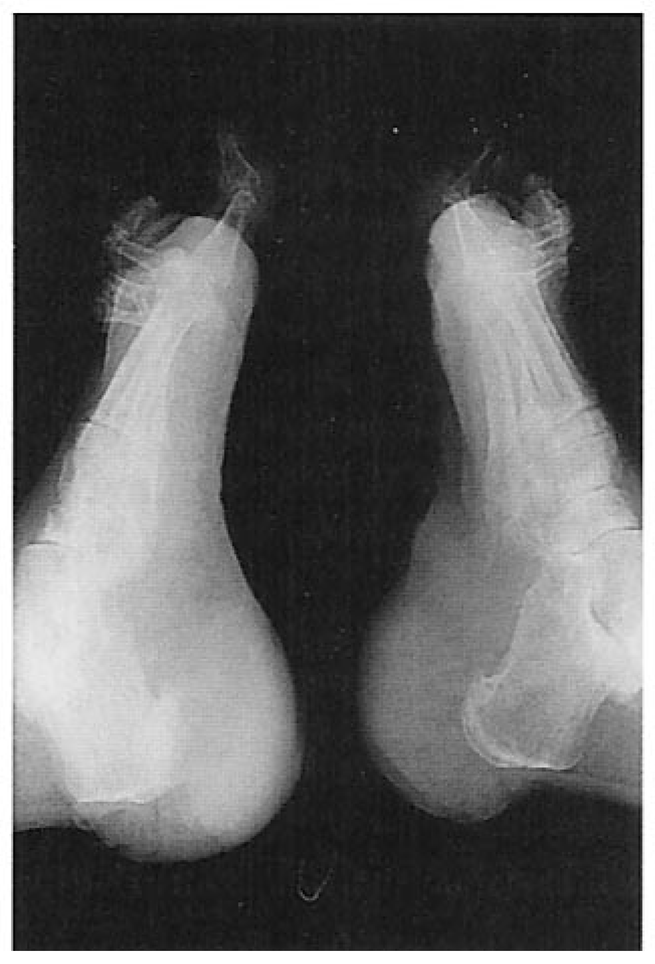
Plain x-rays showed a fluffy periosteal reaction on the plantar aspect of the left calcaneus (
Fig. 3). The grossly enlarged rheumatoid arthritis nodules were visible as the outline of the soft tissue exceeded all normal ranges (
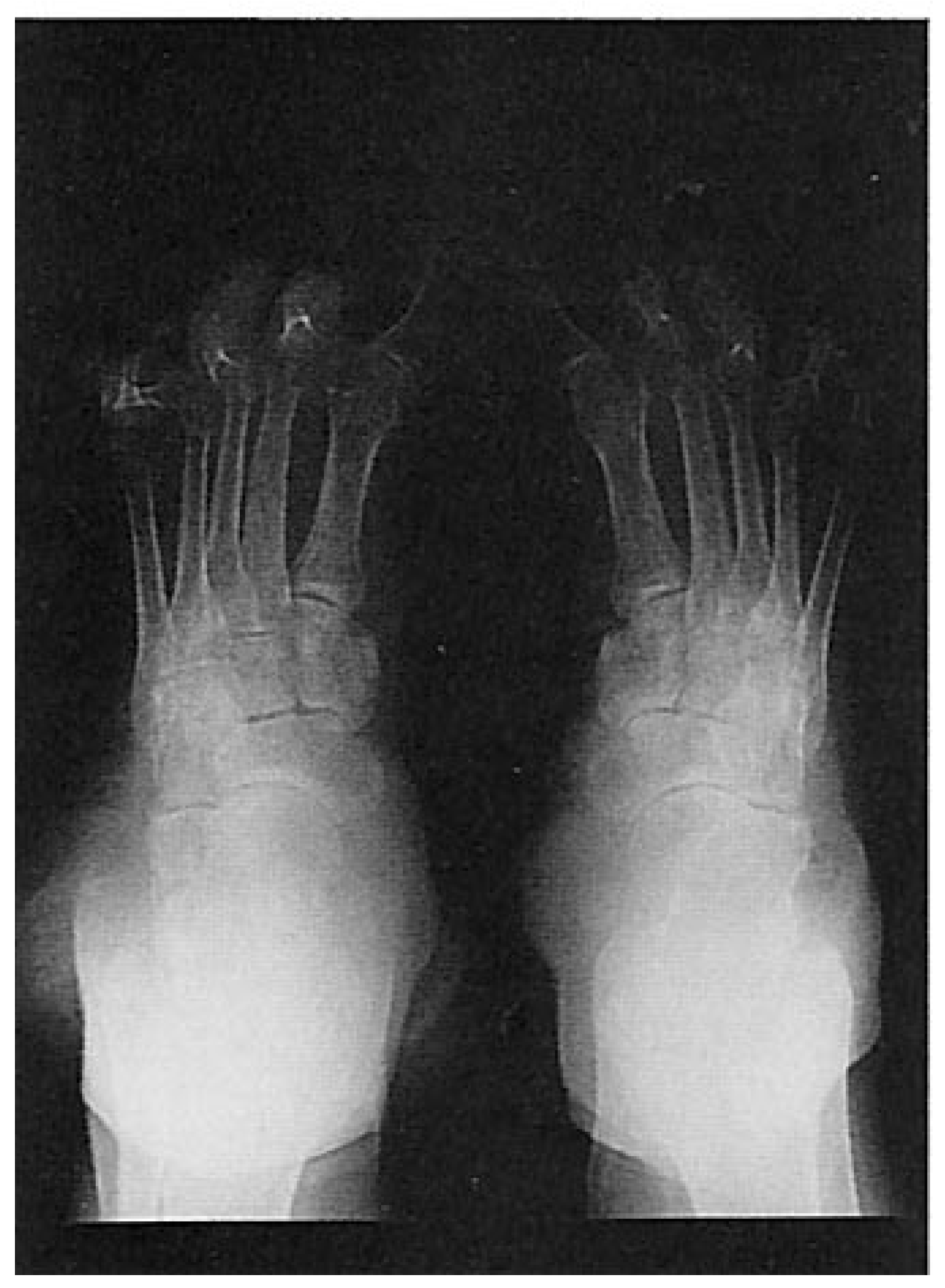
Fig. 4). There was extreme subluxation of the metatarsophalangeal joints (
Fig. 5).
The assessment was severe rheumatoid arthritis nodules that were symptomatic, unsightly, inhibiting walking and use of shoes, and at risk for osteomyelitis. The treatment was to surgically debulk and remodel both heels and to debride any infected bone.
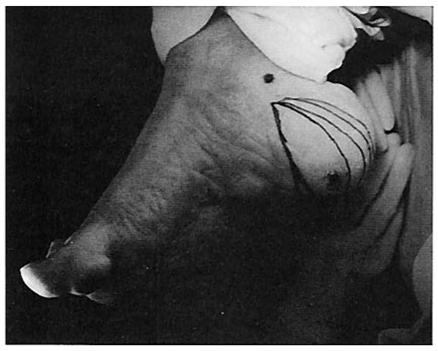
The patient was taken to surgery. Both heels were operated on simultaneously where fusiform incisions were made extending from lateral to medial across the posterior plantar heel flap and across the middle of the plantar aspect of the heel pad. Each incision was approximately 15 cm long (
Fig. 6). The skin wedges were removed
in toto with several large rheumatoid arthritis nodules intact. The dissection was carried down to the fascia on the plantar aspect of the calcaneal tubercles.
The amount of soft tissue removed was significant. All the fragments of soft tissue measured in aggregate from the right heel was 11.5 × 8.0 × 4.0 cm and from the left heel 11.5 × 10.0 × 7.0 cm. A fragment of dense bone from the left heel measured 4.0 × 3.5 × 1.2 cm. The calcanei were inspected and cultured for aerobic, anaerobic, and fungi along with a Gram’s stain. Both wounds were then irrigated with jet lavage. Drains were inserted and the wounds were reapproximated with 3-0 suture for subcutaneous and 3-0 nylon for skin in AO retention suture fashion.
An infectious disease consult was obtained after the cultures were positive indicating the growth of Acinetobacter calcoaceticus (a rare enterococcus). The patient was put on a course of vancomycin and metronidazole for 6 weeks.
The patient continued to use a wheelchair for 2 months postoperatively. This was because her right heel incision failed to unite and continued to drain serosanguinous fluid (
Fig. 7). Repeat triphasic technetium bone scans showed increased uptake on delayed images of both heels. She was then prescribed oral cephradine after a consultation with infectious disease.
Three months later, the patient’s foot was surgically debrided of the wound edges, irrigation, and primary closure of the right heel. After 3 weeks of weekly dressing changes and nonweightbearing, the incision remained closed without any signs of infection. Cultures of the incisions continued to grow A. calcoaceticus. The patient remained on cephradine for 2 weeks and continued dressing changes until the wound healed.
The patient’s left heel healed without complication. She was able to walk on the left foot without pain and wear a custom-molded shoe on that foot. The right heel temporarily healed and later reulcer ated without drainage. The patient continued dressing changes until the ulcer healed. She has minimal pain and is able to walk with molded shoes.
Summary
The presence of subcutaneous nodules in association with rheumatoid arthritis is well documented. In most cases, these nodules occur in association with severe rheumatoid disease. Treatment should be initiated with conservative measures such as custom-molded shoes, nonweightbearing, and oral antibiotic therapy to control infection. The goals of surgery were to alleviate pain, improve function and cosmesis, remove infected bone, and prevent further infection. The surgical sites are completely healed without complications 2 years postoperatively.