Stress fractures are relatively common injuries among athletes, comprising approximately 5% of all sports injuries [
1]. Fitch et al [
2] describe patients who gave a history of insidious onset of vague pain along the dorsum of the foot and ankle or the medial longitudinal arch that is exacerbated by activity and relieved by rest [
3,
4].
Initial treatment of tarsal navicular pain is the conventional management of stress-related bone injuries by avoiding causative stresses, rest with or without a cast, pain control, and the correction of any biomechanical factors. Fitness is maintained by nonweightbearing, and aerobic and anaerobic activity [
2]. Treatment of navicular stress fractures requires 6 weeks in a nonweightbearing cast. If after cast removal there is still tenderness elicited on palpation of the fracture site, then reimmobilization in a nonweightbearing cast is used for an additional 2 weeks. This regimen is followed until there is no tenderness on palpation of the navicular. After 8 to 10 weeks with no radiologic evidence of osseous healing, bone stimulators may be considered.
Although delayed unions and nonunions of stress fractures are relatively rare in athletes, they seem to be on the increase, possibly because of the gradual intensification of training programs at all ages. In many cases, diagnosis is difficult and repeated clinical, radiologic, and isotope examinations are necessary. Stress fractures at risk sites must be kept under careful observation in order to ensure adequate primary healing [
6]. Displacement and nonunion are complications that indicate operative therapy [
2,
6].
There is widespread awareness as to the use of magnetic resonance imaging in evaluating the knee, hip, spine, and other musculoskeletal disorders. Its use in the assessment of foot and ankle disorders is less well known. The intrinsically high soft tissue contrast, the ability to manipulate tissue contrast, and the high sensitivity to marrow-based pathologic conditions give magnetic resonance imaging significant advantages over other imaging techniques [
7].
Previous studies used tomography to make the diagnosis of stress fractures after radiographs and bone scans were performed, but the advent of magnetic resonance imaging has superseded conventional tomography and is now the most valuable aid in diagnosis and assessment.
In a study done by Beltran et al [
8], 85 hips in 49 patients were studied with magnetic resonance imaging, bone scan, and bone marrow pressure. It showed magnetic resonance imaging to be superior to bone scan and bone marrow pressure for the diagnosis of avascular necrosis. Magnetic resonance imaging had a sensitivity of 88.8%, and a specificity of 100%. Bone scan had a sensitivity of 77.7% and a specificity of 75%.
The hallmark of osteonecrosis on magnetic resonance imaging appears to be a focal region of homogenous or nonhomogeneous decrease signal intensity in a subarticular location [
7]. An amorphous pattern of low signal with T1 weighting and high signal with T2 weighting is usually seen in adjacent marrow space and soft tissue [
9]. On magnetic resonance imaging, nondisplaced fractures and stress fractures appear as linear regions of low signal intensity that extend to a cortical margin. Linear regions of high signal intensity may be seen on T2-weighted images.
Plain radiographic changes occur late in the course of the disease; therefore, stress fractures of the tarsal navicular may be overlooked on the initial evaluation. There seems to be an increased correlation with structural findings such as a short first metatarsal, metatarsal adductus, and plantar ward displacement of the talus and navicular with the cuneiforms [
10]. These radiographic findings, along with clinical history and physical examinations, should alert the physician to the possibility of stress fracture. It is the authors’ belief that when radiographs are equivocal, a bone scan should be performed. If the bone scan is positive, magnetic resonance imaging should be done to correlate any clinical suspicions.
Case Report
A 20-year-old male competitive pole vaulter presented to the Sports Medicine Clinic complaining of anteromedial left foot pain of 3 months’ duration. At the time of onset, the patient was training for the upcoming track season. Training consisted of running, sprinting, and pulling up to 100 pounds on a sled. His pain was exacerbated by these activities and relieved by rest. Rest, ice, accommodative strapping, and ultrasound did not improve his symptoms. He returned 2 months later and was also complaining of similar right foot pain. A physical examination showed no evidence of ecchymosis or edema. Pain was localized to the navicular bone bilaterally. Forefoot varus was noted with the left worse than the right. A pronatory gait was also seen on evaluation.
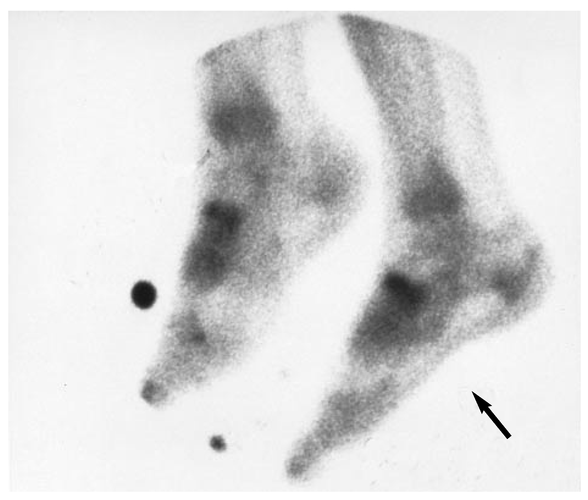
X-rays showed the navicular bones to be slightly sclerotic compared with the adjacent tarsal bones. They revealed a metatarsal protrusion distance of a positive 2 mm bilateral. His metatarsus adductus angle was 25° right and 22° left. Bone scans showed an increased uptake at both navicular bones (
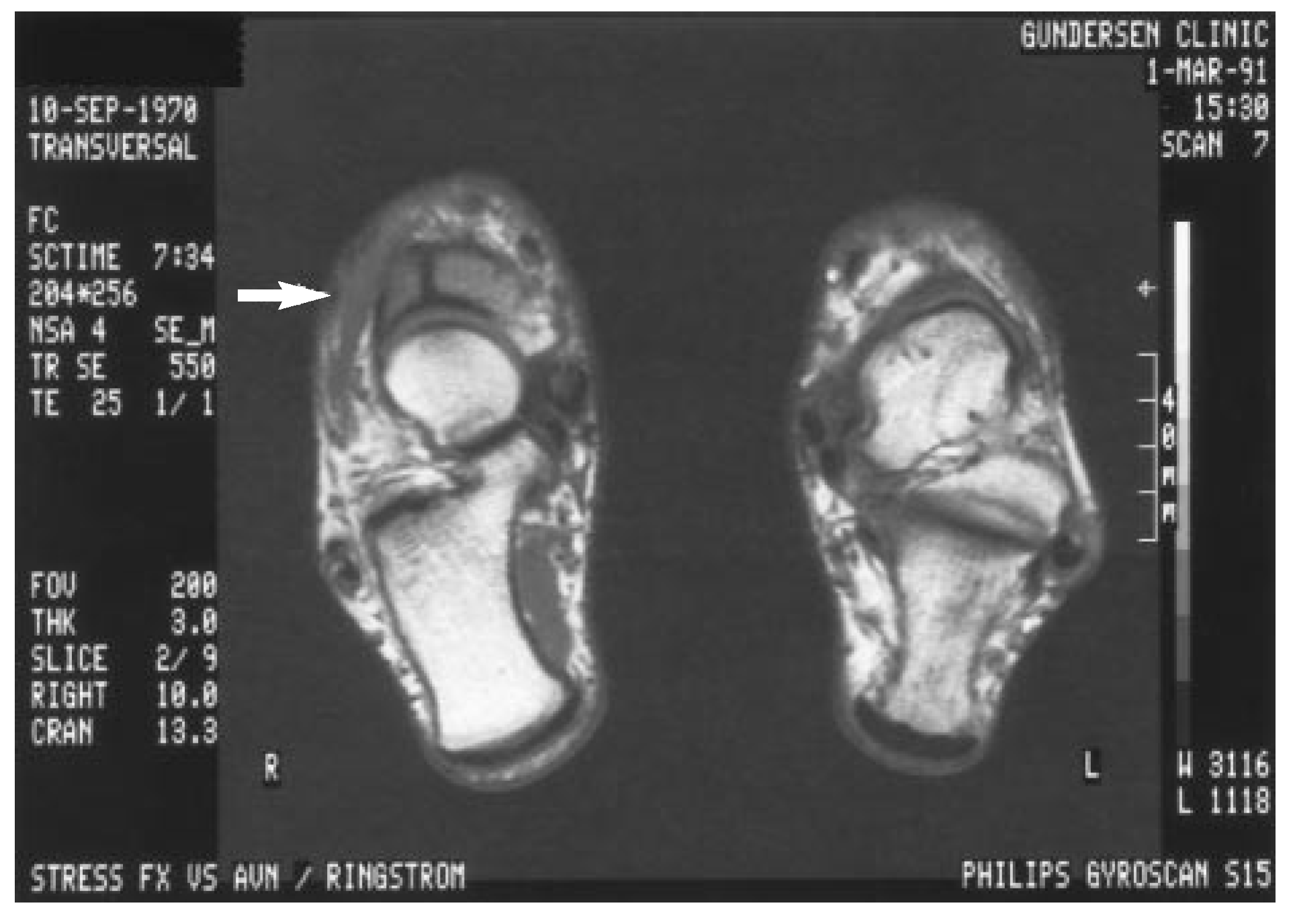
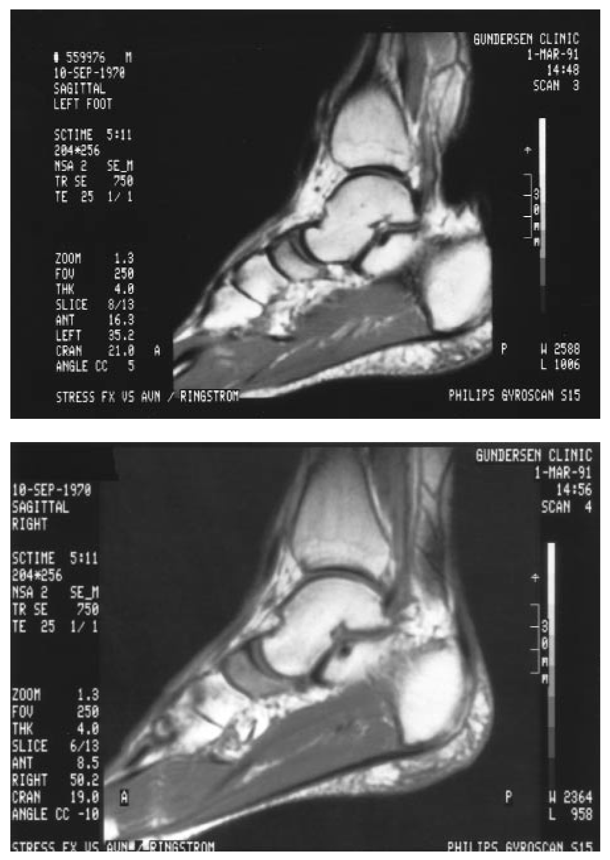
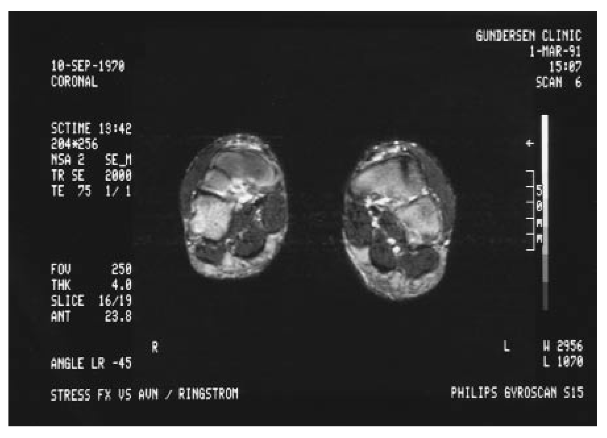
Fig. 1). A magnetic resonance imaging scan was performed, which showed bilateral tarsal navicular stress fractures with associated avascular necrosis (
Figs. 2–5). Complete rest and immobilization were recommended, but the patient wanted to compete in a national track meet 2 weeks later.
Figure 1.
Bone scan of both feet showing increased uptake in the tarsal region.
Figure 1.
Bone scan of both feet showing increased uptake in the tarsal region.
Figure 2.
T1 transversal image showing right navicular fracture and decreased signal activity in tarsal navicular.
Figure 2.
T1 transversal image showing right navicular fracture and decreased signal activity in tarsal navicular.
Figure 3.
Coronal magnetic resonance imaging scan showing left navicular fracture and decreased signal activity in tarsal navicular.
Figure 3.
Coronal magnetic resonance imaging scan showing left navicular fracture and decreased signal activity in tarsal navicular.
Figures 4 and 5.
Sagittal views of right and left feet show decreased signal intensity of both tarsal naviculars.
Figures 4 and 5.
Sagittal views of right and left feet show decreased signal intensity of both tarsal naviculars.
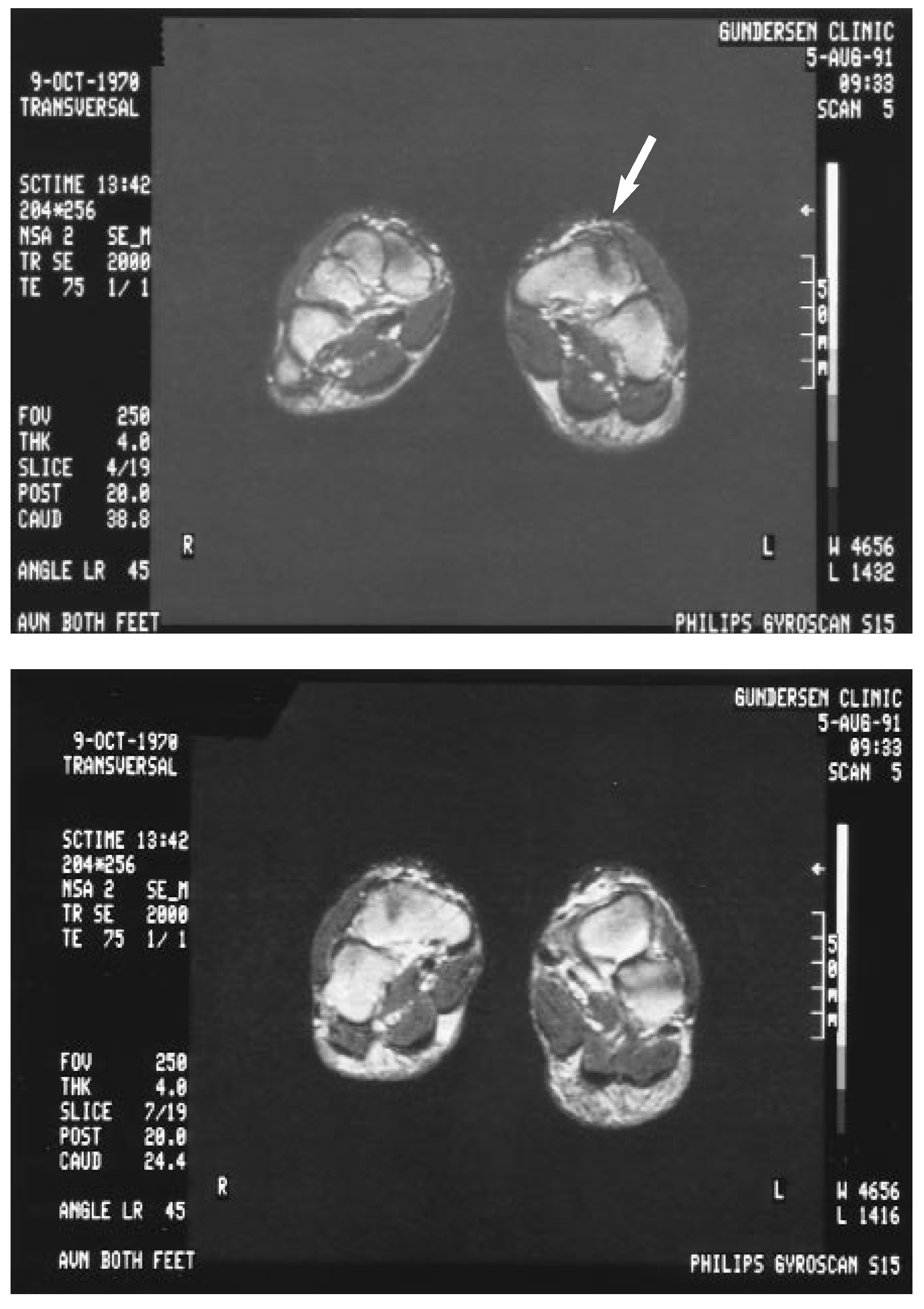
He competed in the meet and his symptoms increased dramatically following the competition. He was then placed in a walker for 4 weeks after which he was asymptomatic. Prescription orthoses were dispensed. A magnetic resonance imaging scan was performed 5 months later which showed improvement of the avascular necrosis, but the fracture lines were still evident (
Figs. 6 and 7). The orthoses gave him significant relief and conservative care was continued. At a collegiate level, the patient was deemed medically ineligible until healing was complete. The patient was offered continued conservative treatment
versus surgical intervention; he chose surgery.
Figures 6 and 7.
Postoperative computed tomography scan 11 months after surgery on right foot and 5 months after surgery on the left foot.
Figures 6 and 7.
Postoperative computed tomography scan 11 months after surgery on right foot and 5 months after surgery on the left foot.
Under general anesthesia, an 8-cm lazy S incision was made over the dorsal aspect of the right navicular from proximal to distal. Vital structures were identified and retracted medially. A transverse periosteal incision was made. The fracture was identified in the lateral one third of the navicular. A dorsal cortical table measuring 6 mm on each side of the fracture and 8 mm from proximal to distal was removed with an oscillating saw and osteotome. Vascular inflow was absent. Approximately 7 to 8 mm3 of cancellous bone was resected with a burr until vascular inflow was noted. An autogenous bone graft from the iliac crest was then packed into the defect. The periosteum was closed over the graft. The cortical tables were not replaced to allow for more vascular ingrowth. The wound was closed in layers and the patient was placed in a below the knee nonweightbearing cast for 6 weeks.
He began using a walker with gradual increase in activity over a 6-week period. Regular shoes were begun at 12 weeks. The patient continued to have pain involving the left foot and wanted surgical intervention. The same procedure was performed on the left foot 7 months later. Excellent relief was obtained from both surgeries. He returned to sports activities and is asymptomatic.
Discussion
Tarsal navicular stress fractures often occur in explosive athletic activities that involve sprinting, jumping, and hurdling [
1,
2,
3,
11]. They have also been reported in mid-distance and long-distance runners and figure skaters. These fractures occur in the sagittal plane and in the middle one third of the bone [
2,
3,
5,
12]. In a study of 78 stress fractures of the navicular by Khan13, he found that with two exceptions, the fractures were in the sagittal plane and involved the central third of the navicular bone.
Microangiograms of fresh cadaver feet showed the tarsal navicular is supplied by small branches of the posterior tibial and anterior tibial arteries. Vessels enter the navicular throughout its nonarticular surfaces and radiate to the center of the bone. Most of the navicular is covered with articular cartilage; therefore, there is only a small area for vessels to enter and leave bone. Transverse sections show that medial and lateral vessels supply the medial and lateral one third of the navicular leaving the center one third relatively avascular [
3].
Short first metatarsals or long second metatarsals were common findings in a series done by Fitch et al [
2] and Pavlov et al [
10]. The consistent site of the fracture in the lateral one half of the bone appears to correspond with the plane of maximum stress, especially during plantarflexion combined with pronation. This would tend to accentuate shear stress because of the greater force being transmitted through the second metatarsal and intermediate cuneiform. This would be increased by metatarsal adduction and pronation [
2].
Pecina et al [
1] found this to be the case with skaters who performed intensive jumping exercises on the inner edge of their skates.
Fitch et al [
2] found that the take-off leg was always the site of fracture. It is the authors’ belief that these stress fractures were a direct result of the patient’s training regimen whereby he would pull a sled loaded with up to 100 pounds. This put excessive stress and pressure on the balls of his feet and eventually led to his stress fracture. The patient’s active lifestyle, competitive nature, and the fact that he did not seek treatment until 3 months after the onset of symptoms led to his avascular necrosis, nonunion, and eventual surgical intervention. The authors found the series by Fitch et al [
2] where they grafted 19 fractures in 18 patients, to be the most acceptable means of surgical correction. Because the navicular is rigidly stabilized by its location in the foot, the extensive network of plantar and dorsal ligaments, and the fact that the size of the navicular does not lend itself to the use of large screws, the authors believe internal fixation is not necessary.
The patient resumed training 3 months after his second surgery. This consisted of biking, leg curls, upper-body weight training, and using a rowing machine. At 5 months, he resumed running. He developed Achilles tendinitis 1 month after he started running. This was resolved with appropriate conservative treatment. Training consisted of multiaxial strengthening and range-of-motion exercises, using a stair-climbing machine, and vaulting 1 day a week and at competitions. The patient went on to complete the entire spring track season of his senior year.
Conclusion
A rare case of bilateral navicular stress fractures has been presented. Early literature suggests the use of tomograms along with standard radiographs and bone scans. Studies have shown magnetic resonance imaging to be superior in the diagnosis of avascular necrosis and in identifying fractures.
Highly motivated athletes continue to train in spite of pain, making early diagnosis difficult. Conservative measures consisting of decreasing activity and immobilization have proven beneficial. Early detection with magnetic resonance imaging will allow conservative treatment to be effective and decrease the risk of avascular necrosis, delayed or nonunion, complete fracture, and the need for surgical intervention.