Case Report

Bidirectional Limb Salvage and Its Impact on Residual Limb Survivorship: Is a Partial Calcanectomy in Ambulatory Midfoot Amputees Worthwhile?

1
Department of Plastic and Reconstructive Surgery, MedStar Georgetown University Hospital, Washington, DC 20007, USA
2
School of Medicine, University of Maryland, Baltimore, MD 21201, USA
3
Department of Plastic Surgery, Georgetown University School of Medicine, Washington, DC 20007, USA
*
Author to whom correspondence should be addressed.
J. Am. Podiatr. Med. Assoc. 2026, 116(4), 56; https://doi.org/10.3390/japma116040056
Submission received: 28 March 2023 / Revised: 10 March 2024 / Accepted: 20 March 2024 / Published: 21 August 2026

Abstract

Partial calcanectomies and midfoot amputation are commonly used functional limb salvaging interventions; however, it is well documented that they independently result in global biomechanical disruption of diabetic foot, further placing the residual limb at risk for limb loss. We retrospectively identified and reviewed 9 ambulatory midfoot amputees (10 feet) who developed heel osteomyelitis, whom were treated with an ipsilateral partial calcanectomy to determine whether the combined anterior and posterior interventions result in additive dysfunction that impacts function or places the residual pedal construct at risk for limb loss.

1. Introduction

Midfoot amputation is a commonly used limb salvage intervention that effectively treats infection, ischemia, and non-healing ulcerations of the forefoot; however, it is well documented that they result in global biomechanical disruption of diabetic foot, further placing the residual limb at risk for limb loss [1,2,3,4,5]. As a result, transfer lesions are common and the creation of a short lever arm for ambulation places patients at increased risk of heel ulceration and increases the rate of development of heel ulceration after midfoot amputation, which can be as high as 47% [6,7,8]. In the setting of calcaneal osteomyelitis, which has the largest amount of morbidity and even mortality associated with its development, the partial calcanectomy (PC) is a well-documented intervention; however, not much is known on whether or not a PC in an already biomechanically compromised and shortened amputated foot increases the risk of limb loss [9,10,11,12,13,14,15,16,17].
Given the biomechanical changes that are independently associated with both midfoot amputation and partial calcanectomy, we seek to determine if the additive and cumulative biomechanical disruption seen in bidirectional limb salvage results in increased rates of failure, loss of ambulatory status, and complications, short and long term.

2. What We Observed

After Institutional Review Board (IRB) approval, we identified nine patients with 10 feet who had Current Procedural Terminology (CPT) code 28120 (partial excision of calcaneus) and 28805 (transmetatarsal amputation) or 28800 (midtarsal amputation) at Georgetown University Hospital. Average age and body mass index (BMI) was 53 years (range, 30 to 66 years) and 28.3 kg/m2 (range, 21.64 to 39.44). Eight patients (88.9%) had diabetes mellitus, five (55.6%) had chronic kidney disease, seven (77.8%) had peripheral arterial disease, and two (22.2%) were active smokers. The average Charlson index, which predicts 10-year survival of patients with multiple comorbidities, was 5.4 (range, 2–10). With respect to level of midfoot amputation, 2/10 feet (20%) were amputated at the Chopart level, 3/10 (30%) at the Lisfranc level, and 5/10 (50%) were at the transmetatarsal level. With respect to laboratory data, the average hemoglobin A1c at the time of the most recent intervention (calcanectomy) was 8.2%.
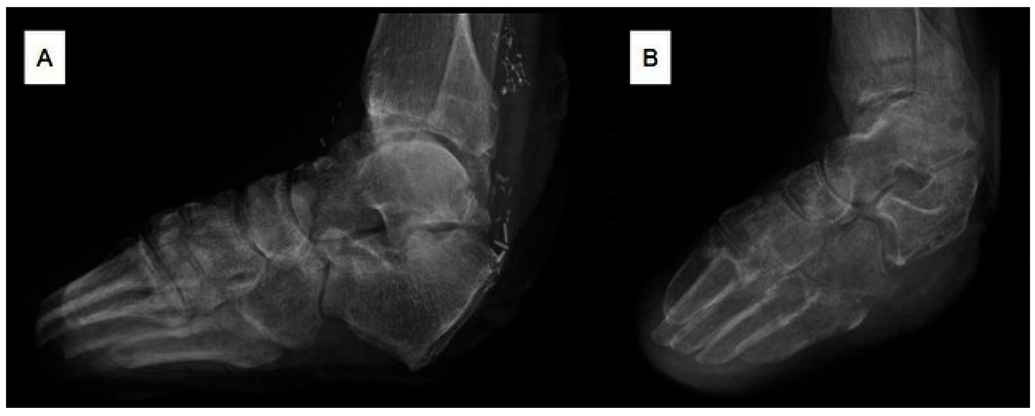
All patients underwent subsequent ipsilateral partial calcanectomy, consisting of traditional calcanectomy or vertical contour calcanectomy (Figure 1), with an average of 24.9 months between index midfoot amputation and partial calcanectomy. All complications and failures were attributed to the poor healing of the heel wound as opposed to failure of the midfoot amputation stump. The only minor complication requiring intervention was dehiscence in two (20%) feet and secondary below-knee amputation (BKA) was performed in two limbs (20%). When evaluating the patients who failed bidirectional interventions, Patients #5 and #9, the average time until BKA was 210 days, average hemoglobin A1c was 6.1%, and average BMI of 7.0 kg/m2. Demographically, both had diabetes and peripheral vascular disease. Patient #5 had a previous history of chronic heart failure and was not a smoker. Patient #9 was a current smoker but did not demonstrate any other serious comorbidities beyond treated hypertension.
At a mean follow-up of 19.5 months (594 days) after the calcanectomy was performed, three (33%) of the previously ambulatory patients were no longer ambulatory at the most recent follow-up, with all three patients who experienced loss of ambulation being patients who underwent bidirectional limb salvage. Of the 66.7% (6/9) of patients who maintained the ability to ambulate, 33.3% (2/6) were BKA patients who ambulated with the assistance of a prosthetic and 66.7% (4/6) were patients who underwent bidirectional limb salvage. Patient demographics are outlined in Table 1.

3. Discussion

In our observation, we posit that bidirectional limb salvage in the bidirectionally threatened limb is certainly possible and worthwhile in the properly selected patient. Unfortunately, when the interventions are performed on the same foot, there does appear to be some consequences with roughly 42.9% (3/7) of our successful cases of bidirectional limb salvage losing their ability to independently ambulate in the postoperative course. Despite the unfortunate loss of ambulation in patients, our findings are comparable to the isolated midfoot amputation and calcanectomy literature with respect to ambulation rates and limb salvage rates [15,18].
In a study in 367 patients by Ordaz et al., the authors found that 56.8% of transmetatarsal amputees were ambulatory after an average of 181 days [18]. In a study by Cates et al., 69% of partial calcanectomy patients were ambulatory at the 1-year mark [15]. The partial calcanectomy and the transmetatarsal amputation are considered to be the most gait preserving amputation levels, which was demonstrated in an article by Brown et al. [19]. In our observation, 3/4 (75%) transmetatarsal, 2/2 (100%) Lisfranc, and 2/3 (67%) of Chopart amputations were ambulatory in the acute postoperative course but one Lisfranc and one Chopart went on to receive a below knee amputation after 162 and 258 days for recurrent infection of heel, respectively. After a follow-up of 19.5 months, there was a 100% (5/5) salvage rate with transmetatarsal, 66% (2/3) salvage rate with Lisfranc, and 50% (1/2) for Chopart amputations. Given the well-documented biomechanical disruption that is associated with Lisfranc and even more so with Chopart amputation, an argument can be made that significant reduction in the pedal lever arm length along with associated loss of vital soft tissue structures places the calcaneal intervention at increased risk for failure due to the relocation to and concentration of plantar pressures on the heel; however, our conclusions warrant further investigation. In our observation, none of the patients developed an equinovarus deformity; however, this may be as a result of the prophylactic release of problematic posterior and medial structures when indicated.
The heel soft tissue envelope is notoriously durable when intact; however, when the envelope is compromised, healing is often difficult despite being perfused by multiple inflow vessels [20]. In our observation, five patients had no three-vessel runoff, two had two-vessel runoff, and one had three-vessel runoff. Unfortunately, none of the five patients with zero-vessel runoff had vessels that were amenable to revascularization but were deemed to have sufficient collateralization. Not surprisingly, the two patients who failed had zero-vessel runoff, which placed them at increased risk for failure. While not always possible, when possible, Cates et al. concluded that the most important vessel for calcanectomy success was the posterior tibial artery; therefore, this artery should be targeted when possible [21].
In the end, with an 80% limb salvage rate and 57.1% rate of ambulation, we propose that bidirectional limb salvage is most appropriate in patients that seek to maintain or improve ambulation and are candidates for the procedure. While the argument can be made that a below the knee amputation is the ideal level of amputation for a patient who seeks to maintain functionality, the previously mentioned study by Ordaz et al. showed that only 30.9% of patients who underwent a below knee amputation maintained gait preservation at an average follow-up of 315 days—making the bidirectional approach certainly worthwhile [18].
While we believe our observations are valuable, our evaluations do have their own inherent limitations. While the development of heel ulcerations after midfoot amputation is relatively common, performing a partial calcanectomy in a midfoot amputee is not ubiquitous, further making large-scale studies and comparisons difficult. Furthermore, it is not clear whether bidirectional limb salvage is superior to the alternative, which is a BKA. While the ambulatory rates of isolated TMAs and calcanectomies are acceptable, it does appear that the addictive effect of the procedures do have their own inherent consequences.

4. Conclusions

In conclusion, the findings of this interesting observation reveal an acceptable rate of limb survivorship and preservation of ambulatory status in patients who underwent bidirectional limb salvage, which is relatively comparable to the independent midfoot amputation and partial calcanectomy literature; however, these findings should raise concern regarding expectations of salvage and ambulation in patients with midfoot amputation requiring subsequent partial calcanectomy all in an effort to reduce morbidity and mortality in this at-risk patient population.

Author Contributions

Conceptualization, C.J.V., N.K.C., K.K.E., C.E.A., J.S.S. and J.N.A.; methodology, C.J.V., N.K.C., H.D.S., K.K.E., C.E.A., J.S.S. and J.N.A.; investigation, C.J.V., N.K.C., H.D.S., K.K.E., C.E.A., J.S.S. and J.N.A.; writing—original draft preparation, C.J.V., H.D.S., K.K.E., C.E.A., J.S.S. and J.N.A.; supervision, K.K.E., C.E.A., J.S.S. and J.N.A. All authors have read and agreed to the published version of the manuscript.

Funding

This research received no external funding.

Institutional Review Board Statement

The study was conducted in accordance with the Declaration of Helsinki and approved by the Institutional Review Board (IRB # 050. 117). Approved 21 August 2022.

Informed Consent Statement

Informed consent was obtained from all subjects involved in the study.

Data Availability Statement

No new data were created or analyzed in this study. Data sharing is not applicable to this article.

Conflicts of Interest

The authors declare no conflicts of interest.

References

  1. Zambetti, B.R.; Stiles, Z.E.; Gupta, P.K.; Stickley, S.M.; Brahmbhatt, R.; Rohrer, M.J.; Kempe, K. Present-day analysis of early failure after forefoot amputation. Surgery 2020, 168, 904–908. [Google Scholar] [CrossRef] [Scilit] [PubMed]
  2. Beaulieu, R.J.; Grimm, J.C.; Lyu, H.; Abularrage, C.J.; Perler, B.A. Rates and predictors of readmission after minor lower extremity amputations. J. Vasc. Surg. 2015, 62, 101–105. [Google Scholar] [CrossRef] [Scilit] [PubMed]
  3. Tokarski, A.R.; Barton, E.C.; Wagner, J.T.; Elliott, A.D.; Simonson, D.C.; Hordyk, P.J.; Rademaker, M. Are Transmetatarsal Amputations a Durable Limb Salvage Option? A Single-Institution Descriptive Analysis. J. Foot Ankle Surg. 2022, 61, 537–541. [Google Scholar] [CrossRef] [Scilit] [PubMed]
  4. Kim, P.J. Biomechanics of the Diabetic Foot: Consideration in Limb Salvage. Adv. Wound Care 2013, 2, 107–111. [Google Scholar] [CrossRef] [Scilit] [PubMed]
  5. Labovitz, J.M.; Day, D. The Biomechanics of Diabetes Mellitus and Limb Preservation. Clin. Podiatr. Med. Surg. 2020, 37, 151–169. [Google Scholar] [CrossRef] [Scilit] [PubMed]
  6. Pollard, J.; Hamilton, G.A.; Rush, S.M.; Ford, L.A. Mortality and morbidity after transmetatarsal amputation: Retrospective review of 101 cases. J. Foot Ankle Surg. 2006, 45, 91–97. [Google Scholar] [CrossRef] [Scilit] [PubMed]
  7. La Fontaine, J.; Brown, D.; Adams, M.; VanPelt, M. New and recurrent ulcerations after percutaneous achilles tendon lengthening in transmetatarsal amputation. J. Foot Ankle Surg. 2008, 47, 225–229. [Google Scholar] [CrossRef] [Scilit] [PubMed]
  8. Holstein, P.; Lohmann, M.; Bitsch, M.; Jørgensen, B. Achilles tendon lengthening, the panacea for plantar forefoot ulceration? Diabetes Metab. Res. Rev. 2004, 20, S37–S40. [Google Scholar] [CrossRef] [Scilit] [PubMed]
  9. Faglia, E.; Clerici, G.; Caminiti, M.; Curci, V.; Somalvico, F. Influence of osteomyelitis location in the foot of diabetic patients with transtibial amputation. Foot Ankle Int. 2013, 34, 222–227. [Google Scholar] [CrossRef] [Scilit] [PubMed]
  10. Mohapatra, A.; Henry, J.C.; Avgerinos, E.D.; Chaer, R.A.; Leers, S.A.; Boitet, A.; Singh, M.J.; Hager, E.S. Heel Wounds Predict Mortality but Not Amputation after Infrapopliteal Revascularization. Ann. Vasc. Surg. 2018, 51, 78–85. [Google Scholar] [CrossRef] [Scilit] [PubMed]
  11. Gaenslen, F. Split-heel approach in osteomyelitis of os calcis. J. Bone Jt. Surg. 1931, 13, 759–772. [Google Scholar]
  12. Bollinger, M.; Thordarson, D.B. Partial calcanectomy: An alternative to below knee amputation. Foot Ankle Int. 2002, 23, 927–932. [Google Scholar] [CrossRef] [Scilit] [PubMed]
  13. Cook, J.; Cook, E.; Landsman, A.S.; Basile, P.; Dinh, T.; Lyons, T.; Rosenblum, B.; Giurini, J. A retrospective assessment of partial calcanectomies and factors influencing postoperative course. J. Foot Ankle Surg. 2007, 46, 248–255. [Google Scholar] [CrossRef] [Scilit] [PubMed]
  14. Elmarsafi, T.; Pierre, A.J.; Wang, K.; Evans, K.K.; Attinger, C.E.; Kim, P.J.; Steinberg, J.S. The Vertical Contour Calcanectomy: An Alternative Surgical Technique to the Conventional Partial Calcanectomy. J. Foot Ankle Surg. 2019, 58, 381–386. [Google Scholar] [CrossRef] [Scilit] [PubMed]
  15. Cates, N.K.; Wang, K.; Stowers, J.M.; Attinger, C.E.; Kim, P.J.; Steinberg, J.S. The Vertical Contour Calcanectomy, an Alternative Approach to Surgical Heel Ulcers: A Case Series. J. Foot Ankle Surg. 2019, 58, 1067–1071. [Google Scholar] [CrossRef] [Scilit] [PubMed]
  16. Cook, H.; Kennedy, C.; Delijani, K.; Popovsky, D.; Elmarsafi, T.; Zarick, C.; Attinger, C.; Steinberg, J. Early Clinical, Functional, and Mortality Outcomes for Heel Ulcers Treated with a Vertical Contour Calcanectomy. J. Foot Ankle Surg. 2022, 61, 117–122. [Google Scholar] [CrossRef] [Scilit] [PubMed]
  17. Cook, H.R.; Cates, N.K.; Kennedy, C.J.; Tefera, E.; Popovsky, D.; Delijani, K.; Kim, P.J.; Attinger, C.E.; Steinberg, J.S. Risks Factors Associated with Major Lower Extremity Amputation After Vertical Contour Calcanectomy. J. Foot Ankle Surg. 2022, 61, 1046–1051. [Google Scholar] [CrossRef] [Scilit] [PubMed]
  18. Ordaz, A.; Trimm, C.; Pedowitz, J.; Foran, I.M. Transmetatarsal Amputation Results in Higher Frequency of Revision Surgery and Higher Ambulation Rates Than Below-Knee Amputation. Foot Ankle Orthop. 2022, 7, 24730114221112938. [Google Scholar] [CrossRef] [Scilit] [PubMed]
  19. Brown, M.L.; Tang, W.; Patel, A.; Baumhauer, J.F. Partial foot amputation in patients with diabetic foot ulcers. Foot Ankle Int. 2012, 33, 707–716. [Google Scholar] [CrossRef] [Scilit] [PubMed]
  20. Faglia, E.; Clerici, G.; Caminiti, M.; Vincenzo, C.; Cetta, F. Heel ulcer and blood flow: The importance of the angiosome concept. Int. J. Low. Extrem. Wounds 2013, 12, 226–230. [Google Scholar] [PubMed]
  21. Cates, N.K.; Pandya, M.; Salerno, N.D.; Akbari, C.M.; Zarick, C.S.; Raspovic, K.M.; Evans, K.K.; Kim, P.J.; Steinberg, J.S.; Attinger, C.E. Evaluation of Peripheral Perfusion in the Presence of Plantar Heel Ulcerations Status After Transmetatarsal Amputation with Achilles Tendon Lengthening. J. Foot Ankle Surg. 2020, 59, 892–897. [Google Scholar] [CrossRef] [Scilit] [PubMed]
Figure 1. Plain film radiographic examples of midfoot amputation (transmetatarsal) with partial calcanectomy (traditional calcanectomy (A) and vertical contour calcanectomy (B)).
Japma 116 00056 g001
Table 1. Summary of patient demographics.
PatientGenderAge at Time of Midfoot Amputation (Years)BMI (kg/m2)Level of AmputationLateralityTime to Calcanectomy (Days)Adjunct ProceduresAmbulatory Postoperatively?Proximal Amputation?Time to BKA (Days)F/u Duration (Days)Diabetic?Hemoglobin A1C (%)PVDCKDCCI
1Male6221.64TMALeft108NoneYesNoN/A1713No8.5YesNo5
1---TMARight53None-NoN/A---YesNo-
2Male6627.72TMALeft36NoneYesNoN/A83Yes7.2YesYes7
3Male3923.6TMARight2105NoneYesNoN/A442Yes12.7YesYes3
4Male6223.53TMARight98NoneNoNoN/A355Yes6.5YesNo10
5Female3939.44LisfrancLeft3044Tendoachilles lengtheningYesYes162187Yes4.1NoYes5
6Female3039.08LisfrancRight889Tibialis anterior tendon lengthening, Achilles tenotomy, peroneus brevis tendon transferNoNoN/A946Yes12.4NoNo2
7Male6024.43LisfrancRight480Achilles tenotomyYesNoN/A529Yes7.5YesYes6
8Male6329.16ChopartRight194Achilles tenotomyYesNoN/A360Yes6.4YesYes7
9Male5625.83ChopartLeft493Achilles tenotomyYesYes258731Yes8.1YesNo4
BKA: below knee amputation; BMI: body mass index; CCI: Charlson Comorbidity Index; CKD: chronic kidney disease; DM II: diabetes mellitus; F/u: follow-up; N/A: not applicable; PVD: peripheral vascular disease; TMA: transmetatarsal amputation.
Disclaimer/Publisher’s Note: The statements, opinions and data contained in all publications are solely those of the individual author(s) and contributor(s) and not of MDPI and/or the editor(s). MDPI and/or the editor(s) disclaim responsibility for any injury to people or property resulting from any ideas, methods, instructions or products referred to in the content.