Article

Early Versus Delayed Plate Fixation in Pilon Fractures—Which Is More Advantageous?


by
İsmail Güzel
1,*,
İbrahim Ulusoy
2,
Mehmet Yılmaz
3,
Mehmet Fırat Tantekin
2 and
Aybars Kıvrak
4
1
Department of Orthopedics and Traumatology, Faculty of Medicine Malatya Turgut Özal University, 44210 Malatya, Turkey
2
Department of Orthopedic Surgery, Selahhadin Eyyubi State Hospital, 21100 Diyarbakır, Turkey
3
Department of Orthopedic and Traumatology, Gaziantep Islamic Science and Technology University, 27010 Gaziantep, Turkey
4
Aybars Kıvrak Orthopedics Clinic, 21100 Adana, Turkey
*
Author to whom correspondence should be addressed.
J. Am. Podiatr. Med. Assoc. 2026, 116(2), 25087; https://doi.org/10.7547/25-087
Submission received: 30 April 2025 / Revised: 8 June 2025 / Accepted: 18 June 2025 / Published: 21 April 2026

Abstract

Background: Pilon fractures refer to distal tibial fractures that may involve extra-articular, partial articular, or complete intra-articular components, most commonly caused by high-energy trauma. The choice between early (<72 h) and delayed (>7 days) surgical fixation significantly impacts clinical outcomes. This study aimed to compare the effects of early vs. delayed plate fixation on fracture healing time, functional outcomes, and complication rates. Materials and Methods: This retrospective study analyzed 80 patients who underwent surgical treatment for pilon fractures between 2018 and 2023. Patients were divided into two groups: early surgery (<72 h, n = 40) and delayed surgery (>7 days, n = 40). Additionally, patients were categorized based on the fixation method: single-plate fixation (n = 40) and double-plate fixation (n = 40). Outcome measures included fracture healing time (weeks), defined as cortical continuity on radiographs; functional outcomes (AOFAS score); and complication rates (infection, malunion, and implant failure). Results: Shorter healing time was observed in the early surgery group (14.2 vs. 16.8 weeks, p < 0.05). Better functional outcomes were recorded in the early surgery group (AOFAS score: 82.3 ± 6.5 vs. 78.1 ± 7.2, p < 0.05). Lower infection rates were noted in the delayed surgery group (7.5% vs. 12.5%, p < 0.05). Double-plate fixation provided better mechanical stability but resulted in higher soft tissue complication rates. Single-plate fixation preserved soft tissue integrity but had higher malunion and implant failure rates. Conclusions: Early surgery is associated with shorter healing time and better functional outcomes, but increased soft tissue complications require careful management. Delayed surgery offers a safer approach for soft tissue healing but may prolong functional recovery. While double-plate fixation ensures greater stability, it may increase soft tissue morbidity, whereas single-plate fixation reduces soft tissue complications but may compromise stability. A personalized surgical approach is recommended for optimal outcomes in pilon fracture management.

1. Introduction

Pilon fractures encompass a spectrum of distal tibial fractures, ranging from extra-articular (43-A) to partial (43-B) and complete intra-articular (43-C) involvement, as classified by the AO/OTA system. They are most frequently caused by high-energy mechanisms such as falls, traffic accidents, and industrial accidents [1]. These fractures pose serious challenges in the treatment process because they often involve the articular surface and are frequently associated with significant soft tissue damage [2]. Treatment planning requires anatomical reduction of the fracture as well as optimization of wound healing by preserving soft tissue integrity [3,4]. Therefore, soft tissue status is as important as the type of fracture in treatment planning [4].
The most widely used systems for classification of pilon fractures are the AO/OTA fracture classification and Ruedi–Allgöwer classification. According to the AO/OTA system, fractures are grouped as extra-articular (43-A), partial intra-articular (43-B), and complete intra-articular (43-C) fractures [5]. The Ruedi–Allgöwer classification is divided into three groups according to the effect of the fracture on the articular surface and the degree of displacement. Type 1 fractures show minimal displacement, whereas type 3 fractures are characterized by severe joint destruction, and they are the most challenging cases for surgical treatment [6,7].
Surgical treatment of pilon fractures is based on the basic principles of anatomical reduction of the articular surface, provision of mechanical stability, and preservation of soft tissue integrity [8]. However, there is still controversy about the appropriate timing of surgery. Early surgical intervention (<72 h) may enable early stabilization of the fracture, resulting in faster union time and better functional outcomes [9]. However, early surgery may be disadvantageous in terms of soft tissue complications and infection risk [10]. Delayed surgery (>7 days) may reduce infection rates by allowing time for soft tissue healing but may prolong the union time and make joint reduction technically more difficult [11].
Methods used for surgical stabilization include open reduction and internal fixation (ORIF), minimally invasive plate osteosynthesis (MIPO), external fixation, and primary arthrodesis [12]. The most commonly preferred method is plate fixation, and this technique can be performed with single- or double-plate application. While double-plate fixation provides more mechanical stability, it may increase soft tissue complication rates. Single-plate fixation may be advantageous in terms of soft tissue protection but may be disadvantageous in terms of stability [13].
In this study, the effects of early and delayed plate fixation on clinical and radiologic outcomes were compared. We also evaluated the differences between single- and double-plate fixation in terms of functional outcomes and complication rates.

2. Materials and Methods

This study was a retrospective review of patients who underwent surgical treatment for a pilon fracture between 2018 and 2023. This study, designed as a single-center and comparative cohort study, aims to evaluate the clinical outcomes of early (<72 h) and delayed (>7 days) surgery. The study protocol was conducted in accordance with the Declaration of Helsinki, and approval was obtained from the İnönü University Clinical Research Ethics Committee (approval no. 2021/2483; approval date 22 December 2021).

2.1. Patient Selection and Grouping

This was a retrospective study, and patient allocation into early or delayed surgery groups was based on the clinical condition of the soft tissues at the time of admission. Early surgery (<72 h) was performed in patients without significant edema, skin compromise, or blistering. Delayed surgery was preferred in the presence of tense swelling, hemorrhagic or serous-filled blisters, or ecchymosis.
The decision between single or double plating was guided by preoperative CT scans and the intraoperative configuration of the fracture. Double plating was selected in cases of extensive medial comminution or instability that could not be managed with a single lateral plate.
Although the numbers of patients in the early and delayed groups and single- vs. double-plating groups appear equal, this distribution was not based on pre-determined matching. Rather, it occurred naturally according to individual patient needs and clinical parameters, and should be interpreted as coincidental.
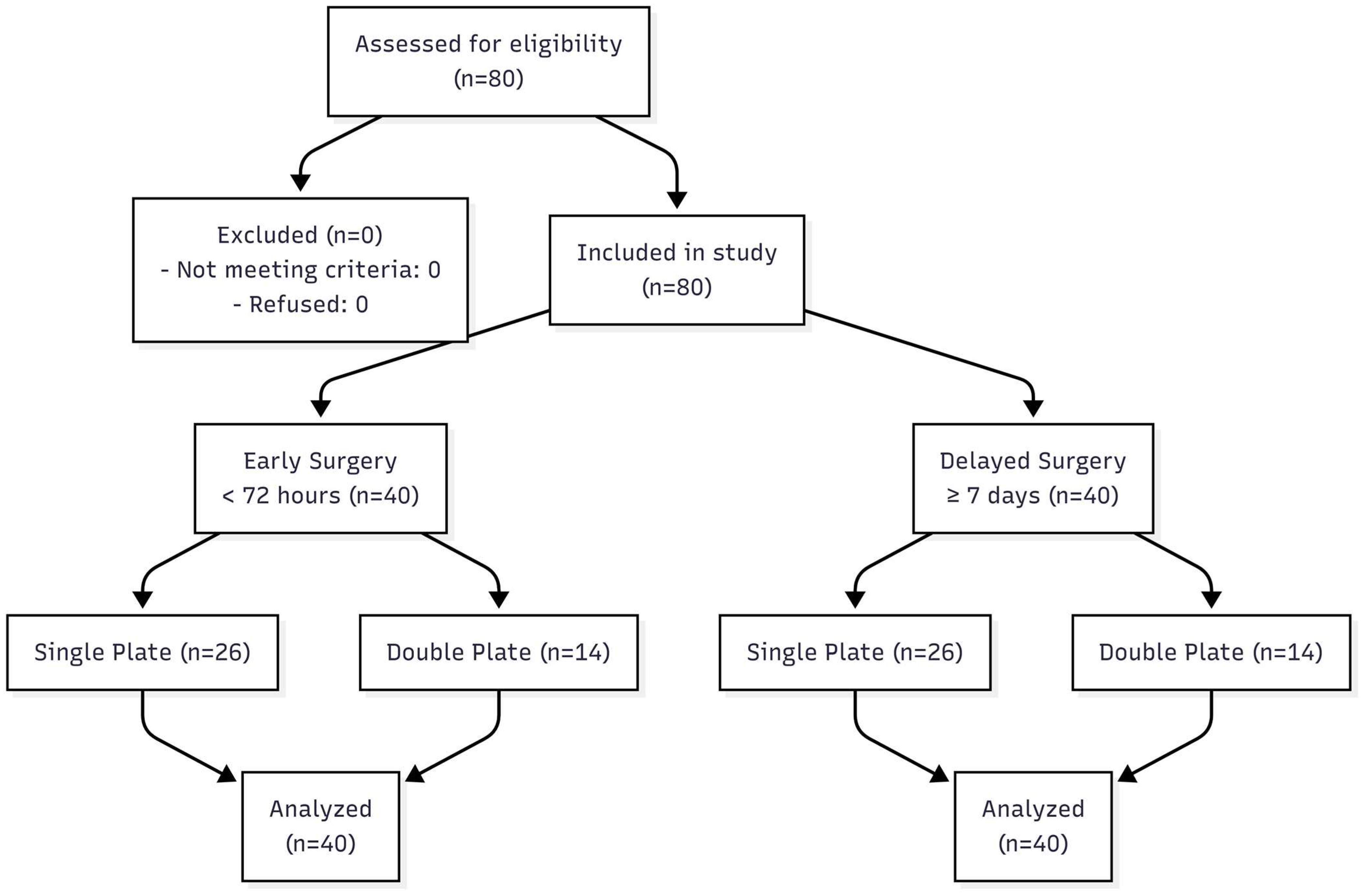
The study included 80 patients who underwent surgical treatment for distal tibial pilon fracture. Inclusion criteria were being 18 years of age or older, having a type 43-B or 43-C pilon fracture according to the AO/OTA classification, and having been treated with open reduction and internal fixation (ORIF). Only patients with a minimum clinical and radiological follow-up duration of 12 months were included in the final analysis. Patients with a previous history of fracture in the same extremity, patients with multi-segmental fractures that could not be reconstructed, patients with primary arthrodesis, and patients with insufficient follow-up data during the study period were excluded. The flow of patient inclusion, surgical timing allocation, and fixation methods is summarized in Figure 1.
Patients were divided into two groups according to the timing of surgery:
  • The early surgery group (<72 h, n = 40).
  • The delayed surgery group (>7 days, n = 40).
Patients were also divided into two subgroups according to the method of fixation:
  • Single-plate fixation (n = 52).
  • Double-plate fixation (n = 28).

2.2. Surgical Techniques

2.2.1. Preoperative Evaluation and Preparation

The study was conducted in a Level 1 trauma center. Although multiple orthopedic surgeons were involved in patient care, all surgical procedures were supervised by senior trauma surgeons and performed according to a standardized protocol defined by the department. All patients were subjected to detailed clinical and radiologic evaluation in the preoperative period. Radiologic evaluation was performed with direct radiographs, computed tomography (CT), and three-dimensional reconstructions when necessary [14]. Patients were evaluated according to the Gustilo–Anderson classification and Tscherne–Oestern closed soft tissue injury classification to determine the soft tissue status [15,16]. Surgical timing was determined based not only on the time elapsed after trauma but also on clinical findings such as regression of local edema, skin wrinkle test, and soft tissue integrity. All patients were clinically evaluated for compartment syndrome in the early period.
Patients in the early surgery group were operated on within the first 72 h if possible. In the delayed surgery group, temporary stabilization was provided with an external fixator in the first stage, and soft tissue status was monitored; open reduction and internal fixation were performed after an average of 8 to 12 days [17].

2.2.2. Surgical Approach

All surgeries were performed by experienced orthopedic surgeons and traumatologists using standard surgical protocols. The surgical approach was determined by considering the fracture pattern and soft tissue status.
  • Anterior midline incision is the most commonly preferred approach [18,19].
  • Anteromedial and anterolateral incisions have been used to aid joint reconstruction in complex fractures [20,21,22].
  • The minimally invasive plate osteosynthesis (MIPO) method has been preferred, especially in multi-segmental fractures [23].

2.2.3. Fixation Methods

Patients were treated with single-plate fixation or double-plate fixation.
Single-plate fixation was achieved with a pilon plate system placed medially or anteromedially [20].
Double-plate fixation was performed using a combination of anteromedial and anterolateral plates to better stabilize the articular surface [24].
The following steps were performed during definitive surgery [21,22]:
  • First, fibula fixation was performed, thus establishing a reference for the reduction of the tibial fracture.
  • Intra-articular fracture fragments were reduced, and temporary fixation was provided with screws.
  • Plate placement was completed, and final fixation was performed.

2.2.4. Postoperative Management and Rehabilitation

All patients were included in the early movement protocol in the postoperative period. Full load was not given for the first 6 weeks, and exercises to maintain knee and ankle range of motion were performed during this period. From the 6th week, a partial load was given, and it was changed to a full load from 8 to 12 weeks according to radiologic union findings [25].

2.3. Evaluation Criteria

The primary outcome measure of the study was determined as bone union time. When cortical continuity and trabecular transition were achieved radiologically, union was considered to have occurred [26].
Secondary evaluation criteria included functional outcomes and complication rates. Functional outcomes were evaluated using the American Orthopaedic Foot and Ankle Society (AOFAS) score at the 12th postoperative month [27,28]. Complications were classified as superficial or deep infection, malunion, implant failure, and late joint degeneration.
SPSS 26.0 software (IBM, Chicago, IL, USA) was used for statistical analysis. Continuous variables are described by mean and standard deviation, while categorical variables are presented as percentages. Compliance with normal distribution was evaluated by the Kolmogorov–Smirnov test. Independent sample t-tests or Mann–Whitney U tests were used for intergroup comparisons. The chi-square test or Fisher’s exact test was applied for categorical variables. Statistical significance was accepted as p < 0.05.
In order to determine the adequacy of the number of patients in the study, power analysis was performed. The analysis was carried out by considering the union time and infection rates in pilon fractures reported in the previous literature. In the analysis performed with 80 patients, 80% power (1-β) and a 5% significance level (α = 0.05) provided a sufficient sample size to evaluate the difference in union time and infection rates [29].

3. Results

The 80 patients included in the study were divided into two groups according to the timing of surgery: early surgery (<72 h, n = 40) and delayed surgery (>7 days, n = 40). The subgroups were reorganized according to the fixation method, with single-plate fixation being more commonly used (65% single plate, 35% double plate), as shown in Figure 1.

3.1. Patient Distribution and Demographic Data

In terms of demographic data, there was no statistically significant difference between the two groups in terms of mean age, gender distribution, and fracture classification (p > 0.05), as shown in Table 1.
Patient distribution is as follows:
The early surgery group (<72 h, n = 40):
  • Single-plate fixation: 26 patients (65%).
  • Double-plate fixation: 14 patients (35%).
The delayed surgery group (>7 days, n = 40):
  • Single-plate fixation: 26 patients (65%).
  • Double-plate fixation: 14 patients (35%).

3.2. Time to Union

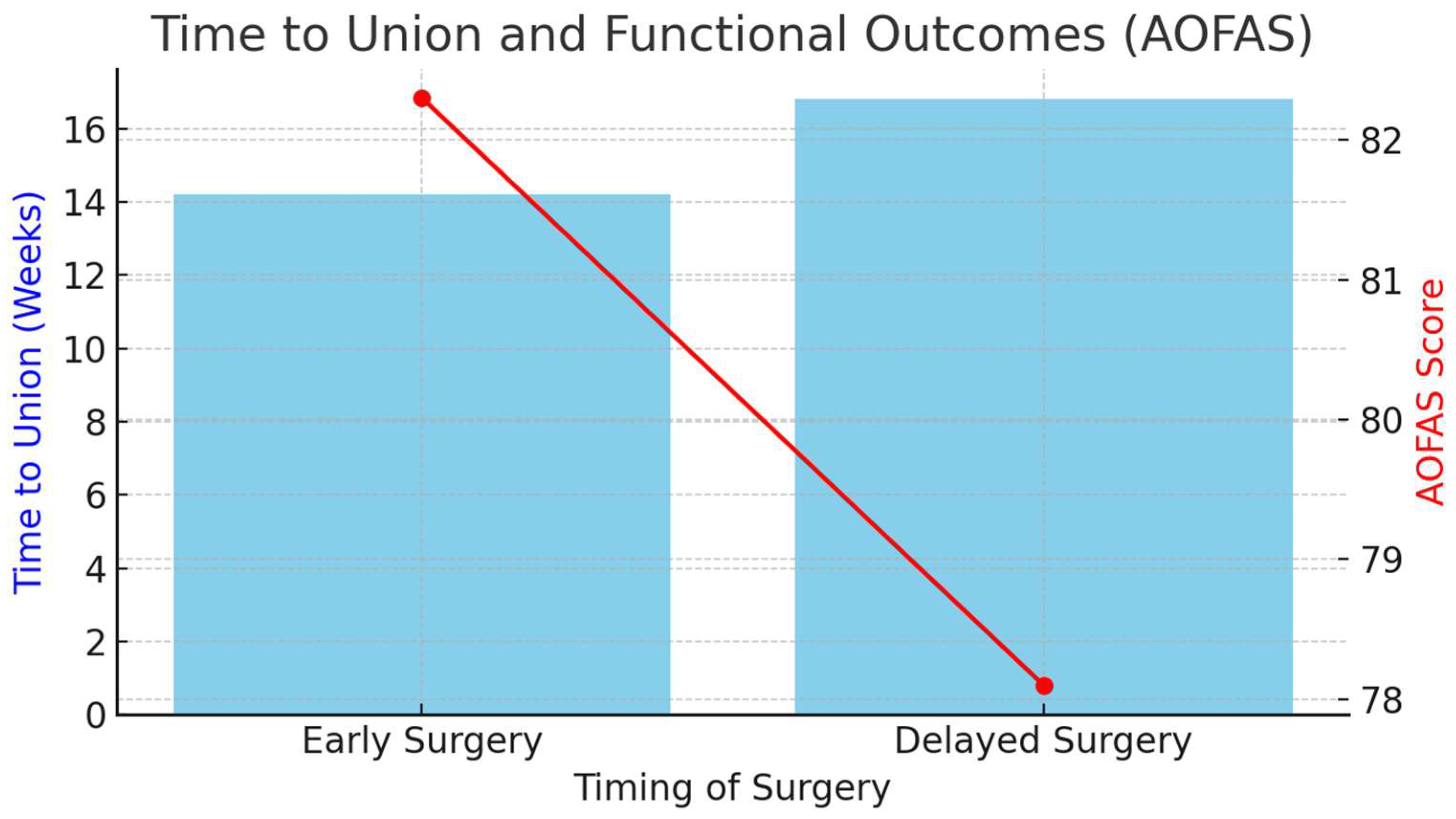
Postoperative radiologic evaluation revealed a significantly shorter union time in the early surgery group (14.2 ± 2.1 weeks vs. 16.8 ± 2.3 weeks, p < 0.05). In the early surgery group, the mean union time was 2.6 weeks shorter, and this difference was statistically significant. Although the union time was longer in the delayed surgery group, the infection rate was lower. As shown in Figure 2, the early surgery group demonstrated better functional outcomes and faster union compared to the delayed group.

3.3. Functional Outcomes

In terms of functional outcomes, better results were obtained in the early surgery group. AOFAS scores measured at 12 months postoperatively were significantly higher in the early surgery group (82.3 ± 6.5 vs. 78.1 ± 7.2, p < 0.05). In 75% of the patients who underwent early surgery, the AOFAS score was 80 and above. In the delayed surgery group, the AOFAS score remained below 75 in 60% of the patients. These data are presented in Table 2.

3.4. Complications

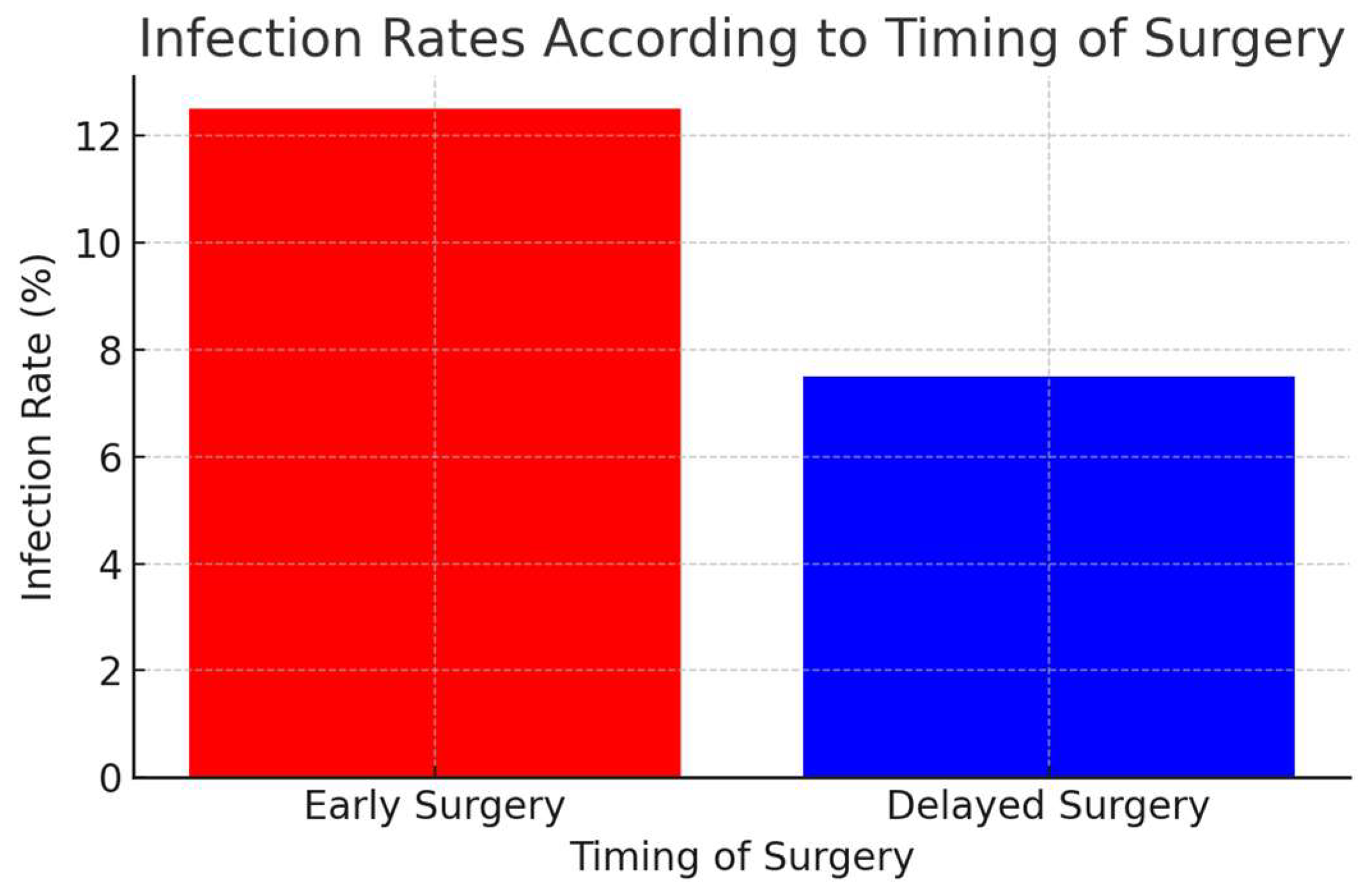
Comparing the two groups in terms of postoperative complications, the infection rate was higher in the early surgery group (12.5% vs. 7.5%, p < 0.05). Superficial infection was observed in four patients (10%) in the early surgery group and in two patients (5%) in the delayed surgery group. The deep infection rate was 2.5% (one patient) in the early surgery group and 2.5% (one patient) in the delayed surgery group. Superficial late wound healing at the edge of the incision was observed in three patients in the early surgery group, and two patients in the delayed surgery group; no deep infection or need for revision occurred in these patients. Acute compartment syndrome did not develop in all patients, and no fasciotomy was performed in any patient. The distribution of infection rates by surgical timing is illustrated in Figure 3.
No statistically significant difference was found in the rate of malunion (p = 0.34). Malunion developed in four patients (10%) in the early surgery group and in three patients (8%) in the delayed surgery group.
No significant difference was found between the two groups in terms of implant failure (p = 0.41). Implant failure was found in two patients (5%) in the early surgery group and one patient (3%) in the delayed surgery group.

3.5. Results of Single-Plate vs. Double-Plate Fixation Outcomes

In terms of fixation methods used in pilon fractures, patients were divided into two groups: single-plate fixation (n = 52) and double-plate fixation (n = 28). Single-plate fixation was more commonly used (65% single plate, 35% double plate). These data are presented in Table 3.
In terms of union time, the mean union time was 14.6 ± 2.1 weeks in patients with double-plate fixation, while the union time was significantly longer in patients with single-plate fixation at 15.8 ± 2.3 weeks (p = 0.048).
In terms of functional outcomes, the AOFAS score was 81.7 ± 7.0 in patients with double-plate fixation and 79.5 ± 6.8 in patients with single-plate fixation, but this difference was not considered statistically significant (p = 0.07).
Considering the infection rates, 11.3% of patients with double plates and 8.7% of patients with single plates developed infections, and no statistically significant difference was found (p = 0.12).
In terms of malunion rates, 8.1% malunion was observed in the double-plate group and 9.6% in the single-plate group, and no significant difference was found between the two groups (p = 0.45).
In terms of implant failure, 3.7% in the double-plate group and 4.8% in the single-plate group developed implant failure, and no statistically significant difference was found (p = 0.38).
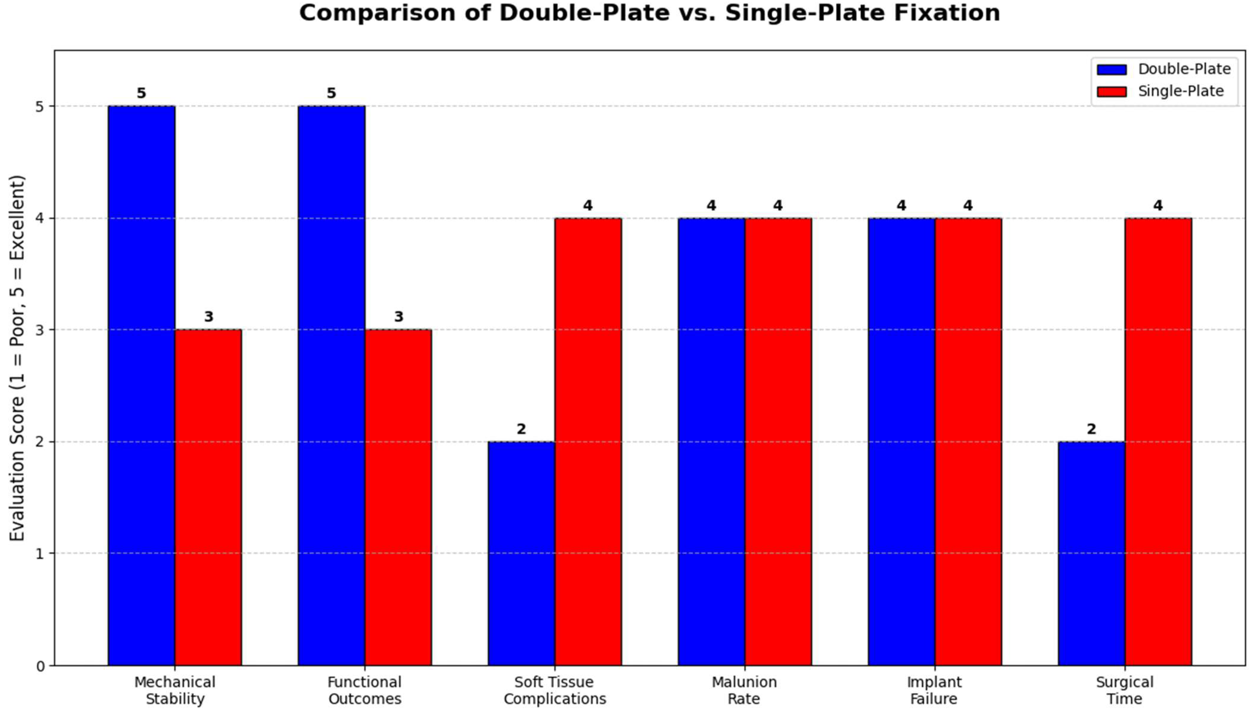
Consequently, although double-plate fixation provides shorter union time and better functional results, it may carry some risk of infection and soft tissue complications. While single-plate fixation offers the advantage of soft tissue preservation, it has the disadvantage of prolonged union time. The comparative effectiveness of single- versus double-plate fixation is summarized in Figure 4.

4. Discussion

In this study, we evaluated the effect of early (<72 h) and delayed (>7 days) surgery on clinical and radiologic outcomes in pilon fractures and compared the advantages and disadvantages of single-plate and double-plate fixation. Our results revealed that early surgery offered shorter union time and better functional outcomes but increased the risk of infection. On the other hand, delayed surgery reduced infection rates by allowing more time for soft tissue healing but resulted in prolonged union time and delayed functional return.
Our study found a significantly shorter union time in the early surgery group (14.2 ± 2.1 weeks vs. 16.8 ± 2.3 weeks, p < 0.05). This result supports that early surgery provides mechanical stabilization faster and shortens the bone healing process. Previous studies have similarly emphasized that early surgery may accelerate fracture healing, but soft tissue management is critical [30,31]. Similarly, Egol et al. found that the union time was 14.5 weeks in patients who underwent early surgery and 16.9 weeks in patients who underwent delayed surgery and that early surgery shortened the union time [9]. However, data reported by Sirkin et al. suggest that there is no significant difference between early and delayed surgery in terms of union time [10].
In terms of functional outcomes, the AOFAS score was found to be significantly higher in patients who underwent early surgery (82.3 vs. 78.1, p < 0.05). This result suggests that early stabilization positively affects the functional return of patients by accelerating mobilization. This result coincides with the results of a study by Patterson and Lou, which showed that patients who underwent early surgery had higher functional scores and an early mobilization advantage [8,32]. However, some studies have reported that delayed surgery did not make a difference in terms of long-term functional outcomes [33,34]. In a study by Wyrsch et al., it was reported that delayed surgery did not adversely affect long-term functional outcomes, but the rehabilitation process took longer [12].
The infection rate was 12.5% in patients who underwent early fixation and 5% in those treated with delayed fixation, and this difference was statistically significant (p < 0.05). This result suggests that early surgery may increase the risk of infection, especially in patients with severe soft tissue damage. In the literature, it has been reported that early surgery may increase infection rates due to high soft tissue stress [35,36]. Therefore, it may be safer to prefer delayed surgery in patients with poor soft tissue status. Sirkin et al. reported that early surgery without waiting for soft tissue healing may increase infection rates [10]. However, Chaundhry and Badavath reported that delayed surgery did not significantly decrease infection rates and that early surgery may still be an appropriate option, especially in multi-segmental fractures [37,38].
Comparing single-plate and double-plate fixation, union time was shorter in patients with double-plate fixation (14.6 vs. 15.8 weeks, p = 0.048), but in terms of functional scores, no significant difference was found between the two groups (p = 0.07). Although double-plate fixation offers better mechanical stability, it may increase the risk of soft tissue complications and infection [39,40]. Single-plate fixation may be a safer option in terms of soft tissue management, but the union time may be longer. Likewise, Blauth et al. demonstrated in their study that double-plate application provides faster union time by increasing mechanical stability but may lead to prolonged surgical time and soft tissue complications [19].
In terms of functional outcomes, the AOFAS score was 81.7 ± 7.0 in patients with double-plate fixation and 79.5 ± 6.8 in patients with single-plate fixation, but this difference was not considered statistically significant (p = 0.07). These results are similar to the data reported in the studies by Tilkeridis and Mair; these studies reported that double-plate fixation increased joint stability but did not significantly differ from single-plate fixation in terms of long-term functional scores [25,41].
A significant result of our study was that there was no significant difference between early/delayed surgery or single-/double-plate fixation in terms of malunion and implant failure (p > 0.05). This suggests that both surgical timings can provide acceptable results with careful planning. However, it should be remembered that surgical techniques should be evaluated on a patient-based basis. Similarly, McFerran et al. reported that the use of a double plate or single plate did not significantly change the malunion rates, but single-plate fixation may be a safer choice in certain fracture types [11].
With respect to infection rates, 11.3% of patients with double plates and 8.7% of patients with single plates developed infections, and no statistically significant difference was found (p = 0.12). In the literature, it has been reported that double-plate applications may increase soft tissue complications [42]. In particular, single-plate applications using minimally invasive techniques have been reported to reduce the risk of infection and accelerate wound healing [23,43].
Soft tissue integrity is one of the main factors determining the success of treatment in pilon fractures. Although early surgery provides advantages in terms of mechanical stabilization and functional results, interventions performed on edematous tissues may cause an increase in wound complications [22,28,42]. A limited number of superficial wound healing delays were observed in the early surgery group in this study, but no deep infection or revision was required. These results support that surgical timing should be planned based not only on the time of trauma but also on soft tissue assessment.
Although compartment syndrome is a significant complication associated with pilon fractures, it was not encountered in any patient in our study. However, this does not eliminate the risk; it emphasizes the need for careful clinical evaluation, especially in young patients with high muscle mass.
This study is one of the limited number of studies directly comparing the effects of surgical timing and fixation methods on clinical outcomes in pilon fractures. The fact that the number of patients was determined based on power analysis increases the reliability of the study in terms of statistical significance. In addition, a homogeneous patient group was established by including patients with AO/OTA 43-B and 43-C fractures.
Our study has some limitations. First of all, access to some patient data may be limited due to the retrospective design. Furthermore, there are certain limitations in terms of patient selection since it is a single-center study. Finally, the follow-up period of 12 months may not be sufficient to evaluate long-term complications. Therefore, the results should be supported by prospective studies with longer follow-up periods.

5. Conclusions

The results of our study suggest that early surgery may shorten union time and accelerate functional return but may carry a higher risk of soft tissue complications. Delayed surgery may favor soft tissue healing by decreasing infection rates but may prolong union time. Double-plate fixation may offer a more stable structure regarding the fixation method, whereas single-plate fixation may offer an advantage regarding the soft tissue management.
Therefore, an individualized approach should be adopted for making surgical decisions in pilon fractures. Delayed surgery may be preferred, especially in patients suffering from severe soft tissue damage, whereas double-plate fixation may be an appropriate option in patients who prioritize joint stability. Not only the fracture pattern but also the soft tissue status and wound healing potential should be taken into consideration during surgical planning.

Author Contributions

Conceptualization, İ.G. and A.K.; methodology, İ.G. and İ.U.; validation, İ.G., İ.U. and M.Y.; formal analysis, İ.G.; investigation, İ.G., İ.U., M.Y., M.F.T. and A.K.; data curation, İ.G.; writing—original draft preparation, İ.G.; writing—review and editing, İ.G., İ.U. and A.K.; supervision, İ.G.; project administration, İ.G. All authors have read and agreed to the published version of the manuscript.

Funding

This research received no external funding.

Institutional Review Board Statement

The study was conducted in accordance with the Declaration of Helsinki and approved by İnönü University Clinical Research Ethics Committee (protocol code 2021/2483; approval date 22 December 2021).

Informed Consent Statement

Written informed consent was obtained from all patients as part of routine clinical practice prior to surgical treatment.

Data Availability Statement

The data that support the findings of this study are available from the corresponding author upon reasonable request. Due to privacy and ethical restrictions, the data are not publicly available.

Conflicts of Interest

The authors declare no conflicts of interest.

References

  1. Mandi, D.M.; Belin, R.P.; Banks, J.; Barrett, B. Pilon fractures. Clin. Podiatr. Med. Surg. 2012, 29, 243–278. [Google Scholar] [CrossRef] [PubMed]
  2. Haller, J.M.; Ross, H.; Jacobson, K.; Ou, Z.; Rothberg, D.; Githens, M. Supination adduction ankle fractures: Ankle fracture or pilon variant? Injury 2020, 51, 759–763. [Google Scholar] [CrossRef] [PubMed]
  3. Luo, T.D.; Pilson, H. Pilon Fracture. In StatPearls; StatPearls Publishing: Treasure Island, FL, USA. [PubMed]
  4. Marsh, J.L.; Saltzman, C.L. Ankle fractures and dislocations. In Rockwood and Green’s Fractures in Adults, 9th ed.; Wolters Kluwer: Philadelphia, PA, USA, 2020. [Google Scholar]
  5. Martin, J.S.; Marsh, J.L.; Bonar, S.K.; DeCoster, T.A.; Found, E.M.; Brandser, E.A. Assessment of the AO/ASIF fracture classification for the distal tibia. J. Orthop. Trauma 1997, 11, 477–483. [Google Scholar] [CrossRef] [PubMed]
  6. Rüedi, T.P.; Allgöwer, M. The operative treatment of intra-articular fractures of the lower end of the tibia. Clin. Orthop. Relat. Res. 1979, 138, 105–110. [Google Scholar] [PubMed]
  7. Luo, T.D.; Eady, J.M.; Aneja, A.; Miller, A.N. Classifications in Brief: Rüedi-Allgöwer Classification of Tibial Plafond Fractures. Clin. Orthop. Relat. Res. 2017, 475, 1923–1928. [Google Scholar] [CrossRef] [PubMed] [PubMed Central]
  8. Patterson, M.J.; Cole, J.D. Two-staged delayed open reduction and internal fixation of severe pilon fractures. J. Orthop. Trauma 1999, 13, 85–91. [Google Scholar] [CrossRef] [PubMed]
  9. Egol, K.A.; Tejwani, N.C.; Walsh, M.G.; Capla, E.L.; Koval, K.J. Predictors of short-term functional outcome following ankle fracture surgery. J. Bone Jt. Surg. Am. 2006, 88, 974–979. [Google Scholar] [CrossRef] [PubMed]
  10. Sirkin, M.; Sanders, R.; DiPasquale, T.; Herscovici, D., Jr. A staged protocol for soft tissue management in the treatment of complex pilon fractures. J. Orthop. Trauma 2004, 18, S32–S38. [Google Scholar] [CrossRef] [PubMed]
  11. McFerran, M.A.; Smith, S.W.; Boulas, H.J.; Schwartz, H.S. Complications encountered in the treatment of pilon fractures. J. Orthop. Trauma 1992, 6, 195–200. [Google Scholar] [CrossRef] [PubMed]
  12. Wyrsch, B.; McFerran, M.A.; McAndrew, M.; Limbird, T.J.; Harper, M.C.; Johnson, K.D.; Schwartz, H.S. Operative treatment of fractures of the tibial plafond. A randomized, prospective study. J. Bone Jt. Surg. Am. 1996, 78, 1646–1657. [Google Scholar] [CrossRef] [PubMed]
  13. Tornetta P3rd Weiner, L.; Bergman, M.; Watnik, N.; Steuer, J.; Kelley, M.; Yang, E. Pilon fractures: Treatment with combined internal and external fixation. J. Orthop. Trauma 1993, 7, 489–496. [Google Scholar] [CrossRef] [PubMed]
  14. Zhao, Y.; Wu, J.; Wei, S.; Xu, F.; Kong, C.; Zhi, X.; Huang, M.; Cai, X. Surgical approach strategies for open reduction internal fixation of closed complex tibial Pilon fractures based on axial CT scans. J. Orthop. Surg. Res. 2020, 15, 283. [Google Scholar] [CrossRef] [PubMed] [PubMed Central]
  15. Gustilo, R.B.; Anderson, J.T. Prevention of infection in the treatment of one thousand and twenty-five open fractures of long bones: Retrospective and prospective analyses. J. Bone Jt. Surg. Am. 1976, 58, 453–458. [Google Scholar] [CrossRef] [PubMed]
  16. Tscherne, H.; Oestern, H.J. A classification of soft-tissue damage related to fractures. Unfallheilkunde 1982, 85, 111–115. (In German) [Google Scholar] [PubMed]
  17. Williams, T.M.; Marsh, J.L.; Nepola, J.V.; DeCoster, T.A.; Hurwitz, S.R.; Bonar, S.B. External fixation of tibial plafond fractures: Is routine plating of the fibula necessary? J. Orthop. Trauma 1998, 12, 16–20. [Google Scholar] [CrossRef] [PubMed]
  18. Rammelt, S.; Bartoníček, J.; Schepers, T.; Kroker, L. Fixation of anterolateral distal tibial fractures: The anterior malleolus. Oper. Orthop. Traumatol. 2021, 33, 125–138. (In English) [Google Scholar] [CrossRef] [PubMed]
  19. Blauth, M.; Bastian, L.; Krettek, C.; Knop, C.; Evans, S. Surgical options for the treatment of severe tibial pilon fractures: A study of three techniques. J. Orthop. Trauma 2001, 15, 153–160. [Google Scholar] [CrossRef] [PubMed]
  20. Pugh, K.J.; Wolinsky, P.R.; McAndrew, M.P.; Johnson, K.D. Tibial pilon fractures: A comparison of treatment methods. J. Trauma 1999, 47, 937–941. [Google Scholar] [CrossRef] [PubMed]
  21. Krettek, C.; Bachmann, S. Pilon fractures. Part 1: Diagnostics, treatment strategies and approaches. Chirurg 2015, 86, 87–104. [Google Scholar] [CrossRef] [PubMed]
  22. Mittlmeier, T.; Wichelhaus, A. Treatment strategy and planning for pilon fractures. Unfallchirurg 2017, 120, 640–647. [Google Scholar] [CrossRef] [PubMed]
  23. Milenković, S.; Mitković, M.; Micić, I.; Mladenović, D.; Najman, S.; Trajanović, M.; Manić, M.; Mitković, M. Distal tibial pilon fractures (AO/OTA type B, and C) treated with the external skeletal and minimal internal fixation method. Vojn. Pregl. 2013, 70, 836–841. [Google Scholar] [CrossRef] [PubMed]
  24. Dai, C.H.; Sun, J.; Chen, K.Q.; Zhang, H.B. Omnidirectional Internal Fixation by Double Approaches for Treating Rüedi-Allgöwer Type III Pilon Fractures. J. Foot Ankle Surg. 2017, 56, 756–761. [Google Scholar] [CrossRef] [PubMed]
  25. Tilkeridis, K.; Iliopoulos, E.; Wall, S.; Kiziridis, G.; Khaleel, A. Severe Pilon Fractures: The Role of Quality of Reduction in Clinical and Functional Outcomes. Cureus 2024, 16, e65245. [Google Scholar] [CrossRef] [PubMed] [PubMed Central]
  26. Fisher, J.S.; Kazam, J.J.; Fufa, D.; Bartolotta, R.J. Radiologic evaluation of fracture healing. Skelet. Radiol. 2019, 48, 349–361. [Google Scholar] [CrossRef] [PubMed]
  27. Kitaoka, H.B.; Alexander, I.J.; Adelaar, R.S.; Nunley, J.A.; Myerson, M.S.; Sanders, M. Clinical rating systems for the ankle-hindfoot, midfoot, hallux, and lesser toes. Foot Ankle Int. 1994, 15, 349–353. [Google Scholar] [CrossRef] [PubMed]
  28. Lu, V.; Zhang, J.; Zhou, A.; Thahir, A.; Lim, J.A.; Krkovic, M. Open versus closed pilon fractures: Comparison of management, outcomes, and complications. Injury 2022, 53, 2259–2267. [Google Scholar] [CrossRef] [PubMed]
  29. Faul, F.; Erdfelder, E.; Lang, A.G.; Buchner, A. G*Power 3: A flexible statistical power analysis program for the social, behavioral, and biomedical sciences. Behav. Res. Methods 2007, 39, 175–191. [Google Scholar] [CrossRef]
  30. Hreha, J.; Metrione, M.D.; Reilly, M.C.; Sirkin, M.S.; Adams, M.R. Perioperative Impact of Early Limited Surgical Intervention to Treat Pilon Fractures. Foot Ankle Int. 2022, 43, 1092–1098. [Google Scholar] [CrossRef] [PubMed]
  31. Bastias, C.; Lagos, L. New Principles in Pilon Fracture Management: Revisiting Rüedi and Allgöwer Concepts. Foot Ankle Clin. 2020, 25, 505–521. [Google Scholar] [CrossRef] [PubMed]
  32. Lou, Z.; Wang, Z.; Liu, C.; Tang, X. Outcomes of tibial pilon fracture fixation based on four-column theory. Injury 2023, 54, S36–S42. [Google Scholar] [CrossRef] [PubMed]
  33. Swords, M.P.; Weatherford, B. High-Energy Pilon Fractures: Role of External Fixation in Acute and Definitive Treatment. What are the Indications and Technique for Primary Ankle Arthrodesis? Foot Ankle Clin. 2020, 25, 523–536. [Google Scholar] [CrossRef] [PubMed]
  34. Liu, J.H.; Zhang, Q.; Wei, G.H.; Liu, L.; Mu, X.; Li, M.L.; Wu, Z.D. A retrospective comparison of double-hooked locking plates versus non-locking plates in minimally invasive percutaneous plate osteosynthesis for the treatment of comminuted distal fibular fractures accompanied by tibial Pilon fractures. J. Orthop. Surg. Res. 2023, 18, 287. [Google Scholar] [CrossRef] [PubMed] [PubMed Central]
  35. Luong, K.; Huchital, M.J.; Saleh, A.M.; Subik, M. Management of Distal Fibular Fractures With Minimally Invasive Technique: A Systematic Review. J. Foot Ankle Surg. 2021, 60, 114–120. [Google Scholar] [CrossRef] [PubMed]
  36. Flores, M.; Ciminero, M.; Kottmeier, S.A.; Botros, D.; Zelle, B.A.; Shearer, D.W. Pilon fractures: Consensus and controversy. OTA Int. 2023, 6, e236. [Google Scholar] [CrossRef] [PubMed] [PubMed Central]
  37. Chaudhry, Y.P.; Papadelis, E.; Hayes, H.; Stahel, P.F.; Hasenboehler, E.A. Fusion versus fixation in complex comminuted C3-type tibial pilon fractures: A systematic review. Patient Saf. Surg. 2021, 15, 35. [Google Scholar] [CrossRef] [PubMed] [PubMed Central]
  38. Badavath, R.R.; Akkala, P.; Aluka, S.K.R.; Patnala, C. Evaluation of Functional Outcome in Surgically Managed Tibial Pilon Fractures. Cureus 2024, 16, e63242. [Google Scholar] [CrossRef] [PubMed] [PubMed Central]
  39. Carter, T.H.; Duckworth, A.D.; Oliver, W.M.; Molyneux, S.G.; Amin, A.K.; White, T.O. Open Reduction and Internal Fixation of Distal Tibial Pilon Fractures. JBJS Essent. Surg. Tech. 2019, 9, e29. [Google Scholar] [CrossRef] [PubMed] [PubMed Central]
  40. Ding, W.; Xu, J.; Zhu, Y.; Nian, S.; Lu, Y.; Zheng, M.; Zhu, X.; Wang, Z.; Ji, F.; Tang, H. Comparative study on the clinical efficacy of small plate assisted anatomic plate and traditional double plate in the treatment of Rüedi and Allgöwer II—III pilon fracture. BMC Surg. 2023, 23, 352. [Google Scholar] [CrossRef] [PubMed] [PubMed Central]
  41. Mair, O.; Pflüger, P.; Hoffeld, K.; Braun, K.F.; Kirchhoff, C.; Biberthaler, P.; Crönlein, M. Management of Pilon Fractures-Current Concepts. Front. Surg. 2021, 8, 764232. [Google Scholar] [CrossRef] [PubMed] [PubMed Central]
  42. Erichsen, J.L.; Andersen, P.I.; Viberg, B.; Jensen, C.; Damborg, F.; Froberg, L. A systematic review and meta-analysis of functional outcomes and complications following external fixation or open reduction internal fixation for distal intra-articular tibial fractures: An update. Eur. J. Orthop. Surg. Traumatol. 2019, 29, 907–917. [Google Scholar] [CrossRef] [PubMed]
  43. Guo, Y.; Tong, L.; Li, S.; Liu, Z. External Fixation combined with Limited Internal Fixation versus Open Reduction Internal Fixation for Treating Ruedi-Allgower Type III Pilon Fractures. Med. Sci. Monit. 2015, 21, 1662–1667. [Google Scholar] [CrossRef] [PubMed] [PubMed Central]
Figure 1. Flowchart of patient inclusion, surgical timing, and fixation type distribution.
Japma 116 25087 g001
Figure 2. Comparison of union time and AOFAS scores between early and delayed surgery groups. Early surgery resulted in shorter union time and higher functional scores.
Japma 116 25087 g002
Figure 3. Infection rates in early and delayed surgery groups. The early surgery group showed a higher superficial infection rate compared to the delayed group.
Japma 116 25087 g003
Figure 4. Comparison of double- versus single-plate fixation in pilon fractures. Double plating was superior in terms of mechanical stability and functional outcomes, whereas single plating showed advantages regarding soft tissue complications and shorter surgical duration. No significant differences were observed between the groups in malunion or implant failure rates.
Japma 116 25087 g004
Table 1. Comparison of demographic and soft tissue characteristics between early and delayed surgery groups.
ParameterEarly Surgery (n = 40)Delayed Surgery (n = 40)p Value
Age (mean ± SD)38.4 ± 10.239.1 ± 9.8>0.05
Gender (Male/Female)28/1226/14>0.05
Fracture Pattern (AO 43-B/43-C)18/2220/20>0.05
Body Mass Index (BMI, mean ± SD)26.3 ± 4.127.1 ± 4.3>0.05
Smoking (%)15 (37.5%)14 (35%)>0.05
Diabetes Mellitus (%)4 (10%)5 (12.5%)>0.05
Mechanism of Injury (Fall/MVA)22/1824/16>0.05
Soft Tissue Injury * (Grade 1/2)27/1329/11>0.05
Wound Closure (Primary/Delayed)36/437/3>0.05
* Soft tissue injury graded using the Tscherne–Oestern classification system for closed fractures. MVA: motor vehicle accident. p < 0.05 was considered statistically significant.
Table 2. Comparison of clinical outcomes between early and delayed surgery.
ParameterEarly Surgery (n = 40)Delayed Surgery (n = 40)p Value
Time to Union (weeks, mean ± SD)14.2 ± 2.116.8 ± 2.3<0.001
AOFAS Score (mean ± SD)82.3 ± 6.578.1 ± 7.20.013
Superficial Wound Problems (n, %)3 (7.5%)2 (5.0%)>0.05
Deep Infection (n, %)00-
Compartment Syndrome (n)00-
Malunion/Nonunion (n, %)2 (5.0%)1 (2.5%)>0.05
Implant Failure (n, %)1 (2.5%)1 (2.5%)>0.05
AOFAS: American Orthopaedic Foot and Ankle Society Score. p < 0.05 was considered statistically significant.
Table 3. Comparison of clinical outcomes between single- and double-plate fixation.
ParameterSingle Plate (n = 52)Double Plate (n = 28)p Value
Time to Union (weeks, mean ± SD)15.8 ± 2.314.6 ± 2.00.048
AOFAS Score (mean ± SD)79.3 ± 7.481.2 ± 6.70.07
Superficial Wound Problems (n, %)3 (5.8%)2 (7.1%)>0.05
Deep Infection (n, %)00-
Compartment Syndrome (n)00-
Malunion/Nonunion (n, %)2 (3.8%)1 (3.5%)>0.05
Implant Failure (n, %)1 (1.9%)1 (3.5%)>0.05
AOFAS: American Orthopaedic Foot and Ankle Society Score. p < 0.05 was considered statistically significant.
Disclaimer/Publisher’s Note: The statements, opinions and data contained in all publications are solely those of the individual author(s) and contributor(s) and not of MDPI and/or the editor(s). MDPI and/or the editor(s) disclaim responsibility for any injury to people or property resulting from any ideas, methods, instructions or products referred to in the content.