Dog bites to humans are considerable in number; the American Veterinary Medical Association reports 4.5 million dog bites take place in the US, with 800,000 of those necessitating medical care [
1]. The literature is robust with reports and studies on animal bites including dogs, cats, and rats [
2,
3,
4]. These primarily discuss the resulting infections that manifest from those bites. Given the issues seen in persons with diabetes, as far as susceptibility to infection, many of the reports discuss impact with diabetes or include subjects with diabetes [
5,
6,
7,
8,
9].
What is uncommon is when a canine becomes attracted to necrotic/infected tissue and proceeds to “debride” or masticate the tissue. A canine’s instinctive behavior, coupled with an incredible sense of smell and an individual with severe neuropathy, often sound asleep, makes this possible.
Instances of this taking place are mentioned anecdotally by students on rotation and podiatric colleagues, but overall awareness within the provider community seems lacking. Especially rare are published articles in the literature dealing with mastication of the feet by canines. Indeed, only four publications that address this are found: two from podiatric journals, one from an orthopedic journal, and one from a plastic surgery journal.
Nearly all of the cases found in the literature, as well as the web-based stories, follow the pattern of a person with diabetes and associated severe neuropathy, who experiences the loss of digits or more on their feet due to a nonaggressive canine that is attracted to the scent of necrotic tissue.
Canine Olfactory System
What allows a canine to detect a preclinical infection is their remarkable olfactory system. Indeed, the literature is replete with studies that demonstrate the incredible acuity canines have in detecting odors [
10,
11,
12,
13]. An olfactory epithelium that is 20 times larger than humans [
10,
12], and has 44 times more scent receptors than humans, results in a canine sense of smell that is up to 100,000 times more acute than humans [
12,
14].
Diabetes Alert Dogs
Although this article addresses a “misuse” of the canine sense of smell, canines have successfully been used for tasks that greatly aid society such as detecting explosives, finding lost persons, and early detection of many diseases and abnormal blood sugar levels [
15].
Medical Conditions Detectable by Canines
Nelson and Wiles [
16] have presented an excellent update on the status of canines detecting a variety of cancers. What seems to be at the heart of this ability is that humans produce and shed huge numbers of volatile organic compounds (VOCs) that can represent a variety of disease conditions. Canines have been trained to recognize many of these VOCs that are found to be definitive for specific disease states. Some specific cancers canines have been trained to detect, as noted in their article, include ovarian, lung, and prostate cancers. In addition to diabetes, as is discussed in this manuscript, canines are able to detect COVID-19 [
16]. Other cancers that canines have been trained to detect, include cancer of the cervix, breast, bladder, and stomach, as well as melanoma [
17].
Another medical condition that canines are reportedly exceptional at detecting is seizures. Trained seizure-alerting dogs are able to signal their owner that a seizure is imminent, thus the owner can take protective actions. Similar to diabetes alert dogs, seizure-alerting dogs detect not only metabolic changes represented by VOCs but also behavioral changes in their owner (facial expressions and body language) [
18].
Reported Cases of Canines Masticating on the Lower Extremity
Regarding canine debridement of infected tissue in an unaware person with diabetes and neuropathy, there are only a few cases reported in the literature that reflect this scenario [
19,
20,
21,
22]. All of these involved loss of a digit or hallux in a sleeping person with diabetic neuropathy. Aside from the several case studies, no actual research on this phenomenon exists. One case of unprovoked mastication by a feline of a neuropathic foot due to diabetes was found [
23].
These episodes are not exclusive to persons with diabetic neuropathy, as Lovett et al [
24] describe a person with neuropathy due to spina bifida losing a digit to canine mastication. In addition to the published cases noted above, numerous anecdotal accounts on media sites can be found [
25,
26,
27,
28,
29].
Instances of this occurrence had been reported to the authors by students at the Arizona College of Podiatric Medicine while on clinical rotations. Follow-up with residents and attendings led to the three case studies presented below.
Case Study Patient A
A 49-year-old male with type 2 diabetes presented to the emergency department at Covenant Health Care after a traumatic amputation to their right hallux. The patient relates that his spouse awoke in the middle of the night due to a “crunching” noise and observed their dog chewing on his toe. His medical history also includes peripheral neuropathy, hypertension, and chronic obstructive pulmonary disease. The patient relates that he is a smoker with a 30-pack-year history. The patient’s vitals were recorded in the emergency department. His blood pressure was 161/75, temperature was 97.9°F, heart rate was 95 beats per minute, and respiratory rate was 20 breaths per minute. Laboratory results showed a white blood cell count of 9.82 10
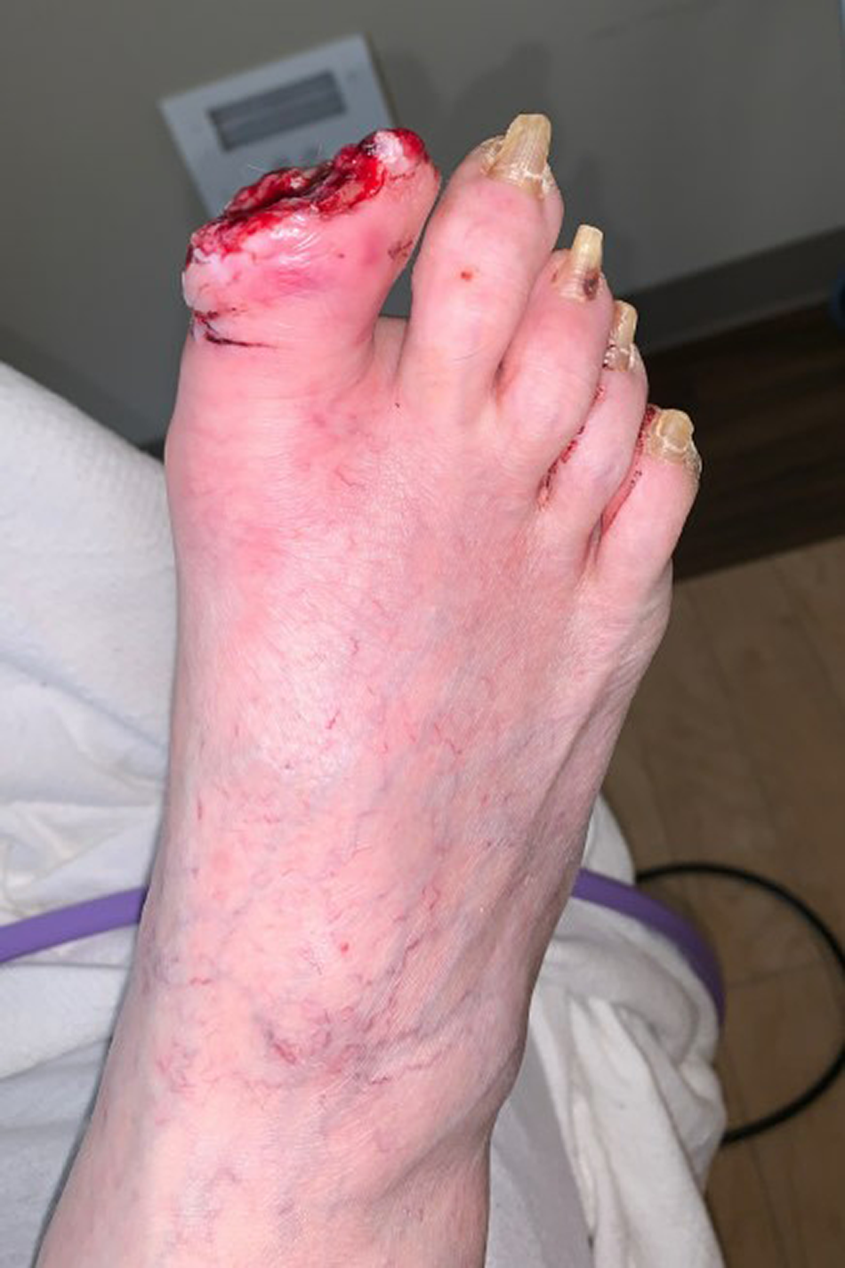
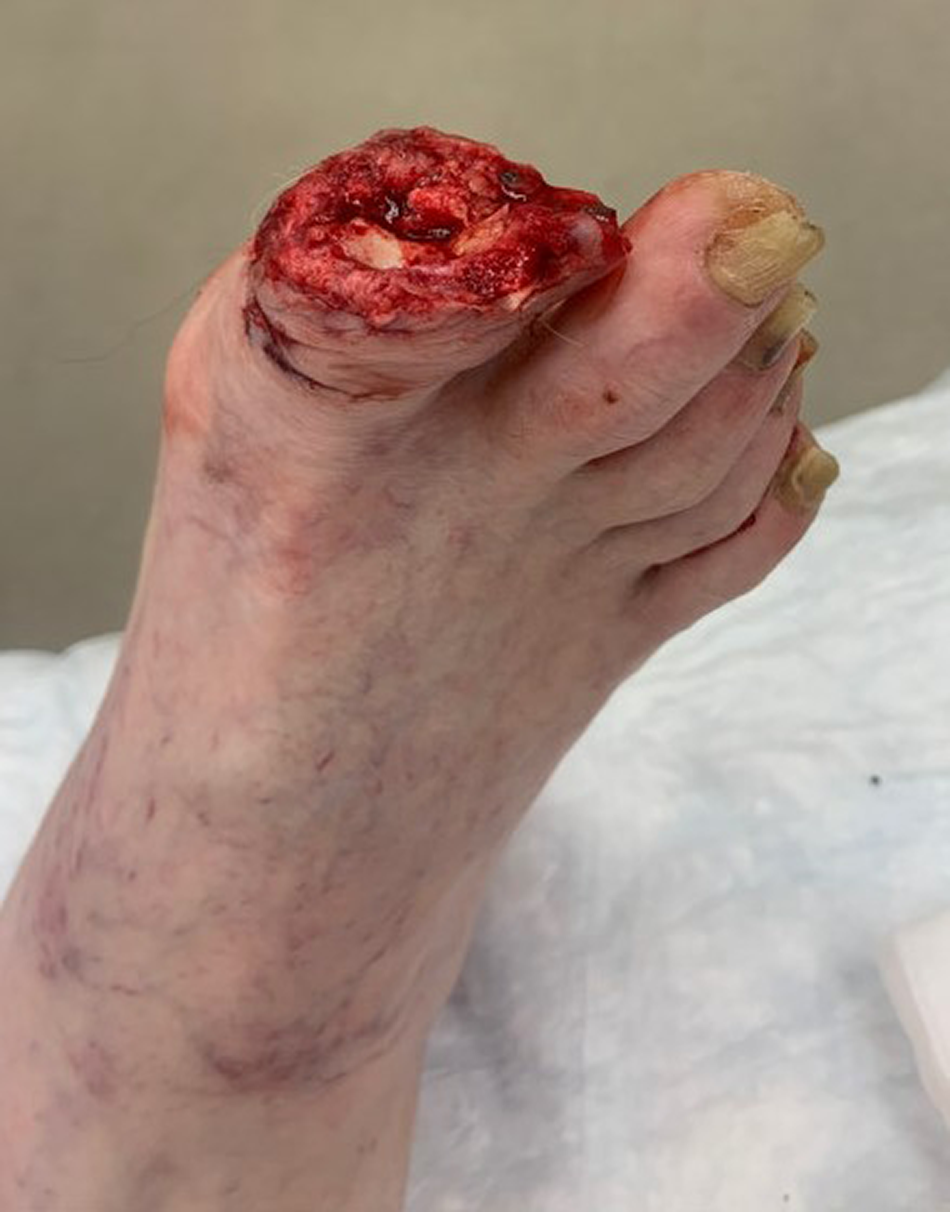
9/L, glucose at 108 mg/dL, erythrocyte sedimentation rate at 42 mm/hr, and C-reactive protein at 2.73 mg/L. Upon vascular examination, pedal pulses were palpable, pedal hair was absent, and capillary refill time was brisk to remaining digits. Neurologic examination revealed that epicritic sensation was absent via light touch. The right hallux was examined and found to have a full thickness traumatic amputation at the interphalangeal joint. The proximal phalanx was exposed through the wound bed with mild peri-wound edema. There was no drainage or malodor present (
Figs. 1 and
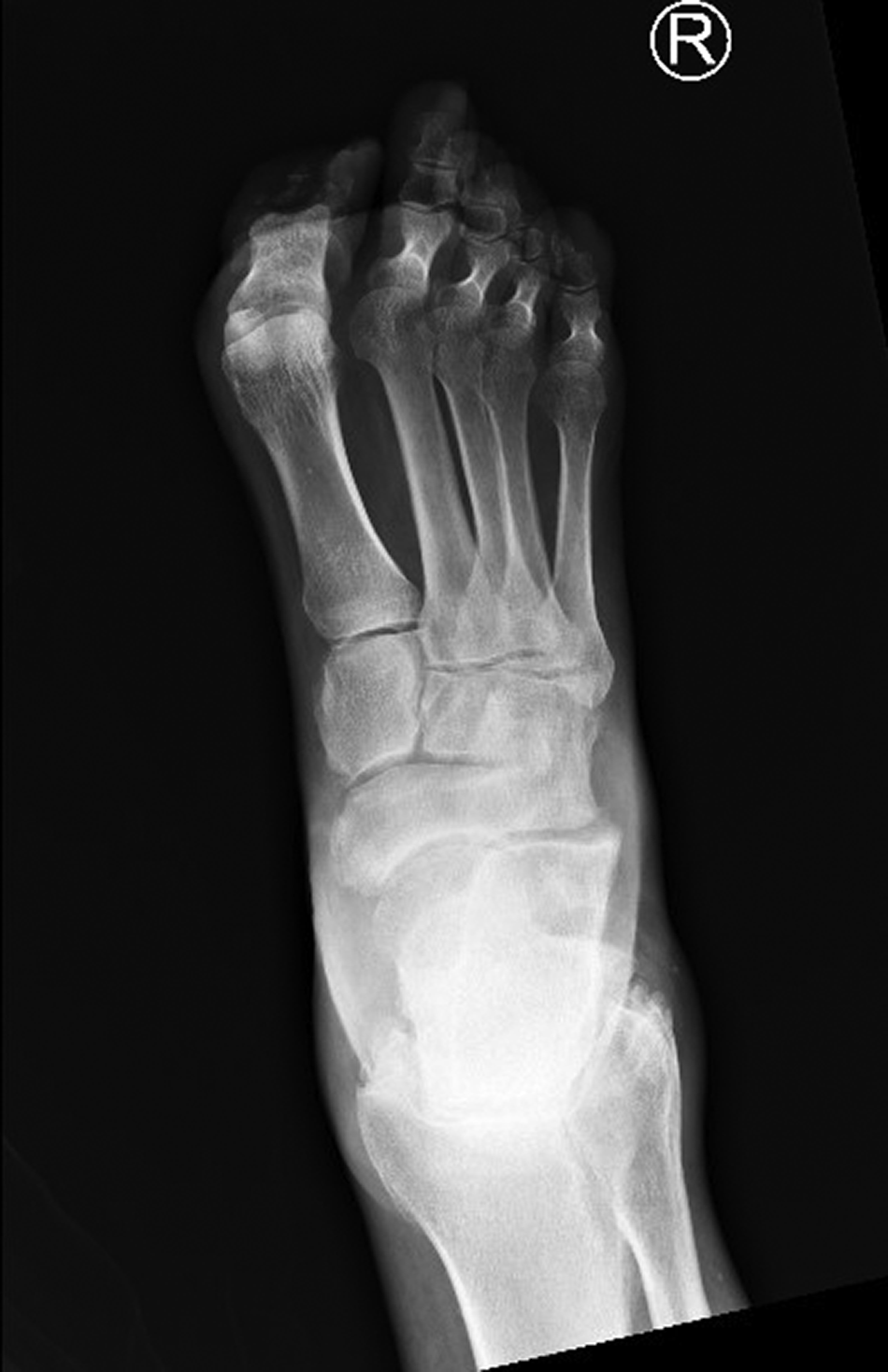
2). Plain radiographs demonstrated that only small fragments of the distal phalanx remained (
Fig. 3).
Figure 1.
Anteroposterior photograph (distalproximal view) of the right hallux of Patient A, partially consumed by canine.
Figure 1.
Anteroposterior photograph (distalproximal view) of the right hallux of Patient A, partially consumed by canine.
Figure 2.
Anteroposterior phototgraph of the right hallux of Patient A, partially consumed by canine.
Figure 2.
Anteroposterior phototgraph of the right hallux of Patient A, partially consumed by canine.
Figure 3.
Preoperative anteroposterior radiograph of the right foot of Patient A.
Figure 3.
Preoperative anteroposterior radiograph of the right foot of Patient A.
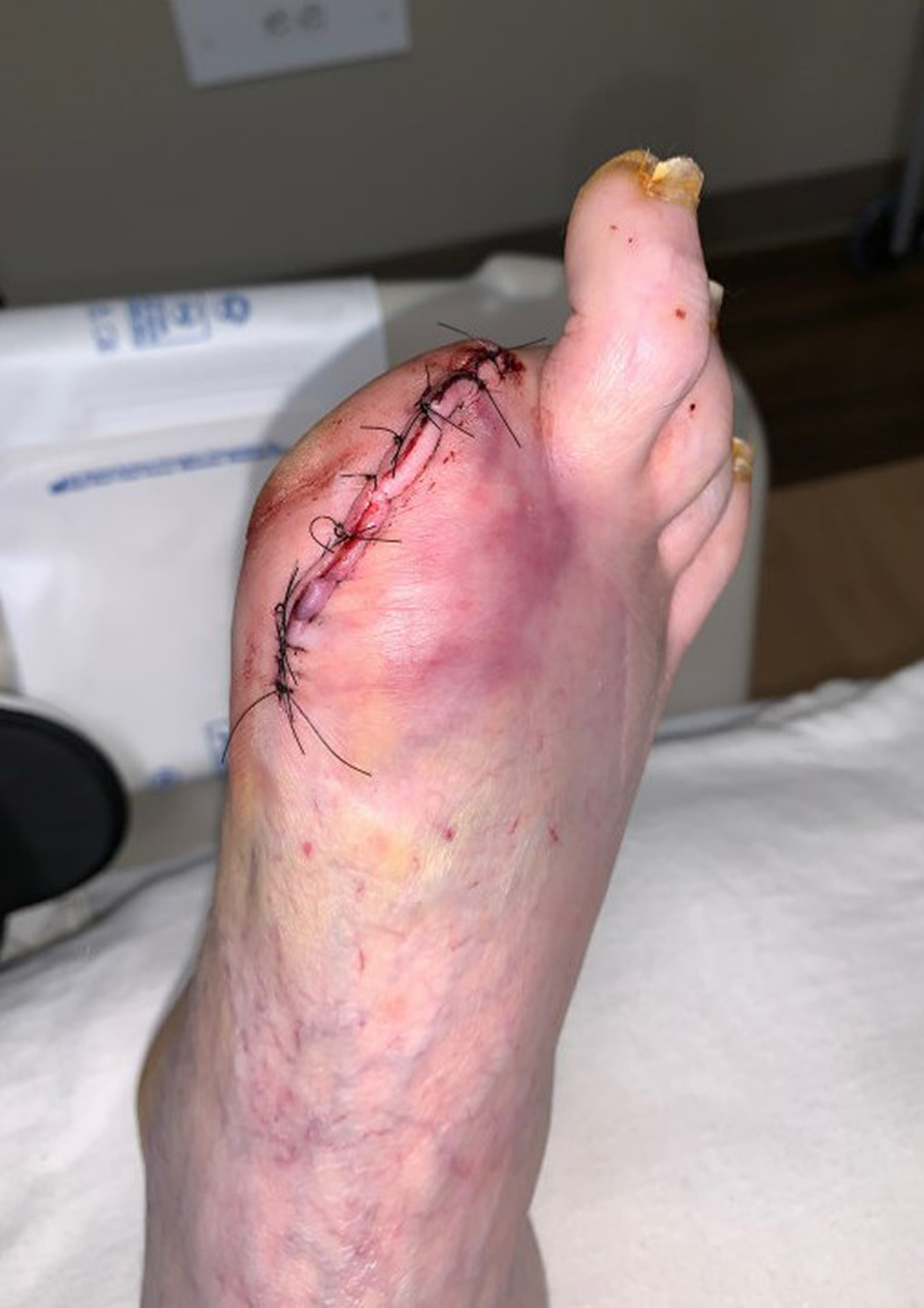
The wound was flushed with 3 L of sterile saline and the patient was placed on ampicillin/sulbactam antibiotic therapy in the emergency department. The next day, the patient went to the operating room for right hallux/partial first ray amputation due to soft-tissue deficit. Amputation level was determined on extent of damaged and dysvascular tissue (
Figs. 4 and
5). The wound was dressed with dry sterile dressings and ace wrap. Nonweightbearing status in a postoperative shoe was undertaken until suture removal. Oral doxycycline was ordered for 10 days postoperative. Healing was uneventful. Regarding follow-up, the patient remained in the hospital due to concerns of the dog repeating behavior. On conclusion of the hospital stay the patient was transferred to a skilled nursing facility where he followed-up for routine care.
Figure 4.
Postoperative view of incision site on Patient A.
Figure 4.
Postoperative view of incision site on Patient A.
Figure 5.
Postoperative anteroposterior radiograph of right foot of Patient A.
Figure 5.
Postoperative anteroposterior radiograph of right foot of Patient A.
Case Study Patient B
A 54-year-old male with type 2 diabetes presented to the emergency department of St. Mary’s Hospital with a chief complaint that his dog was chewing on his toe. His medical history also includes peripheral neuropathy and a left above-the-knee amputation. The patient reports that he is a current smoker. The patient relates that he woke up in the morning to his dog chewing on his toe. He states that prior to this episode, his dog had licked his wound on that toe. In the emergency department, temperature was 98.2°F, blood pressure was 126/84, and the patient was tachycardiac (heart rate, 98/min) with a respiratory rate of 18/min. Laboratory results included a random glucose of 187 mg/dL and white blood cells at 10.8 109/L.
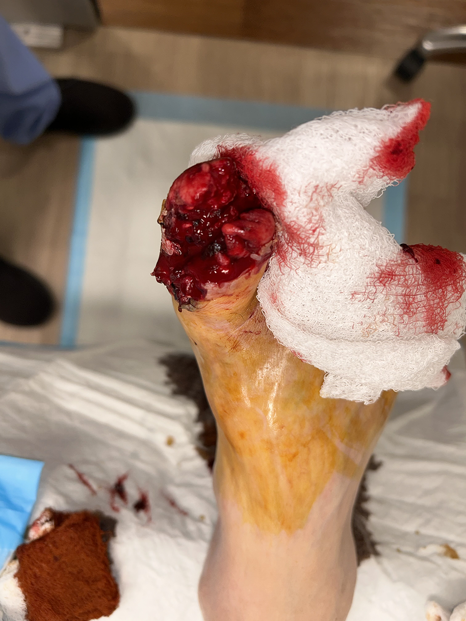
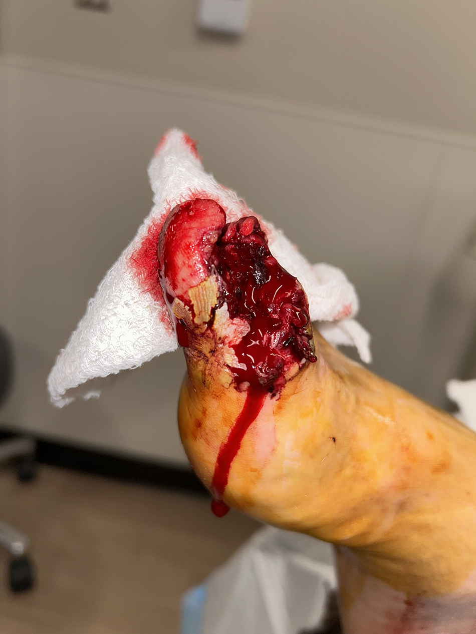
Upon physical examination, the right foot revealed a wound to the sub first metatarsal head and a full thickness ragged wound of the right hallux. The peri-wound revealed several small puncture wounds. The distal and proximal phalanx were exposed in the wound bed. The lateral one-third of the nail bed to the right hallux was present with complete avulsion of the nail plate (
Figs. 6 and
7). No ischemic changes were noted. Upon vascular examination, pedal pulses were palpable and capillary refill time was brisk to distal digits. The right forefoot had edema present with tenderness to palpation. Neurologic examination revealed that epicritic sensation diminished via light touch to the right foot.
Figure 6.
Anteroposterior photograph of the initial wound to the right hallux of Patient B.
Figure 6.
Anteroposterior photograph of the initial wound to the right hallux of Patient B.
Figure 7.
Lateral view of the initial wound to right hallux of Patient B.
Figure 7.
Lateral view of the initial wound to right hallux of Patient B.
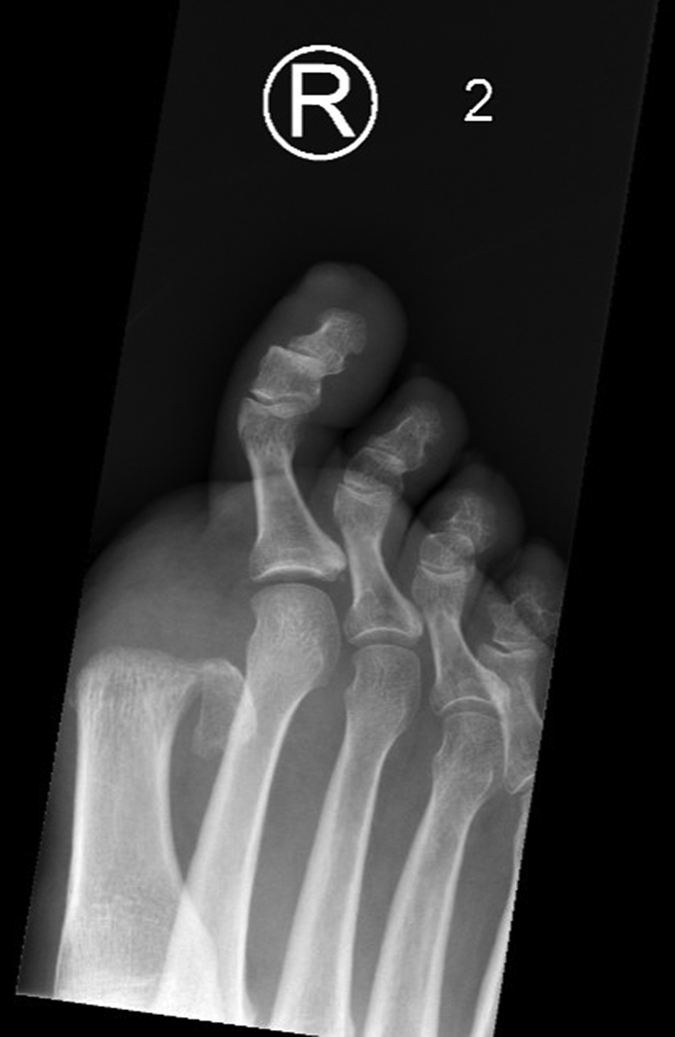
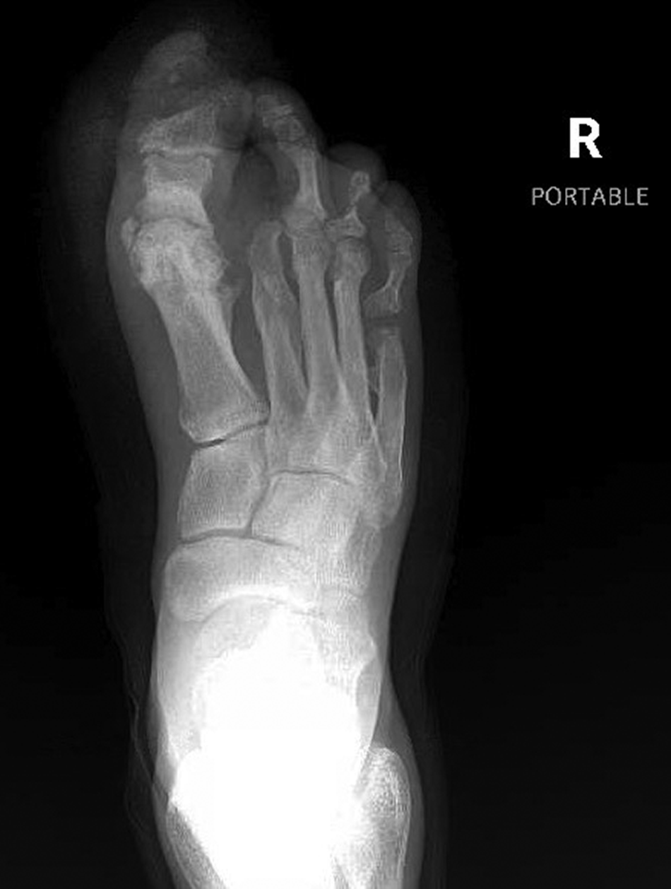
Plain radiographs demonstrated that a portion of the distal phalanx remained (
Fig. 8).
Figure 8.
Preoperative anteroposterior radiograph of the right foot of Patient B.
Figure 8.
Preoperative anteroposterior radiograph of the right foot of Patient B.
The patient was placed on ampicillin/sulbactam in the emergency department and went to the operating room the following morning for right hallux amputation. Amputation level was determined on extent of damaged and dysvascular tissue. The wound was dressed with dry sterile dressings and ace wrap. Nonweightbearing status in a postoperative shoe was undertaken until suture removal. Healing was uneventful. He was discharged with a 10-day supply of amoxicillin and clavulanate potassium. A wound culture was performed and grew beta hemolytic streptococcus. Healing and suture removal were uneventful, but the patient was subsequently lost to follow-up.
Case Study Patient C
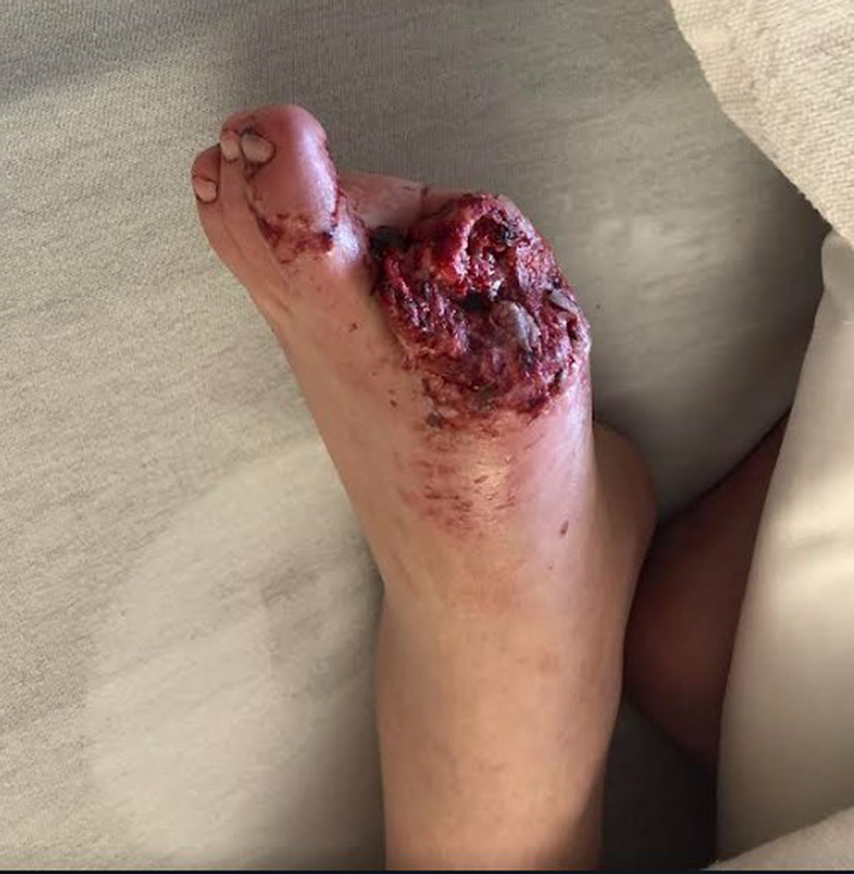
A 43-year-old paraplegic male presented to the emergency department of St. Mary’s Hospital with a chief complaint of noticing his left great toe was missing. His medical history also includes obesity, hypertension, and obstructive sleep apnea. The patient relates that he went to let out his 11-month-old puppy in the middle of the night and noticed his toe was missing. It is unknown if wound was present prior to amputation by the dog. In the emergency department, his temperature was 97.8°F; blood pressure, 119/86 mm Hg; heart rate, 130 beats per minute; and a respiratory rate of 16 breaths per minute. No labs were ordered. The wound was cleansed with 3 L of normal saline and dressed in saline, wet to dry. Tdap immunization was given in the emergency department. Vascular examination revealed palpable pedal pulses. Physical examination revealed a traumatic amputation of the left hallux at the base with course margins and peri-wound edema (
Fig. 9). A simple laceration was noted on the second digit from a dog bite. The patient started on vancomycin and went to the operating room that night for a left hallux/partial first ray resection amputation. Amputation level was determined on extent of damaged and dysvascular tissue. The wound was dressed with dry sterile dressings and an ace wrap. He was discharged with a 10-day supply of amoxicillin and clavulanate potassium. Due to the patient being paraplegic, weightbearing status was irrelevant. Healing and suture removal were uneventful.
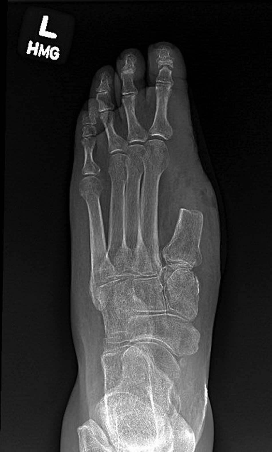
Figure 10 demonstrates the mid-shaft first-ray resection anteroposterior radiograph.
Figure 9.
View of traumatic amputation to Patient C’s left hallux at the base with course margins.
Figure 9.
View of traumatic amputation to Patient C’s left hallux at the base with course margins.
Figure 10.
Postoperative anteroposterior view of the left foot of Patient C.
Figure 10.
Postoperative anteroposterior view of the left foot of Patient C.
Discussion
All of the cases presented here consistently follow the pattern seen in prior reports: a digit or portion thereof is discovered missing, or patient or family member finds a canine actively masticating the patient’s foot. The cases all result in debridement and amputation, and fortunately, none became seriously infected, a major concern with this presentation.
As with prior reports, the authors want to express an urgent message that persons with diabetes and neuropathy who own canines should be especially wary of this possibility and take appropriate steps to prevent it from happening. These individuals must be very careful as per interactions with pets, especially with regards to sleeping arrangements. Wounds should be well covered/protected, and some have even recommended no pets at all [
19,
21].
As strange and unseemly as this phenomenon may appear, the authors feel it is worthwhile to alert those who treat patients with diabetes to the possibility that persons with diabetes and neuropathy are at risk of pet canine “debridement” of an infected wound. Of course, the primary concern here is a secondary infection from the “debridement” in a person not only already at a very increased risk for infection but also for a more serious infection. It would seem this is not comparable to maggot debridement seen in wound care.
Possible Use of Canines for Infection Alert
Can this instinctive but destructive behavior be used in a fashion similar to a diabetes alert dog to alert an owner with diabetes to an early infection, that is, to train the canine pet of a person with diabetes to alert the owner if they detect an impending infection rather than “debride” the wound? At first glance one would think the person with diabetes should themselves be able to determine if an infection is developing, but many of these patients live alone or cannot see their feet or suffer other disabilities making a proper exam difficult. By alert we certainly don't mean to begin “debriding” the wound but to sit, bark, lay down, or signal in another safe manner. The patient could then endeavor to see a professional for immediate evaluation and care. Indeed, the ability of a canine to detect infection well before a human is incredible owing to the remarkable olfactory system of the dog.
Cambau et al [
30] performed a recent extensive review on studies that evaluated a canines’ ability to detect a variety of infectious diseases. This review, looking at numerous infectious disease conditions, did not mention soft-tissue infections specifically. But in noting what the canine is actually detecting, that is VOCs of specific microorganisms, it is easy to extrapolate that an infected ulcer would be especially detectable [
30]. Therefore, it would seem that using a canine’s incredible sense of smell to detect the onset of an ulcer well before it was clinically apparent would be a terrific tool for early detection. Indeed, a trained clinician in the office may similarly detect early changes, especially if able to use technology such as temperature sensors; however, it is important to consider that the canine would be assessing a person with diabetes who may be unable to inspect their foot due to physical or visual issues. A canine that would sniff the patient’s foot and alert by sitting, barking, laying down, or otherwise signaling, would be an early indication to seek professional help promptly.
Conclusions
Subtle changes that occur in the human body, such as inadequately controlled glucose fluctuations, bacterial infections, and a variety of disease states can be detected by canines. With the evidence gathered from clinical reports and anecdotal readings, it is apparent that dogs can detect infection or necrotic tissue. Upon detection, canines are drawn to masticate the tissue leading to premature amputations of the lower extremity. Diabetic patients with associated peripheral neuropathy are predisposed to canine mastication of their lower extremity, which puts them at risk of premature amputation, infection, and death. After analysis of the literature, there is no evidence that suggests the canine’s actions were out of aggression. A canine’s ability to detect an infection prior to clinical presentation may be used as a preventive measure.
Thus far, this topic is rarely mentioned in the literature. While raising awareness of this possible canine behavior is a primary objective of this article, it is equally important to instruct providers caring for individuals with diabetes—particularly podiatrists—to inform their dog-owning patients with neuropathy to cover their feet while asleep.