Infection of the foot and ankle places patients at risk for amputation, which then places patients at risk for increased morbidity and mortality [
1]. Although major amputation does have some functional benefits in certain patient populations, limb salvage in comorbid populations should be attempted when reasonable, especially given underwhelming ambulatory and mortality data [
2,
3]. In recent years, orthoplastic and podoplastic reconstruction has seen an increase in use and value for limb salvage in the diabetic population [
4,
5]. Through the use of osseous and soft-tissue reconstruction, lower-extremity surgeons can reduce the morbidity and mortality associated with limb loss. Common tools that are used in the orthoplastic/podoplastic approach include the flap (locoregional and free) and osseous stabilization through amputation or use of fixation, internal or external. The tissue flap is the workhorse for orthoplastic/podoplastic intervention that allows clinicians to create an environment conducive to healing and provide a predictable level of soft-tissue coverage [
6–
14]. Despite the improved outcomes associated with flap use, flaps in the foot and ankle are often at risk for failure due to increased exposure to biomechanical forces secondary to the numerous mobile structures, such as joints and tendons, that are found in the region [
15–
17]. Consequently, the protection of soft-tissue reconstruction with the use of external fixators was first described by Nappi and Drabyn [
18] in 1983 and is recommended for all transarticular flaps, free or local, for the purpose of immobilizing the targeted joint and, in turn, preventing biomechanical forces across the flap interface to allow for a static environment that optimizes flap take [
16,
18–
20]. Ever since, flap-frame immobilization (FFI) with the use of an external fixator has become the standard of care for flap immobilization, shear force reduction, and off-loading; however, not much is known about the effect that FFI duration and timing may have on outcomes. Furthermore, there is a significant need for information because a great deal of the current literature focuses on the use of external fixation for the purpose of orthoplastic reconstruction in the setting of trauma as originally described by Levin [
21] as opposed to the diabetic population that requires podoplastic reconstruction, which are two drastically different populations. When evaluating the literature, there seems to be a significant amount of heterogeneity in terms of timing of FFI, which ranged from 3 weeks as described by Sagebien et al [
22] in 2007 to as high as 9.5 months as described by Lowenberg et al [
23].
Given that there are no data describing the optimal duration and timing of FFI in patients who undergo flap reconstruction, we sought to provide preliminary data on outcomes and complications. Given the potential for increased morbidity and mortality associated with failed limb salvage along with the clinical importance of limb preservation, obtaining a better understanding of and optimizing the limb salvaging intervention is paramount. We suspected that increased immobilization would result in the increased opportunity for flap maturation and healing, which, in turn, would result in increased rates of limb salvage and a reduction in complications.
Results
Initial medical record review identified 20 consecutive patients who underwent flap-frame reconstruction and were treated by the podiatric and plastic surgeons at Georgetown University Hospital over a targeted period from August 1, 2018, to March 31, 2023. Subsequent review revealed that 18 of the medical records met the inclusion criteria and two were excluded because of the lack of a targeted follow-up duration. The two medical records that were excluded were locoregional flaps; however, the patients had follow-up of 69 and 97 days.
With respect to the 18 remaining medical records (15 males, three females) that met the inclusion criteria, the mean patient age was 55.5 years (range, 40–75 years) and the mean body mass index (the weight in kilograms divided by the square of the height in meters) was 29.6 (range, 20.2–43.3). Sixteen of the 18 patients (88.9%) were diabetic; six (33.3%) had chronic kidney disease, with half undergoing dialysis; and seven (38.9%) had peripheral vascular disease. The mean Charlson Comorbidity Index was 4.7 (range, 1–7), and one mortality occurred 643 days after FFI due to unrelated causes. The mean defect size at the time of flap inset was 110.9 cm2 (range, 36–500 cm2). Thirteen feet (72.2%) had wounds that were plantar, and the remaining five were in the nonplantar region. Eleven of the wounds (61.1%) were in the midfoot region, and seven (38.9%) were in the rearfoot region (all of the heel wounds). Three patients had an antecedent guillotine/open midfoot amputation (two transmetatarsal and one Chopart) that required flap coverage, and the remaining four amputations were fifth-ray resections. Three patients (16.7%) had Charcot’s neuroarthropathy that required surgical intervention at the time of flap-frame intervention.
Regarding hematologic data, the average hemoglobin A1c level at the time of flap inset was 7.9% (range, 5%–14%), and the average albumin level was 3.3 g/dL (range, 2.0–3.9 g/dL). Eight of 16 patients (50%) had angiographically evident three-vessel, six (37.5%) had two-vessel, and two (12.5%) had one-vessel runoff, which was determined by a single vascular surgeon in our department. Two patients did not have a recorded angiogram that allowed for the characterization of inflow patterns.
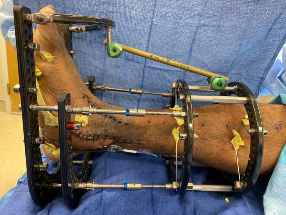
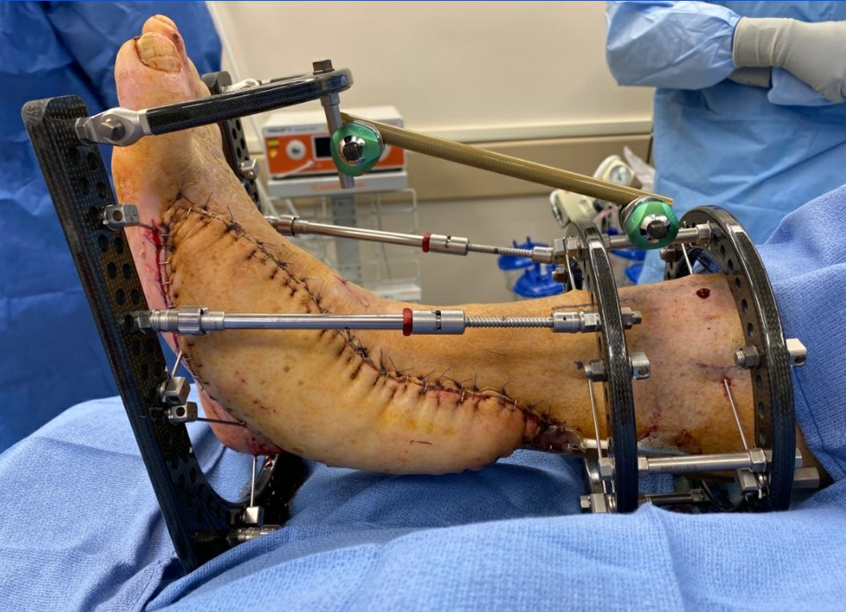
All of the patients were treated with a multiplanar circular external fixator for a mean of 39.4 days (range, 20–77 days). No instances of frame- or flap-related complications requiring premature removal of external fixator were noted. Seventeen of the circular frames (94.4%) were static, with no plans for gradual correction, and one construct was dynamic, with gradual compression achieved through the frame due to the presence of concomitant Charcot’s neuroarthropathy before staged and eventual internal fixation (5.6%). Of note, 66.7% of Charcot’s reconstructions had internal fixation placed at the time of FFI. The mean time between flap inset and subsequent application of the external fixator was 8.2 days (range, 0–48 days). Sixteen limbs (88.9%) were treated with a four-ring construct (
Fig. 1), and the remaining two (11.1%) with a three-ring construct (
Fig. 2). The average ring diameter was 159.7 mm (range, 140–180 mm), an average of 8.7 skinny wires were used (range, 7–9), along with an average of 2.2 olive wires (range, 0–4) and 0.5 half pins (range, 0–4). In total, seven frame-related complications occurred in four patients (22.2%). Five instances of pin-site infections were observed in three patients (16.7%), equating to a 1.7% pin-site infection rate relative to the overall number of pin sites (n = 302). One instance of wire breakage was noted, and the wire was subsequently removed without compromising the overall construct stability, and, last, there was one case of chronic sinus tract formation where a previous fixator half pin was placed that was subsequently treated with operative debridement and local wound care.
Figure 1.
Intraoperative photograph demonstrating a four-ring external fixator construct for the purpose of off-loading an anterolateral thigh flap.
Figure 1.
Intraoperative photograph demonstrating a four-ring external fixator construct for the purpose of off-loading an anterolateral thigh flap.
Figure 2.
Intraoperative photograph demonstrating a three-ring external fixator construct for the purpose of off-loading an anterolateral thigh flap.
Figure 2.
Intraoperative photograph demonstrating a three-ring external fixator construct for the purpose of off-loading an anterolateral thigh flap.
Eleven flaps (61.1%) were fasciocutaneous, and the remaining seven (38.9%) were vascularized muscle flaps (MFs). Overall, the most common flap was the anterolateral thigh flap, which was the only type of fasciocutaneous flap used (
Figs. 1 and
2). The most common MF was the vastus lateralis MF (
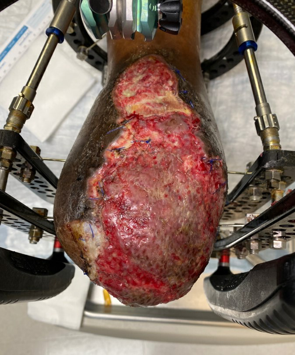
Fig. 3), which was used in 22.2% of the cases (n = 4). Other MFs that were used were the latissimus dorsi MF (n = 2) and the abductor hallucis MF (n = 1). All of the MFs required subsequent skin grafting that was performed at a mean of 9 days (range, 0–49 days) after flap inset.
Figure 3.
Clinical photograph showing a free vastus lateralis muscle flap, the most common muscle flap in the cohort, to cover an open Chopart amputation.
Figure 3.
Clinical photograph showing a free vastus lateralis muscle flap, the most common muscle flap in the cohort, to cover an open Chopart amputation.
Overall, a 66.7% limb salvage rate (12 of 18 patients) was observed, with mean follow-up of 2.4 years, or 892.6 days (range, 222–1,555 days). All of the failures resulting in a BKA were attributed to recurrent infection (three of six patients) or hardware complications (three of six patients), which resulted in a BKA a mean of 123 days (range, 33–227 days) after external fixator removal. No instances of major flap complications resulting in total flap loss were observed, resulting in 100% flap survival; however, there was a 38.9% rate of minor flap complications (seven of 18 patients) requiring a return to the operating room. The most common minor flap complication was dehiscence of the flap margins, which occurred in six flaps (33.3%); however, all were managed with operative debridement or local wound care, and one complication was partial split-thickness skin graft necrosis (80% graft take) in the MF group that required reapplication of a split-thickness skin graft without incident.
The data set was divided into two groups based on successful limb salvage and failed limb salvage resulting in a BKA. In addition, the data were chronologically subdivided into equal tertiles (six patients per tertile) based on time spent in the external fixator. The mean duration of FFI in the successful limb salvage group was 40.6 days (range, 20–77 days), and 37.2 days (range, 22–56 days) for the failed limb salvage group, with an average delay between flap and frame application of 9.7 days (range, 0–48 days) and 5.3 days (range, 0–10 days), respectively. The mean defect size at the time of flap inset for the limb salvage group was 70.4 cm2 (range, 36–120 cm2) and for the failure group was 191.8 cm2 (range, 40–500 cm2). When evaluating the tertiles, there was a 50% failure rate (three of six patients) in the upper tertile, a 100% success rate in the middle tertile, and, again, a 50% failure rate (three of six patients) in the bottom tertile. The mean defect size was 127.7 cm2 (range, 42–322 cm2) in the upper tertile, 63.8 cm2 (range, 36–108 cm2) in the middle tertile, and 141.2 cm2 (range, 40–500 cm2) in the lower tertile. The mean duration of FFI in the upper tertile was 58.3 days (range, 44–77 days), in the middle tertile was 35.8 days (range, 29–44), and in the lowest tertile was 24.2 days (range, 20–28 days). The time between flap inset and frame application was 5.67 days (range, 0–9 days) in the upper tertile, 11.8 days (range, 0–48 days) in the middle tertile, and 7.2 days (range, 0–13 days) in the bottom tertile. Regarding the number of minor flap–related complications (N = 7), 0%, 57.1% (n = 4), and 42.9% (n = 3) complication rates were observed in the upper, middle, and lower tertile, respectively. Last, frame-related complications occurred at a rate of 5.6% (n = 1), 5.6% (n = 1), and 11.1% (n = 2) in the upper, middle, and lower tertiles, respectively.
Discussion
External fixation has proved itself to be a valuable asset in podoplastic reconstruction because of its ease of use and inherent stability, which is advantageous when used in combination with soft-tissue reconstruction in regions of increased biomechanical forces. Despite the literature being plentiful about the use of free and locoregional flaps for the purpose of flap-based limb salvage, the literature is nonexistent on the purposeful examination of the timing that FFI may have on outcomes and complications, and in many instances, complications are not mentioned at all [
25–
31].
In 2008, Lowenberg et al [
19] evaluated ten patients who spent a mean of 12 weeks in a circular external fixator and demonstrated a 20% complication rate. Again, in 2008, Clemens et al [
16] described the use of FFI with the use of multiplanar external fixation in 12 patients who were off-loaded for a mean of 66 days and reported a 50% complication rate. In 2022, Jafari Kafiabadi et al [
30] conducted a pilot study in 26 patients who underwent FFI with the use of a circular external fixator and concluded that the use of an Ilizarov external fixator resulted in a 0% total flap failure rate, but there was no mention of minor flap complications outside of swelling [
30]. The largest cohort to date describing FFI secondary to trauma requiring orthoplastic reconstruction is an article by Hollenbeck et al [
32] in which the average time spent in a frame across 62 patients was 6.9 months, and a 24% complication rate was noted; however, it is not clear whether outcomes in relatively healthy trauma patients can be translated to comorbid diabetic patients [
32]. Overall, we reported a flap complication rate of 38.9% and a limb salvage rate of 66.7% in patients who were immobilized for a mean of 39.4 days, which is relatively comparable with the wide range of reported complication rates in the scant literature in which complications and timing were both reported [
16,
19,
32].
Interestingly, when the data were chronologically divided, we observed a bimodal distribution of failures and a unimodal distribution of flap complications, which could be an argument for a potential “Goldilocks zone” of duration of FFI when it comes to reducing flap-related complications. The upper tertile, or the group with the longest duration of FFI, demonstrated a 0% flap-related complication rate but a 50% limb salvage rate. This counterintuitive increase in failures with increased time spent in an external fixator is likely related to the increased complexity and locoregional trauma associated with the type of cases that required an increased amount of time in the frame, such as concurrent osseous reconstruction or open amputation, which consisted of one open midfoot amputation, one concurrent midfoot Charcot’s reconstruction, and a previous partial calcanectomy that occurred before free flap inset for the treatment of calcaneal osteomyelitis. The middle tertile demonstrated a limb salvage rate of 100% and a flap-related complication rate of 57.1%, which is most comparable with the findings by Clemens et al [
16]. Although it is not clear why the middle tertile demonstrated the largest number of complications, we posit that this trend may be related to the timing between flap inset and frame application as the average time between these two critical events was 1.5 times longer than the time between events compared with the upper and lower tertiles. Consequently, it is likely that the delayed elimination or neutralization of biomechanical forces across the flap interface during the acute phases of healing may negatively impact healing and result in a higher rate of flap complications [
33,
34]. In the lower tertile, the limb salvage rate was 50%, along with a 42.9% flap-related complication rate after a mean duration of FFI of 24.2 days. As was the case in the upper tertile, the failures were cases that required increased metabolic demand for healing, with one open midfoot amputation, one concurrent ankle Charcot’s reconstruction via internal fixation, and a non-Charcot’s ankle arthrodesis with the use of internal fixation.
Although external fixation is certainly useful for podoplastic intervention, it is not without inherent morbidity. We reported a frame-related complication rate of 22.2%. The use of an external fixator has inherent disadvantages and can result in pin-site infection rates as high as 38%, as demonstrated by Gopal et al [
35]; however, the use of external fixation, or more specifically, a circular external fixator, is advantageous because it allows for increased rates of ambulation during treatment and increased visibility of the flap for observation and monitoring [
30,
35]. Aside from the articles by Clemens et al [
16], Jafari Kafiabadi et al [
30], and Lowenberg et al [
19], we found it difficult to effectively compare the present results with the literature because almost all of the flap-frame literature describes the use of FFI in the setting of traumatic open fractures and wounds, and the literature that does discuss the use of FFI in a comparable diabetic cohort often describes the use of a delta external fixator for strict off-loading, which does not have the inherent benefits of a circular external fixator in terms of possible ambulation and overall construct stability [
36–
38].
Overall, the present findings hint that the use of external fixation should be considered especially when a flap is placed in the foot and ankle region. Historically, external fixation is considered when a flap crosses major joints (ie, the ankle joint), but we posit that the effects of tendinous structures and multiple smaller joints are additive and should be immobilized in any circumstances, especially if the flap is on the plantar foot. The result of our observation hints that failure of podoplastic intervention may likely be related to the size of the targeted defect and the magnitude of concurrent reconstruction, both soft tissue and osseous, as opposed to FFI duration, which results in an increased metabolic demand for sufficient healing in a comorbid population that may not be able to meet those requirements for healing, as evidenced by a mean Charlson Comorbidity Index score of 4.7. Furthermore, the presence of Charcot’s neuroarthropathy and open amputation seems to have an effect on the podoplastic limb salvage rate. In the present study, 66.7% of the cases (four of six) that resulted in a BKA were in the setting of Charcot’s neuroarthropathy (two of six) that required osseous intervention or open amputation (two of six). In the absence of open amputation or Charcot’s neuroarthropathy, the limb salvage rate was noted to be 85.7% (12 of 14 patients). It is well documented that the presence of Charcot’s neuroarthropathy places patients at increased risk for proximal amputation, as demonstrated by Elmarsafi et al [
39] in 2019, and findings by Berceli et al [
40] support the conclusion that open amputation is a potential risk factor for limb salvage failure in podoplastic intervention.
Despite an interesting bimodal distribution of failures that does not seem to be related to the use of external fixation, we believe that the timing and duration of FFI is most impactful on the rate of complications as opposed to limb salvage rates. The reduction of time spent in an external fixator may likely result in an increased rate of complications secondary to the reduced time for flap neovascularization. According to the literature, neovascularization peaks in healthy tissue 7 to 10 days after flap inset, and it is reasonable to assume that the neovascularization in diabetic patients occurs well after the first week and may reach a stable plateau at an average of 35 days, further explaining the drastic drop in flap-related complications in the upper tertile [
33,
34]. As a result, we believe that immobilization with an external fixator should optimally occur simultaneously or within 7 to 10 days to allow for maximization of neovascularization through the reduction of biomechanical forces and, in turn, the reduction of dehiscence complications. If simultaneous application of the external fixator is not possible, then we recommend placement of a splint or immobilizing dressing with strict nonweightbearing until a frame is applied. This conclusion is largely supported by the rate of complications in patients who underwent same-day flap and frame application as one of the flap-related complications (14.3%) was in a patient who underwent simultaneous flap and frame application within the same day, whereas the average delay between flap and frame application for the remaining 85.7% of complications (six of seven) was 16 days. Even more, based on our observation, when possible, a longer duration of FFI seems to be most beneficial for flap healing because flap healing and revascularization can take up to 1 year to allow full incorporation free of any dependence of the vascular pedicle and anastomosis [
41–
43]. Given the small sample size and retrospective nature of this observational case series, generalizable conclusions are weaker in strength, and it is unclear whether the conclusions are significant from a statistical perspective, which we regrettably were not able to achieve due to low power. Also note that the purpose of this evaluation was to generate hypotheses for which future studies of larger volume may lend statistical strength to our observations. Regardless, we believe that this attempt to establish a preliminary relationship between timing and duration of FFI will benefit future studies and can impact clinical decision making. As a result of these preliminary findings, at Georgetown University Hospital, the targeted duration of FFI is typically around the 28- to 35-day mark. Future studies of greater size and volume are needed to help clinicians better understand the effectiveness of FFI in the hopes of increasing the rates of limb preservation and preventing the morbidity associated with major lower-extremity amputation.
In conclusion, based on these findings and the current literature, we believe that knowledge of the effect of external fixator timing is imperative to optimize outcomes in podoplastic and flap-based limb salvage. Regardless, we conclude that timing and duration of FFI is impactful, especially considering the well-documented morbidity that is associated with free flap failure and subsequent limb loss.