The plantar fascia is an essential anatomical structure that is responsible for stabilizing the foot during gait. It is a band of connective tissue comprising three bands that originate at the medial tubercle of the calcaneus and fan distally to insert into the base of each proximal phalanx of the digits [
1]. The plantar fascia tenses during the terminal stance to toe-off phases of gait, and this tension elevates and reinforces the medial longitudinal arch, which in turn allows the foot to function as a rigid lever for forward propulsion.
Degeneration of the plantar fascia, known as plantar fasciitis (PF), is a common problem that an estimated one in ten people will experience in their lifetime [
2]. Plantar fasciitis is caused by overuse, resulting in repetitive microtrauma and degeneration of the fascia at its calcaneal insertion. Lemont et al [
3] looked at 50 cases of PF that had been treated with heel spur surgery and failed to find histologic evidence of inflammation. Histologically, the condition is considered fasciosis [
3,
4].
The etiologies of PF are numerous and may include, but are not limited to, excessive pronation of the foot, trauma, and excessive running [
5]. Patients classically present with a complaint of heel pain that is worse with first steps in the morning, known as poststatic dyskinesia. The pain usually improves as activity continues but can return at the end of the day. On clinical exam, the plantar medial tubercle of the calcaneus is maximally tender to palpation at the insertion of the plantar fascia. Risk factors for PF include tightness of the Achilles tendon, obesity, pes planus, overpronation, pes cavus, overuse, and prolonged periods of weightbearing.
Radiographic imaging is typically the initial study performed out of practicality; however, radiographs offer only minimal findings suggestive of PF. These findings are limited to thickening of the plantar fascia or heel spurs, which can often be incidental rather than pathognomonic of PF. Radiographs are useful to rule out osseous lesions, trauma, or underlying abnormality or deformity that could be causing heel pain. Musculoskeletal ultrasound and magnetic resonance imaging are more accurate at depicting the soft tissue and its associated pathology but are usually not the initial studies performed because of cost or availability.
In gait, the heel serves as the first point of contact of the body with the ground and is the primary absorber of the ground reaction force. The calcaneus is supported and cushioned during the heel strike phase of gait by the calcaneal fat pad (CFP), which serves as a load bearer and shock absorber and can minimize superstructural impact because of its unique dynamic, viscoelastic structure [
6,
7]. It is estimated that the CFP absorbs between 20% and 25% of the shock of heel strike [
8]. It is composed of a bilayer of closely packed fat chambers enclosed by elastin fibers and circumferential collagenous septa [
9]. In a study by Ledoux and Blevins [
10], the CFP, when subjected to variable loads, was found to exhibit a prolonged relaxation time accompanied by an increase in the modulus of elasticity and a decrease in energy dissipation. The force-dissipating effects of the heel fat pad were observed to be critical for reducing the effect of loads absorbed by the calcaneus and surrounding structures [
7,
10]. In patients with decreased CFP thickness, the phasic activity of the soleus has been shown to increase from its normal 40% of the gait cycle to 60% of the gait cycle, indicating that it is contracting for almost the entire stance phase of gait [
8].
In previous studies, it has been reported that the CFP thickness in healthy adults is approximately 1.5 to 1.8 cm [
6,
11]. There are many different things that can contribute to a reduction in CFP thickness, such as aging, obesity, running repetitively on hard surfaces, diabetes, rheumatoid arthritis, and corticosteroid injections [
9]. Significant alterations in the histomorphology of the CFP involve incrassation of septa, disorganization of septa caused by breaking of collagen bundles and fragmentation of elastin strands, and relative shrinking of adipocytes [
12]. These alterations subsequently result in further modifications of biomechanical properties, causing increased stiffness of septa, decreased damping ability, and increased vulnerability to tissue injury. In older patients, histology of the CFP shows atrophy [
4].
It has been suggested in the literature that individuals with decreased CFP thickness or degeneration of the CFP often present with plantar heel pain [
6,
9]. The CFP is a closely related anatomical structure to the plantar fascia, overlying the inferior and posterior aspects of the calcaneus, as well as the proximal plantar fascia [
9].
There is currently limited research delving into the relationship between PF and the CFP. A study by Osborne et al [
13] concluded that the key radiologic features for diagnosing PF on a lateral nonweightbearing radiograph are thickening of the plantar fascia at the origin >0.5 mm and an abnormal-appearing CFP. The CFP is usually triangular in appearance and is seen immediately deep to the plantar fascia at the calcaneal origin. It becomes narrowed or absent on radiographs in individuals with PF. Although the mechanism is unknown, it may be associated with mechanical and inflammatory mechanisms related to chronic PF. This study, however, did not mention or measure the CFP thickness, and the radiographs were nonweightbearing.
A study by Rome et al [
14] concluded that thicker heel pads might be a contributing factor in the development of plantar heel pain. The authors suggested that the increased CFP thickness in patients with plantar heel pain may represent the accumulation of repetitive microtrauma and consequent inflammatory edema. By contrast, a study by Belhan et al [
15] measured CFP in patients with PF through ultrasound and identified that a decrease in CFP thickness existed in patients with PF.
We hypothesized that if the CFP thickness is reduced, there may be an increased incidence of PF secondary to decreased support and shock absorption to the heel and foot. Thus, individuals with decreased CFP thickness or damage to the CFP would be more susceptible to heel and arch pain. The purpose of this study was to determine if a relationship exists between radiographic CFP thickness and the incidence of PF.
Materials and Methods
This study was conducted at the Foot Center of New York and was approved by the institution’s institutional review board. This research was designed as a retrospective study and involved 20 patients. The study cohort was made up of ten patients with a provisional clinical diagnosis of PF who met the specific study inclusion criteria. The diagnosis of PF in this group was based on patient history, clinical examination, and weightbearing radiographs. The inclusion criteria for the study cohort were patients with a diagnosis of PF, patients aged 18 to 70, and patients with a weightbearing lateral radiograph in the AccuVueCloud software (Radmedix, Kettering, Ohio). The exclusion criteria for the study cohort were 1) patients with a nonweightbearing lateral radiograph in the AccuVueCloud software, 2) obese patients with a body mass index ≥35 kg/m2, 3) patients with a history of nerve impingement or tarsal tunnel syndrome, 4) patients with a prior forefoot/rearfoot surgery, 5) patients with a history of fibromyalgia, 6) patients with a history of chronic regional pain syndrome, 7) patients who were not within the age range of 18 to 70, 8) pregnant patients, and 9) patients with a history of amputation of any part of the foot.
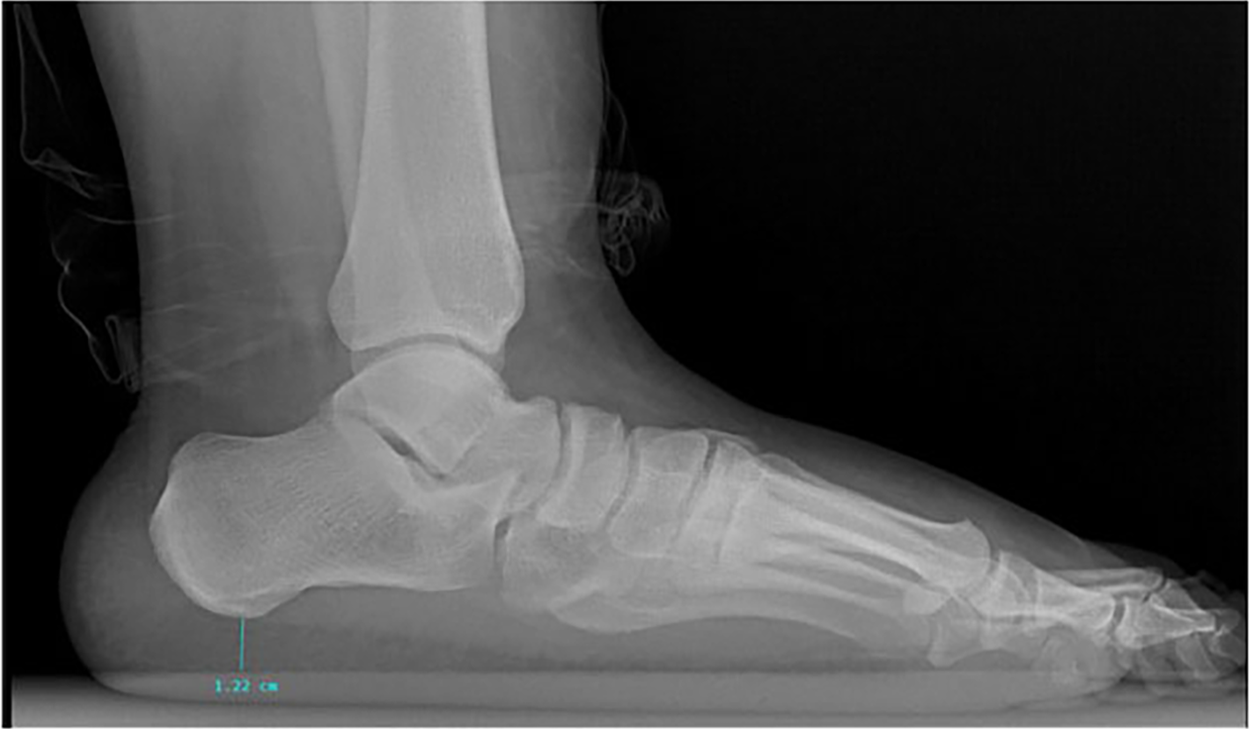
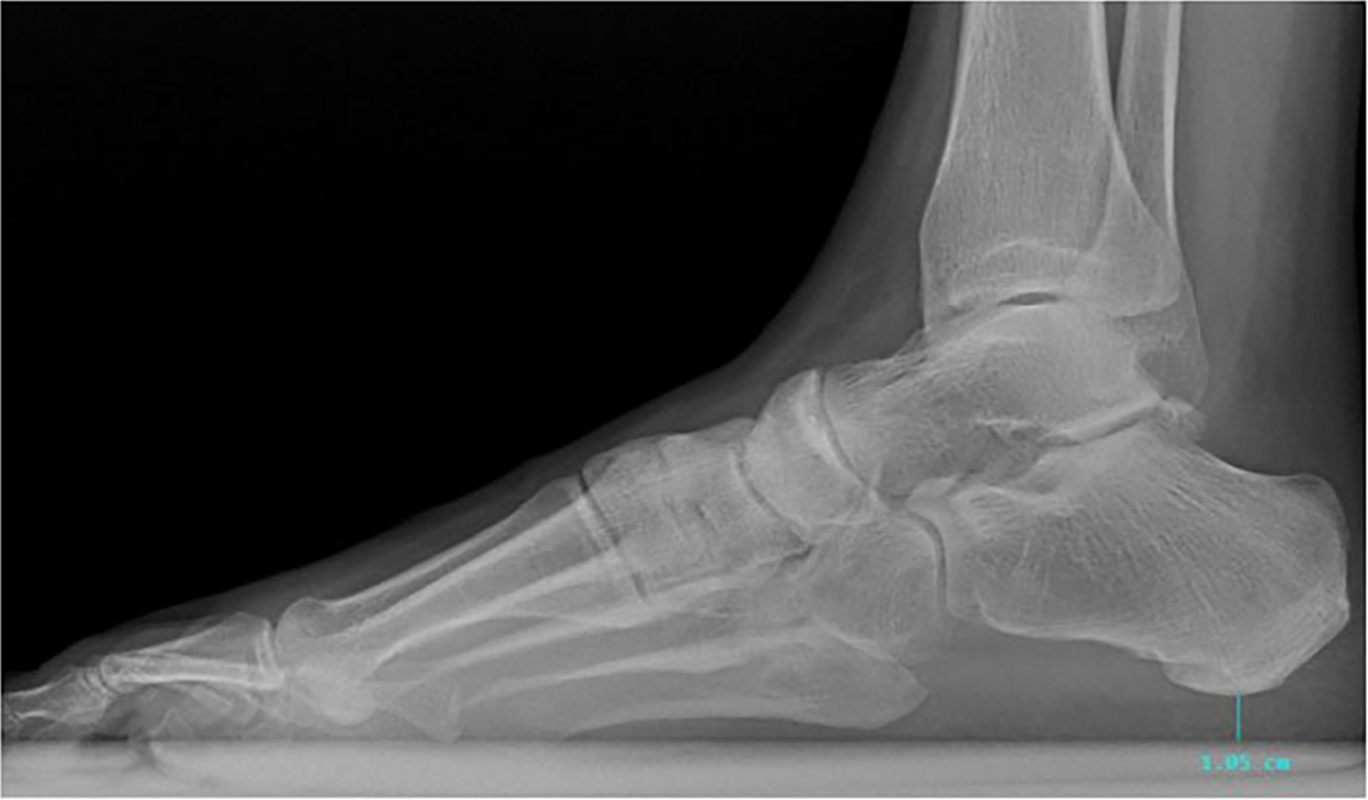
The control group was made up of ten Foot Center of New York patients who met the proper inclusion criteria, which included having no known history of plantar fascia pathology, age 18 to 70, and having weightbearing lateral radiographs in the AccuVueCloud system. The exclusion criteria for the control group were the same as those for the study group. Intrarater reliability was maintained by measuring the CFP thickness using the ruler tool in the AccuVueCloud radiography software from the same points of reference. The thickness of the fat pad was measured from the most inferior point of the plantar tuberosity of the calcaneus to the most superior point on the imaging plate. Visual examples of the CFP thickness using the ruler tool and reference points for the control group and the study group can be seen in
Figures 1 and
2, respectively. The radiographic CFP thickness was then compared (
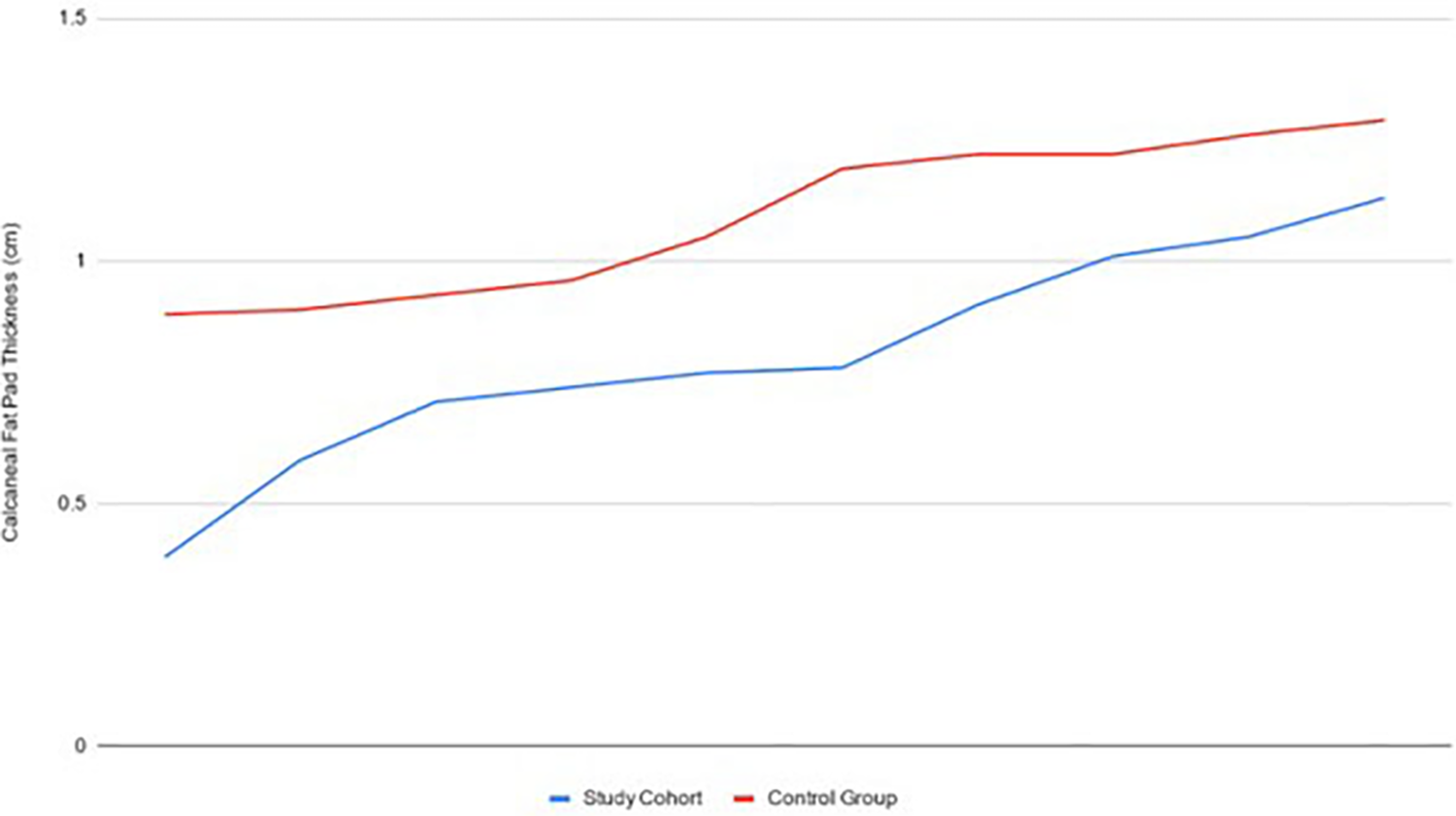
Fig. 3) between the study group and the control group using statistical analysis.
Figure 1.
Example of how measurements of the thickness of the calcaneal fat pad were taken on lateral weightbearing radiographs of a patient from the control group.
Figure 1.
Example of how measurements of the thickness of the calcaneal fat pad were taken on lateral weightbearing radiographs of a patient from the control group.
Figure 2.
Example of how measurements of the thickness of the calcaneal fat pad were taken on lateral weightbearing radiographs of a patient from the study group.
Figure 2.
Example of how measurements of the thickness of the calcaneal fat pad were taken on lateral weightbearing radiographs of a patient from the study group.
Figure 3.
Comparison of the calcaneal fat pad thickness of the study cohort (blue) and the control group (red), depicting less thickness in patients in the study cohort.
Figure 3.
Comparison of the calcaneal fat pad thickness of the study cohort (blue) and the control group (red), depicting less thickness in patients in the study cohort.
Results
Statistical analysis was performed after gathering the data of the control and study groups and finding the averages of both groups. The average of the control group after measuring the CFP on ten different radiographic images of patients who met the inclusion criteria was 1.091 cm. The average of the study group after measuring the CFP of ten different radiographic images of patients who met the inclusion criteria was 0.808 cm. A t test was then conducted to determine if there was a significant difference between the means of the two groups, and a P value of 0.0045 was obtained.
Discussion
In the literature, it has been reported that the CFP thickness in healthy adults is approximately 1.5 to 1.8 cm [
6,
11]. This reported range differs slightly from our study, in that our control group CFP thickness was comparatively lower, at 1.1 cm. One reason to suggest this difference in results could be related to foot positioning at the time of the radiograph. Our study was performed retrospectively, so there was no way to control positioning of the foot at the time of the lateral radiograph. Moving forward, controlling the position of the foot during radiographs to ensure proper angle and base of gait would create more consistent, reliable, and reproducible results. Another reason for the difference could be that in our study we measured the distance between the most inferior portion of the plantar tuberosity of the calcaneus and the most superior point on the imaging plate, whereas the study by Campanelli et al [
11] measured posterior to the plantar tuberosity. Another consideration we did not take into account with our results was the difference in CFP thickness measurements between males and females.
Our study results varied considerably from the study by Rome et al [
14], which measured CFP thickness in an athletic population (runners). The CFP thickness in the group without heel pain was 0.502 cm, the CFP thickness in the control group (considered not runners) was found to be 0.505 cm, and those with plantar heel pain had the thickest measurement of 0.575 cm. The authors concluded that thicker heel pads might be a contributing factor in the development of plantar heel pain and suggested that the increased CFP thickness in patients with plantar heel pain may represent the accumulation of repetitive microtrauma and consequent inflammatory edema. Our study results likely varied as a mechanism of the patient population, the variation between ultrasound in the study by Rome et al and radiography in ours, and foot positioning.
The study by Belhan et al [
15] that measured CFP thickness in patients with PF using ultrasound had results similar to our study, showing a decrease in CFP thickness in patients with PF. In that study, patients with heel pain had a CFP thickness of 1.945 cm (range, 1.2–2.9 cm), whereas patients without heel pain had a CFP thickness of 1.994 cm (range, 1.3–2.9 cm). A key factor in the variation in results that could explain the authors’ overall larger values for CFP thickness is that the measurements were not taken with patients weightbearing, but rather with constant pressure applied to the ultrasound probe.
In our study, patients in the study group had an average CFP thickness of 0.808 cm, whereas patients in the control group had an average CFP thickness of 1.091 cm. The results from our study show that there is a relationship between the CFP and PF, with decreased CFP thickness observed in patients with PF. Radiographs, typically the first imaging modality ordered in the clinical setting, have not been used before to analyze the relationship between PF and decreased CFP thickness. Our study shows that measuring the CFP thickness on lateral radiographs could be a useful diagnostic tool and help guide early treatment protocols for PF.
Conclusions
Calcaneal fat pad pathology is a major contributor to abnormal shock and overuse injury in the lower extremities [
8]. Our study suggests that there is a relationship between CFP thickness and the incidence of PF. Plantar fasciitis is usually diagnosed clinically based on a detailed history and physical examination. Radiographs are typically the first imaging modality used, but prior to our study, they lacked usefulness in confirming a diagnosis of PF. Using the ruler tool on a lateral weightbearing radiograph to measure the CFP reference points suggested in this study may help confirm the diagnosis of PF in a symptomatic patient or identify patients at risk.
A CFP measurement of 0.81 cm (approximately the average thickness of the fat pad in the study group) or less on a weightbearing lateral radiograph would be consistent with a decrease in CFP thickness and concerning for CFP degeneration as a causative factor in PF symptoms. This finding could help with early identification and treatment of PF and improve prognosis. Additionally, considering predisposing factors in CFP atrophy/degeneration such as age, weight, employment, athletic activity, and medical history (diabetes, rheumatoid arthritis) could help with early preventative measures to preserve normal shock absorption as well as diagnosis and treatment. Prophylactic measures in high-risk populations should be taken to preserve the architecture and density of the CFP. Artificial external shock-absorbing materials in footwear should be encouraged [
8].
A limitation of our retrospective study is that foot position during radiograph examination was not controlled. The study could be improved by controlling interrater reliability between researchers measuring fat pad thickness. In addition, age, foot type, and occupation are variables that can be considered when assessing factors contributing to a diagnosis of PF. Our study and results were limited by the small number of participants examined. The relationship between the CFP thickness and PF needs further evaluation with more robust studies in the future. The specificity and sensitivity of ultrasonography as well as digital calipers to measure the CFP thickness may yield stronger evidence in support of our study results.