Circular ring external fixation has become increasingly common since it was first introduced by Gavriil Ilizarov in the 1950s, with a variety of applications in the foot and ankle. Complex or open fractures of the tibial pilon, ankle, calcaneus, and Lisfranc joint have all been treated successfully [
1,
2,
3,
4,
5,
6]. Other indications include ankle and subtalar joint arthrodesis, management of osteomyelitis, off-loading wounds, correction of congenital or acquired deformities, and reconstruction of the Charcot foot and ankle [
7,
8,
9,
10,
11,
12,
13,
14,
15,
16,
17,
18,
19,
20,
21,
22]. The use of circular ring external fixation devices allows the foot and ankle surgeon to address a variety of pathologies that would be difficult to manage with traditional internal fixation alone. It has the potential advantages of allowing less invasive approaches, monitoring of soft tissues and local wound care, adequate fixation of osteopenic bone, and gradual correction of deformity. There are several complications that can occur with the use of circular ring external fixation of the foot and ankle, including neurovascular injury, edema, traction injuries, and wire or pin-site irritation, infection, or breakage. Of all of the complications, those involving wires or pins are by far the most common. Complication rates can be high in external fixation, but the literature is limited to small case series. To our knowledge, this study is the largest review of complications associated with circular ring external fixation to date.
Of all of the indications for circular ring external fixation in the foot and ankle, its use for management of the Charcot foot or ankle deformity has become common among foot and ankle surgeons, with dozens of published studies. These patients are generally considered high-risk surgical candidates, with many of them having diabetes, poor glycemic control, neuropathy, peripheral artery disease, or immunopathy. We hypothesized that patients with higher body mass index (BMI; calculated as the weight in kilograms divided by the square of the height in meters), peripheral artery disease, and worse glycemic control would have more complications. In these scenarios, the application of circular ring external fixation is often considered preferable to traditional internal fixation for management of neuropathic fractures, ulceration, and osteomyelitis. It has been used as the only method of fixation, as part of a staged management with subsequent internal fixation, and with concomitant use of internal fixation [
16,
18,
19,
20,
23,
24,
25,
26].
Circular ring external fixation has been shown to be successful in the treatment of foot and ankle pathology in diabetic patients with multiple comorbidities and soft-tissue compromise, but the procedures are still technically demanding, and complications are common. Definitions and rates of complications vary widely in the literature, with complication rates as high as 1.0 to 1.7 per patient [
8,
27]. However, most of these are considered pin-related complications [
7,
19,
22,
27,
28,
29]. Despite the significant number of reports on the use of circular ring external fixation for foot and ankle pathologies, they are generally small, retrospective studies, and only a handful specifically investigate their complications [
22,
27,
30]. The heterogeneity among the small studies at present led us to investigate the complications encountered at our institution. The primary goal of this study was to identify complications associated with circular ring external fixation, and the secondary aim was to identify any independent risk factors associated with them.
Methods
Institutional review board approval was obtained, and patient medical records were reviewed from March 1, 2006, to December 31, 2016, for patients who were surgically treated with circular ring external fixation. The only inclusion criterion was undergoing circular ring external fixation. The only exclusion criterion was incomplete medical record information. Medical records were searched using
Common Procedural Terminology code 20692 (application of multiplanar external fixation). After eliminating all of the pin-to-bar fixators, there were 113 consecutive procedures identified that were performed by the senior author (P.R.B.). Thirteen procedures were excluded based on incomplete medical records, giving a total review of 100 procedures in 99 patients. Their demographic and clinical characteristics are presented in
Table 1. To identify any independent risk factors associated with complications, the clinical characteristics and indications for circular ring external fixation were compared in patients with and without complications.
Table 1.
Patient Demographic and Clinical Characteristics and Indications for Procedures.
Table 1.
Patient Demographic and Clinical Characteristics and Indications for Procedures.
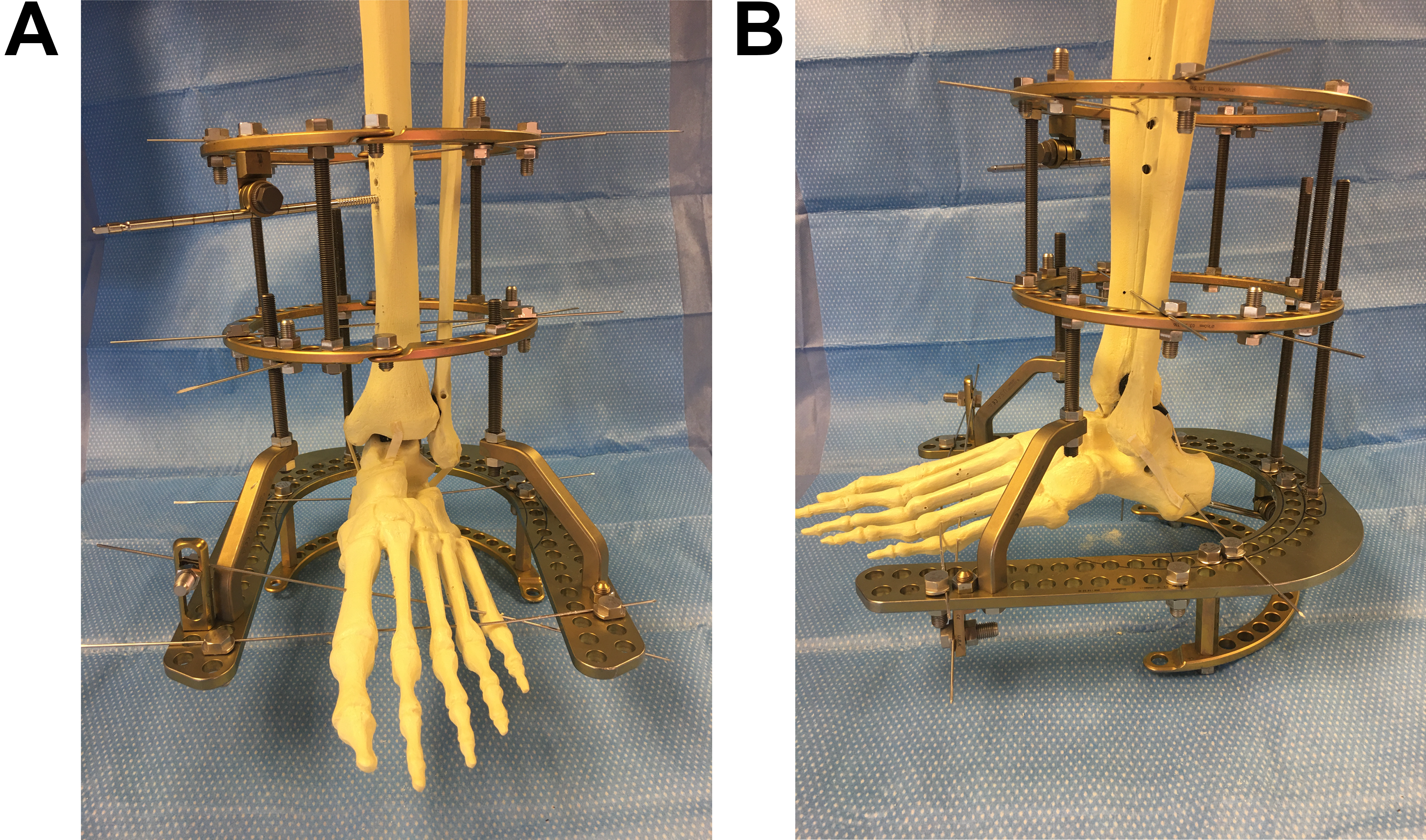
The surgical method, frame construct, number of wires versus half-pins, pin-site care, and weightbearing status were individualized according to treatment goals, patient requirements, and available supplies. Our constructs were mostly standardized to increase efficiency. The standard frame construct used a tibial block with two rings, four fine wires, and one half-pin. For hindfoot/ankle pathologies we used a foot plate with two fine wires in the forefoot and usually two fine wires or a half-pin in the calcaneus (
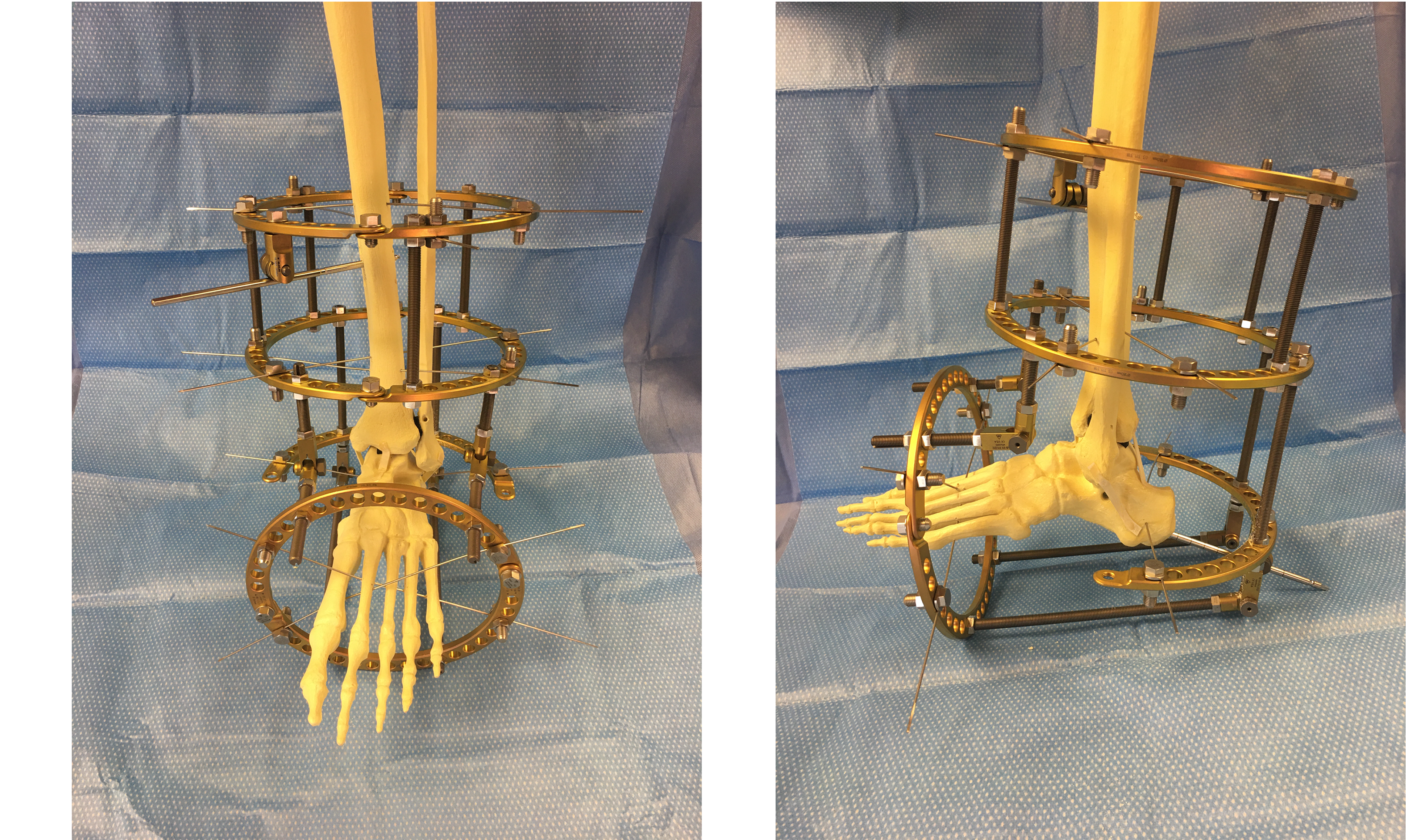
Fig. 1). For midfoot pathologies we used a foot ring with two fine wires in the foot and a half-pin in the calcaneus (
Fig. 2). Fine wires and half-pins were inserted according to anatomical safe zones with a wet lap sponge, pulsed drilling, and copious irrigation to minimize thermal necrosis. Wires were tensioned with a dynamic wire tensiometer or by Russian technique. Pin-site dressings evolved over the years of treatment from various patches to xeroform and dry gauze.
Figure 1.
Anterior (A) and lateral (B) views of a typical construct for hindfoot or ankle pathologies with a tibial block and foot plate. Seven fine wires and two half-pins were used.
Figure 1.
Anterior (A) and lateral (B) views of a typical construct for hindfoot or ankle pathologies with a tibial block and foot plate. Seven fine wires and two half-pins were used.
Figure 2.
Anterior (A) and lateral (B) views of a typical construct for midfoot or forefoot pathologies with a tibial block and foot ring. Seven fine wires and two half-pins were used.
Figure 2.
Anterior (A) and lateral (B) views of a typical construct for midfoot or forefoot pathologies with a tibial block and foot ring. Seven fine wires and two half-pins were used.
Patient demographics, duration of treatment, and complications were recorded by multiple investigators (C.D.B., N.S.P., P.R.L.). Complications were divided into minor and major complications. A minor complication was an event that did not require a change in the treatment course and included wire irritation or drainage, mild infections treated with oral antibiotics, and loosening that did not require frame revision or return to the operating room. Major complications required change in treatment plans and included wire breakage, frame shifting, or infected wires/pins requiring exchange, revision, or early removal of the frame.
Continuous variables are summarized with means and standard deviations. Categorical variables are summarized with frequencies and percentages. Categorical variables were tested for an association with presence of a complication with the χ2 or Fisher exact test, and continuous variables were tested with the two-sample t test. Logistic regression analysis estimated the odds ratio with a 95% confidence interval controlling for predictor variables of overall complications. The BMI was dichotomized into obese (BMI ≥ 30) and nonobese (BMI < 30) categories. An independent investigator performed all of the data analysis using a statistical software program (IBM SPSS Statistics for Windows, Version 24.0; IBM Corp, Armonk, New York).
Results
For all of the patients, the mean age was 55.5 years and the mean BMI was 34.0. Sex and laterality were approximately equal, with 56 males and 45 left-sided procedures. Diabetes was prevalent (68%), but active tobacco use (36%) and nonpalpable pulses (18%) were less common. The mean duration of the external fixation construct was 55.2 days. The indications for the use of circular ring external fixation included infection or wound 72 times and Charcot’s deformity 66 times. Nonunion or malunion and cavus or planus deformities were each managed eight times. There were four cases of fracture management and one case each of arthrodiastasis and arthritis.
Overall, 61 patients experienced 67 frame-related complications (
Table 2). Forty-six of the complications were minor: 27 wire irritations, two half-pin irritations, 16 wire infections treated with oral antibiotics alone, and one superficial dressing ulcer that resolved with local wound care. Twenty-one major complications occurred: nine wire infections and one half-pin infection treated with oral or intravenous antibiotics and frame revision, four cases of aseptic loosening or shifting requiring revision, three cases of broken skinny wires (five wires total), and one case each of revision for edema, tibia fracture, deep venous thrombosis, and pulmonary embolism. Both patients with deep venous thrombosis/pulmonary embolism were on anticoagulation prophylaxis at the time of the incident, and both recovered uneventfully. Three patients died of sudden cardiac death during the study period. One patient died with the fixator on and mild active wire infection, and one patient died with the fixator on and no evidence of infection. One patient died immediately after removal of the fixator and had no signs of infection. One patient underwent elective below-the-knee amputation but had no frame complications. The three deceased patients and the one who underwent elective amputation were not included in the total complication count because they were not directly attributable to a complication encountered with the external fixation.
Table 2.
Complications Experienced by 61 of the 99 Study Patients
Table 2.
Complications Experienced by 61 of the 99 Study Patients
The BMI was the only preoperative patient characteristic to demonstrate a statistically significant association with increased fixator-associated complications (P < .001). When BMI was dichotomized at 30 for obese (n = 65) versus nonobese (n = 35) patients, the obese group had 3.2 times the odds of having a complication as nonobese patients (95% confidence interval, 1.4–7.6). The presence of diabetes mellitus or Charcot’s arthropathy trended toward an increased rate of complications, although these did not meet statistical significance (P = .05 and P = .12, respectively). The indication nonunion/malunion was somewhat protective against complications, trending toward significance as well (P = .05).
Discussion
The present findings are consistent with the wide range of complications reported in the literature and with the frequency of overall complications; most were minor (46 of 67 [69%]), and 58 of 67 (87%) were associated with wires or pins.
One of the most comprehensive reviews of complications involving circular ring external fixation of the foot and ankle was a retrospective review of 56 patients comparing outcomes in diabetic and nondiabetic patients [
27]. This study demonstrated that diabetes and male sex were significantly associated with complications, with a sevenfold increase in risk of complications in diabetic patients (odds ratio = 7.35) and a relatively smaller 78% risk reduction for females (odds ratio = 0.22). The present study trended toward increased complications in the diabetic cohort but did not reach significance (
P = .05) and did not demonstrate any association between sex and complications.
Increased BMI has been demonstrated to increase the risk of complications in salvage arthrodesis of the ankle using circular ring external fixation as well [
7]. Zarutsky et al [
7] reviewed 43 patients, and the major complication rate was 51.2%. Diabetes mellitus, smoking, and increased BMI had relatively higher complication rates, but they did not perform any formal statistical analysis. The present study supports their findings, with obesity demonstrating a 3.2 times increased risk of complications and diabetes mellitus trending toward increased complications. However, we did not demonstrate an association between smoking and complications. This may be attributed to the fact that we did not evaluate for outcomes of nonunion or malunion over an extended follow-up period and instead focused on complications while the external fixator was in place or in the immediate perioperative period. Obesity may contribute to complications in several ways. Wire placement is more difficult in larger legs, and there may be more soft-tissue mobility despite a stable frame, creating a more inflammatory response at pin sites. In addition, our frames are all designed to be nonweightbearing constructs, which is likely to be more difficult to achieve in the obese patient cohort, although we were unable to evaluate this.
Charcot’s arthropathy is one of the more frequent reasons for the use of circular ring external fixation in the foot and ankle, and despite the correlation between diabetes mellitus and Charcot’s arthropathy, neither Wukich et al [
27] nor Zarutsky et al [
7] suggested the presence of Charcot’s deformity as a predictor of complications, which is in agreement with the findings from the present study. Rogers et al [
22] retrospectively reviewed 15 patients who underwent a Charcot reconstruction with external fixation and had 62.5% of patients experience a serious complication. Similarly, Grant et al [
31] reported 34 complications in 50 patients undergoing staged Charcot’s reconstruction with external fixation, including one unstable pseudarthrosis, 13 pin-tract infections, nine wound dehiscence, eight osteomyelitis, one deep venous thrombosis, and two amputations. The present study supports these findings with an overall complication rate of 62% (61 of 99), with the Charcot group experiencing a similar complication rate of 67% (44 of 66).
This study is limited in several ways. The retrospective design of the study is inherently flawed and carries a level of both misclassification and selection bias. To our knowledge, this is the largest review specific to complications associated with circular ring external fixation of the foot and ankle, but we are still limited by sample size, and it is unlikely that we had adequate power to identify all of the risk factors for complications. Follow-up is also limited in this study because we chose to focus on the time with the external fixator and the immediate perioperative period. This obviously does not represent any delayed complications, such as possible osteomyelitis, delayed union, or nonunion. However, the standard protocol involved a staged approach with the introduction of internal fixation, which would be a significant confounding variable in any delayed complications. In addition, heterogeneity in the literature about what constitutes minor versus major complications makes it somewhat difficult to directly compare the present outcomes with much of what is currently published.
In conclusion, complications are common when using circular ring external fixation in the foot and ankle but are not necessarily universal, as some researchers have suggested. These are mostly minor complications associated with fine wires and pins, but major complications still exist, so the surgeon should be comfortable managing all of these complications. There is significant heterogeneity in the literature, but diabetes mellitus, BMI, sex, and smoking status may all impact complication rates despite BMI being the only one to reach statistical significance in the present study.