Next Article in Journal
Ankle Arthroscopy in Early-Stage Ankle Tuberculosis A Case Report
Previous Article in Journal
Effects of Home-Based Telerehabilitation on Foot Intrinsic Muscle Activity and Thickness in Individuals With Flat Feet: A Preliminary Study
 
 
Journal of the American Podiatric Medical Association is published by MDPI from Volume 116 Issue 1 (2026). Previous articles were published by another publisher in Open Access under a CC-BY (or CC-BY-NC-ND) licence, and they are hosted by MDPI on mdpi.com as a courtesy and upon agreement with American Podiatric Medical Association.
Case Report

Rare Case of Osteomyelitis Caused by Achromobacter xylosoxidans

by
Christopher P. Girgis
*,
Tyler L. Coye
,
Elizabeth Ansert
,
Amanda L. Killeen
and
Peter A. Crisologo
University of Texas Southwestern Medical Center, Department of Plastic Surgery, 5323 Harry Hines Blvd F4.310, Dallas, TX 75390
*
Author to whom correspondence should be addressed.
J. Am. Podiatr. Med. Assoc. 2025, 115(1), 22213; https://doi.org/10.7547/22-213
Published: 1 January 2025

Abstract

Achromobacter xylosoxidans is a rare opportunistic aerobe that has rarely been documented in the literature as a causative agent for osteomyelitis. It can present with antibiotic resistance, making treatment without operative management challenging. We present the case of a 51-year-old male with a past medical history of uncontrolled type 2 diabetes. The patient presented to a clinic with a grade 2 infection to the right fifth digit, as defined by the International Working Group on the Diabetic Foot, and was subsequently admitted for intravenous antibiotics and operative management. Magnetic resonance imaging revealed abnormal signal of the phalanges of the fifth toe with sparing of the base of the proximal phalanx, consistent with osteomyelitis. An arthroplasty of the digit yielded pathology consistent with acute osteomyelitis and culture of Achromobacter xylosoxidans. Surgical debridement and culture-directed antibiotic therapy resulted in clinical cure. In chronic diabetic foot ulcerations, osteomyelitis should be considered, and Achromobacter xylosoxidans should be acknowledged as a potentially resistant organism. Therefore, it is paramount that a multidisciplinary approach is used when treating this condition.

Diabetic foot ulcers (DFU) are a common complication of diabetes, with a global prevalence of 6.3%, and are the leading cause of nontraumatic lower-limb amputations [1,2]. Osteomyelitis in the diabetic foot is a serious complication that increases the risk of prolonged exposure to antibiotic therapy, amputation, and extended hospitalization [3,4]. Given the difficulty of treating osteomyelitis in the foot, multi-drug resistant organisms are of additional concern. Liu et al [5] report the most common multi–drug-resistant organisms in the diabetic foot are A baumannii, S aureus, S epidermidis, Streptococcus spp and E coli. Given the variety of organisms that cause diabetic foot osteomyelitis, it is vital to identify and treat accordingly.
Achromobacter xylosoxidans is a nonfermentative aerobic gram-negative rod [6]. This organism has been documented to cause bacteremia, skin and soft-tissue infections, and osteomyelitis [6,7]. In the lower extremity, this organism has been documented as the causative agent of osteomyelitis in the tibia, metatarsal, fibula, calcaneus, hallux, talus, navicular, and cuneiform [8,9,10,11,12,13]. To the best of our knowledge, this is the ninth documented case of A xylosoxidans being the causative agent for osteomyelitis. This is the fifth documented case of pedal A xylosoxidans osteomyelitis and the first documented case of lesser digit A xylosoxidans osteomyelitis.

Case Report

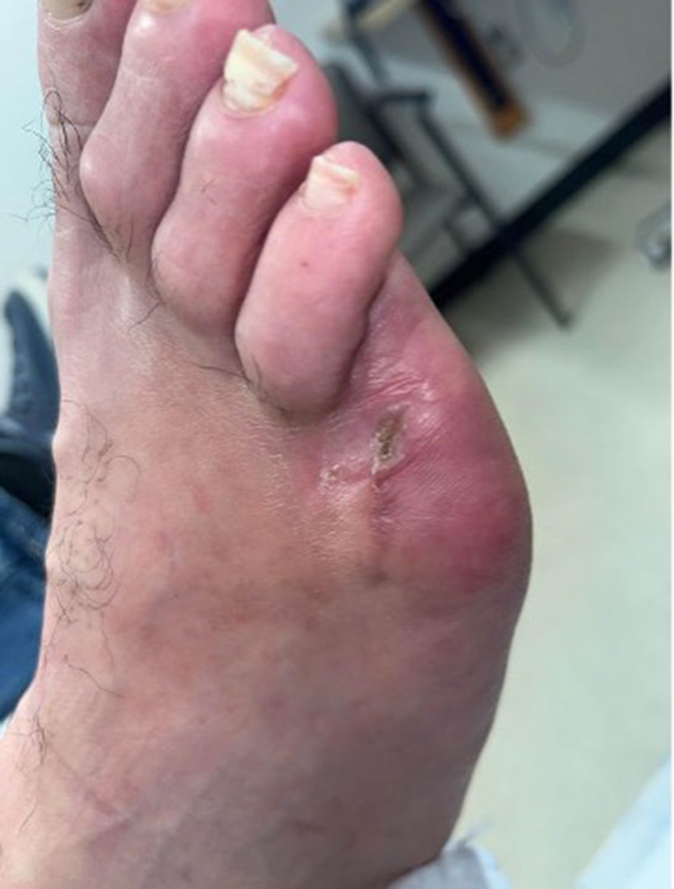
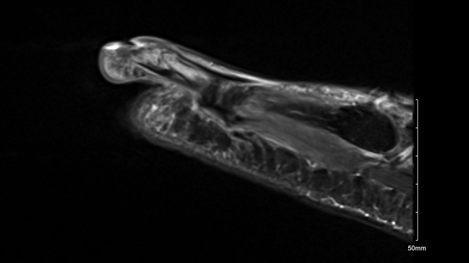
A 51-year-old Hispanic male, with past medical history of type 2 diabetes mellitus, hyperlipidemia, and tobacco use, was evaluated in the clinic with a moderate right foot infection per the International Working Group on the Diabetic Foot guidelines, centralized to an ulcer on the medial fifth digit with cellulitis extending greater than 2 cm (Figure 1) [14]. The patient was admitted to the hospital for surgery and parenteral antibiotics. There was no leukocytosis (white blood cell count 8.85 x 10⁹ L), and inflammatory markers were not elevated (C-reactive protein 0.3 mg/dL, erythrocyte sedimentation rate was 5 mm/hr). Due to bone exposure, magnetic resonance imaging was performed, which showed an abnormal signal in the phalanges of the fifth toe with sparing of the base of the proximal phalanx, consistent with osteomyelitis (Figure 2).
Figure 1. Initial Clinical Presentation showing moderate infection centralized to an ulcer on the medial fifth digit with cellulitis extending greater than 2 cm.
Japma 115 22213 g001
Figure 2. Magnetic resonance imaging of fifth digit showing an abnormal signal in the phalanges of the fifth toe with sparing of the base of the proximal phalanx, consistent with osteomyelitis.
Japma 115 22213 g002
The patient did not have palpable pedal pulses. Ankle brachial indices showed noncompressible vessels and triphasic waveforms for the posterior tibial and dorsalis pedis arteries with right toe pressures of 21 mm Hg. An angiogram showed prominent posterior tibial artery runoff with a short segment occlusion at the ankle. The peroneal and anterior tibial arteries were occluded in the mid segment. Angioplasty of the posterior tibial artery was performed, giving the patient one vessel run-off to the foot.
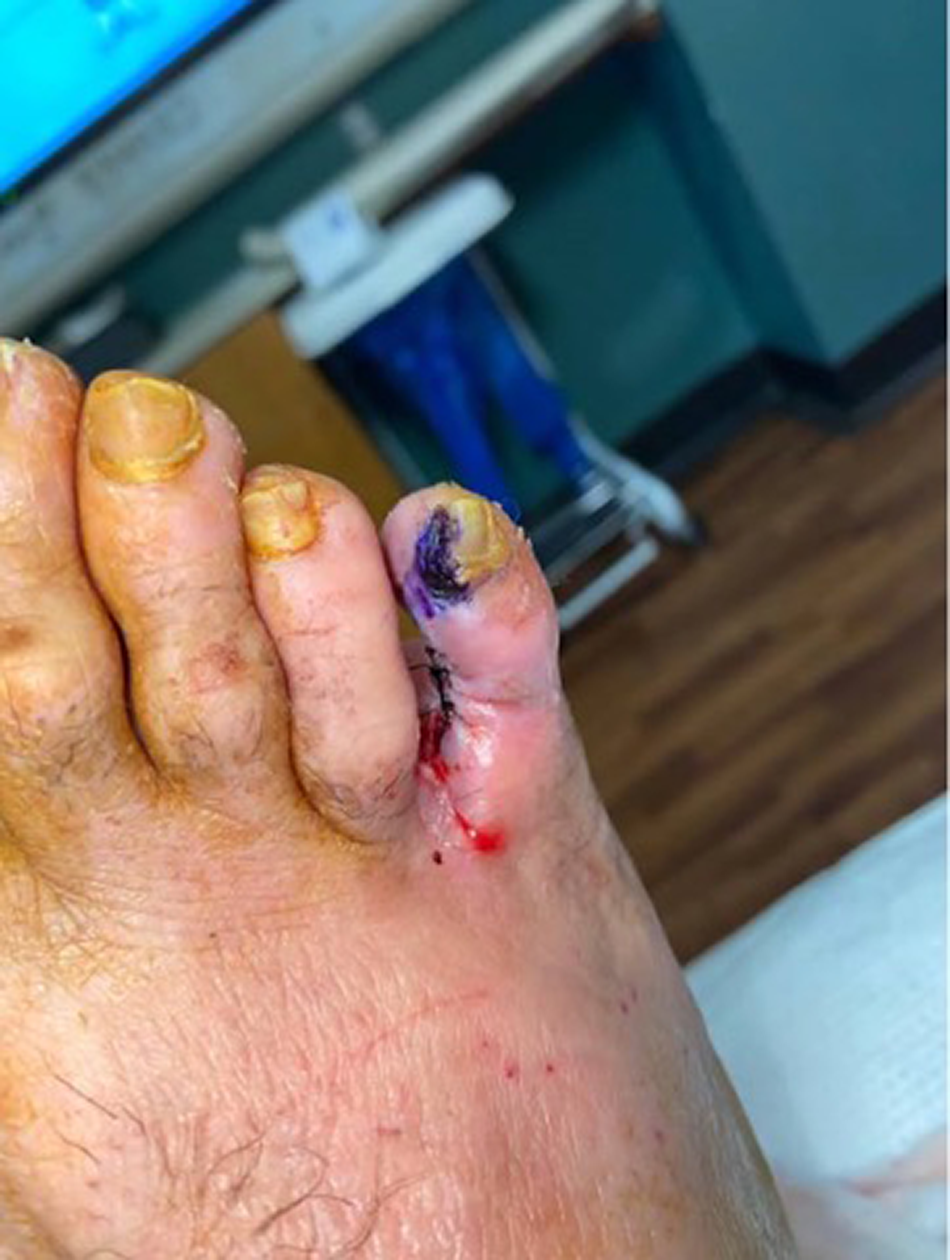
The patient preferred to save the digit if possible, and thus underwent a right fifth digit arthroplasty. Histology was indicative of “bone and cartilage with acute and chronic osteomyelitis” and light growth of A xylosoxidans was cultured from the bone. Figure 3 demonstrates the site 1 day postoperatively. Table 1 reports the susceptibilities of the isolated bacteria in this case.
Figure 3. Postoperative photo showing the site 1 day after fifth digit arthroplasty.
Japma 115 22213 g003
Table 1. Susceptibilities of the Bacterium Isolated in This Case.
Japma 115 22213 i001
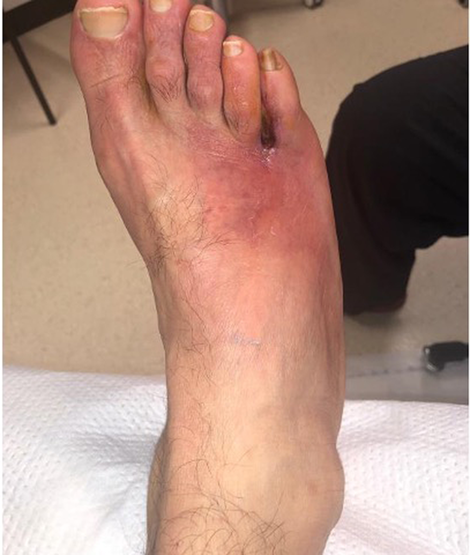
Medical versus surgical management of this patient’s osteomyelitis was reviewed in detail with him. After weighing the various benefits, alternatives and risks of each option, the patient elected to proceed with a fifth digit amputation for definitive source control. The clean cultures following this procedure revealed no further growth of the previously isolated A xylosoxidans bacterium. Most importantly, there was complete resection of infected bone, confirmed by histologic examination. The patient was seen 74 days after the index surgery noted to have healed uneventfully (Figure 4).
Figure 4. Postoperative photo on day 74 after fifth digit amputation.
Japma 115 22213 g004

Discussion

A xylosoxidans is an aerobic gram-negative rod commonly found in contaminated water and soil [15]. Similar to other opportunistic pathogens, it is resistant to many hospital disinfectants. As a result, it is commonly associated with nosocomial infections [16,17]. From our literature review, this is an organism that infects immunocompromised hosts; given the patient’s uncontrolled diabetes, this may have made the patient susceptible to A xylosoxidans osteomyelitis. Further, the genus is particularly susceptible to antibiotic resistance due to their unique ability to encode an apparatus such as aminoglycoside modifying enzymes, beta lactamases, as well as active efflux pumps [18,19]. To our knowledge, there have been nine published cases that have documented A xylosoxidans osteomyelitis in the literature (Table 2). Six of these followed a penetrating trauma, puncture wound, or surgery. In our case, the A xylosoxidans was likely inoculated by direct extension from this patient’s chronic wound.
Table 2. Summary of Current and Past Cases.
Japma 115 22213 i002
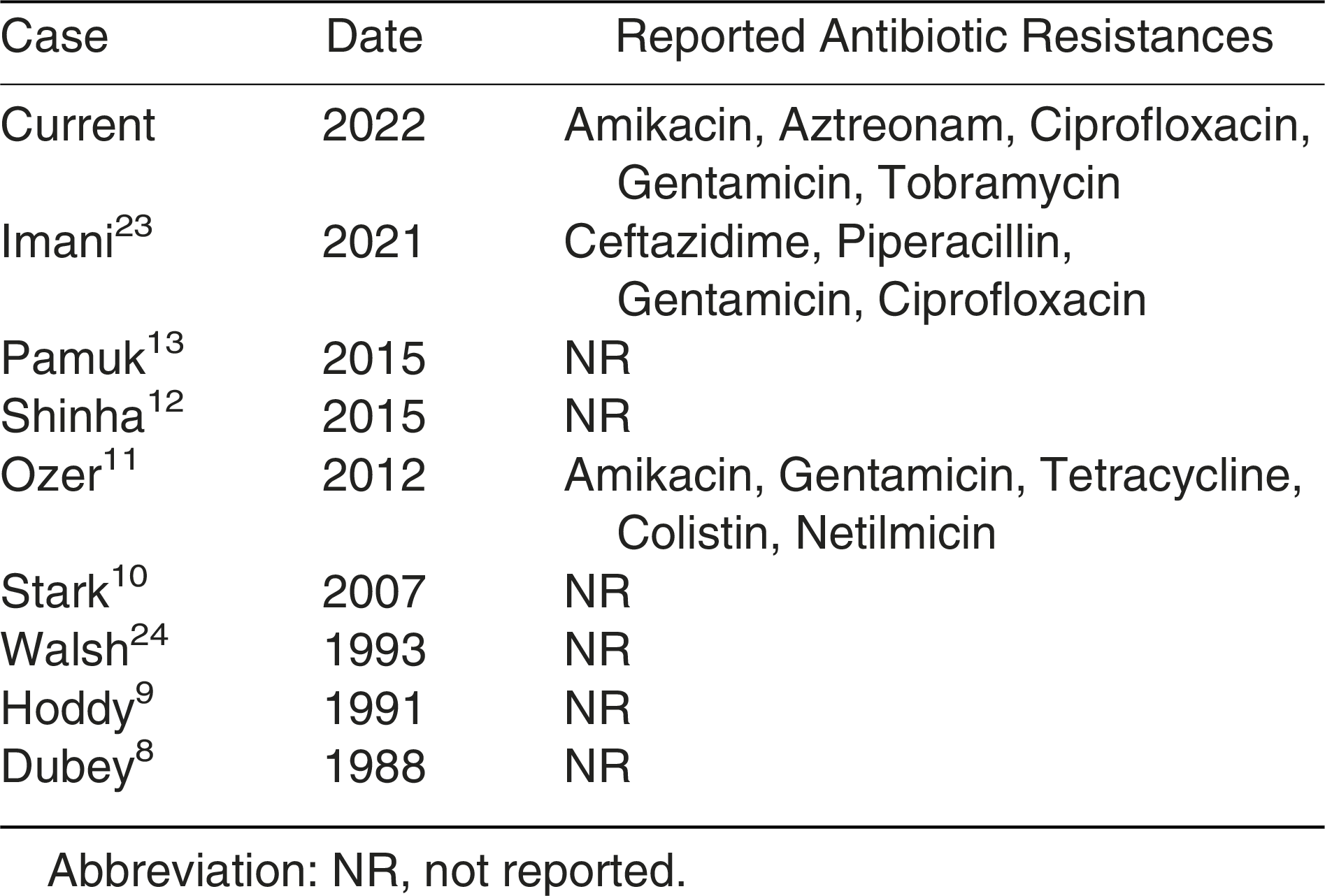
It has been documented that A xyloxidans species are resistant to multiple antibiotics including cefoxitin, cefotaxime, aztreonam, and aminoglycosides [12]. A summary of antibiotic susceptibilities in this case is reported in Table 1. A summary of antibiotic resistance and successful therapies from current and past cases are documented in Table 2 and Table 3. It is notable that many prior studies did not report antibiotic susceptibilities from isolated A xylosoxidans. Including ours, only three papers have reported antibiotic resistance, and of these, A xylosoxidans is most often resistant to amikacin, ciprofloxacin, and gentamicin. It has previously been reported that this bacterium is usually susceptible to trimethoprim-sulfamethoxazole, antipseudomonal penicillin, ceftazidime, cefoperazone, and carbapenems [12]. In our case, it was susceptible to levofloxacin, which was the therapeutic antibiotic selected. Since the patient underwent surgical resection of the underlying infected phalanges, antibiotics were discontinued 2 weeks after the right fifth toe amputation.
Table 3. Summary of Antibiotic Resistance from Current and Past Cases.
Japma 115 22213 i003
This case is pertinent to the treatment of the diabetic foot for several reasons. First, it is important to monitor nonhealing wounds for osteomyelitis. Since uncontrolled diabetes is a factor contributing to an immunocompromised host, diabetic soft-tissue infections and osteomyelitis can be caused by atypical organisms that may be best treated surgically or medically, depending on the antibiotic susceptibilities [20,21]. Secondly, as with other aspects of diabetic foot care, a multidisciplinary team is necessary to manage the complexities of this type of osteomyelitis. The team needs to consist of a skilled musculoskeletal radiologist, infectious disease specialist, vascular surgeon, internist, and podiatrist [22].

Conclusions

Osteomyelitis caused by A xylosoxidans is a rare clinical situation, illustrated by the existence of only nine cases documented in the literature. In recalcitrant diabetic foot ulcers, it is critical to have a suspicion of osteomyelitis. This case highlights the importance of having a high index of suspicion for osteomyelitis in chronic diabetic foot ulcerations and emphasizes the importance of having a multidisciplinary team to effectively manage individuals with chronic diabetic foot ulcerations and infections.

Financial Disclosure

None reported.

Conflict of Interest

None reported.

References

  1. Zhang P, Lu J, Jing Y, et al: Global epidemiology of diabetic foot ulceration: a systematic review and meta-analysis (dagger). Ann Med 49: 106, 2017.
  2. Moulik PK, Mtonga R, Gill GV: Amputation and mortality in new-onset diabetic foot ulcers stratified by etiology. Diabetes Care 26: 491, 2003.
  3. Lavery LA, Crisologo PA, La Fontaine J, et al: Are we misdiagnosing diabetic foot osteomyelitis? Is the gold standard gold? J Foot Ankle Surg 58: 713, 2019.
  4. Krentz AJ AP, Basu A, Kilvert A, et al: Morbidity and mortality associated with diabetic foot disease: A 12-month prospective survey of hospital admissions in a single UK centre. The Foot 7: 144, 1997.
  5. Liu X, Ren Q, Zhai Y, et al: Risk factors for multidrug-resistant organisms infection in diabetic foot ulcer. Infect Drug Resist 15: 1627, 2022.
  6. Gomez-Cerezo J, Suarez I, Rios JJ, et al: Achromobacter xylosoxidans bacteremia: a 10-year analysis of 54 cases. Eur J Clin Microbiol Infect Dis 22: 360, 2003.
  7. Crosby MD, Petropolis AA, Mackey VT, et al: An unusual skin infection with Achromobacter xylosoxidans. Cutis 106: 2110, 2020.
  8. Dubey L, Krasinski K, Hernanz-Schulman M: Osteomyelitis secondary to trauma or infected contiguous soft tissue. Pediatr Infect Dis J 7: 26, 1988.
  9. Hoddy DM, Barton LL: Puncture wound-induced Achromobacter xylosoxidans osteomyelitis of the foot. Am J Dis Child 145: 599, 1991.
  10. Stark JJ: Alcaligenes xylosoxidans osteomyelitis without trauma in a patient with Good's syndrome. Eur J Intern Med 18: 447, 2007.
  11. Ozer K, Kankaya Y, Baris R, et al: Calcaneal osteomyelitis due to Achromobacter xylosoxidans: a case report. J Infect Chemother 18: 915, 2012.
  12. Shinha T, Oguagha IC: Osteomyelitis caused by Achromobacter xylosoxidans. IDCases 2: 11, 2015.
  13. Pamuk G, Aygun D, Barut K, et al: Achromobacter causing a thrombophlebitis and osteomyelitis combination: a rare cause. BMJ Case Rep 2015: bcr2015210718, 2015.
  14. Monteiro-Soares M, Russell D, Boyko EJ, et al: Guidelines on the classification of diabetic foot ulcers (IWGDF 2019). Diabetes Metab Res Rev 36: e3273, 2020.
  15. Igra-Siegman Y, Chmel H, Cobbs C: Clinical and laboratory characteristics of Achromobacter xylosoxidans infection. J Clin Microbiol 11: 141, 1980.
  16. Marion-Sanchez K, Olive C, Platon MG, et al: Achromobacter xylosoxidans in hospital environments: still waters run deep! Trans R Soc Trop Med Hyg 114: 470, 2020.
  17. Pereira RH, Leao RS, Carvalho-Assef AP, et al: Patterns of virulence factor expression and antimicrobial resistance in Achromobacter xylosoxidans and Achromobacter ruhlandii isolates from patients with cystic fibrosis. Epidemiol Infect 145: 600, 2017.
  18. Hu Y, Zhu Y, Ma Y, et al: Genomic insights into intrinsic and acquired drug resistance mechanisms in Achromobacter xylosoxidans. Antimicrob Agents Chemother 59: 1152, 2015.
  19. Doi Y, Poirel L, Paterson DL, et al: Characterization of a naturally occurring class D beta-lactamase from Achromobacter xylosoxidans. Antimicrob Agents Chemother 52: 1952, 2008.
  20. Berbudi A, Rahmadika N, Tjahjadi AI, et al: Type 2 Diabetes and its impact on the immune system. Curr Diabetes Rev 16: 442, 2020.
  21. Tardáguila-García A, Sanz-Corbalán I, García-Alamino JM, et al: Medical versus surgical treatment for the management of diabetic foot osteomyelitis: a systematic review. J Clin Med 10: 1237, 2021.
  22. Musuuza J, Sutherland BL, Kurter S, et al: A systematic review of multidisciplinary teams to reduce major amputations for patients with diabetic foot ulcers. J Vasc Surg 71: 1433, 2020.
  23. Imani S, Wijetunga A, Shumborski S, et al: Chronic osteomyelitis caused by Achromobacter xylosoxidans following orthopedic trauma: A case report and review of the literature. ID Cases 25: e01211, 2021.
  24. Walsh JW, Hoffstad OJ, Sullivan MO, et al: Association of diabetic foot ulcer and death in a population-based cohort from the United Kingdom. Diabet Med 33: 1493, 2016.