Up to 25% of adults with diabetes develop an ulcer affecting the foot during their lifetime, of which approximately half are complicated by infection of the underlying bone [
1]. This diabetic foot osteomyelitis (DFO) often manifests as persistent ulceration, redness, swelling, and bony lesions on standard radiographs. The management of DFO is difficult, often failing to result in long-lasting success, and costly [
2]. Among those who undergo minor amputation, many will later require some type of revision surgery [
3,
4,
5,
6,
7,
8]. This revision might be necessitated by clinical or microbiological recurrence, the onset of ischemia, or the development of mechanical problems. These may lead to another episode of DFO in the same, or the neighboring, anatomical area [
9]. Hence, surgeons treating these infections must consider not only how best to perform surgery that is curative but also how to avoid potential postoperative mechanical problems.
In the past decade, an increasing number of minor amputations were performed at a level selected to preserve as much of the foot as possible [
10]. This is largely determined by the extent of radiologic bone lesions, whether the status of the underlying tissues will allow for primary wound closure, and the arterial perfusion to the foot [
11,
12,
13,
14,
15]. For each topographic area of the foot and ankle, there are distinct amputation levels described in the literature, as summarized in the
Atlas of Amputations and Limb Deficiencies [
10]. Besides those amputation levels, foot and ankle amputations can be divided into transarticular and transosseous amputation types [
10]. In transarticular amputations, the entire bone is resected without exposure of cancellous bone. Because the remaining bones in the stump are covered by cartilage and cortical bone, the skeleton is “sealed off” against infection by potential pathogens from the soft tissues [
16]. The question of whether to remove the cartilage to minimize the risk of remaining infection is controversial. One author states that when undertaking toe disarticulation, they preserve the articular cartilage in hopes that it would remain “resistant to infection” [
17]. In contrast, Atnip [
18] advocates for removing the cartilage during metatarsophalangeal joint disarticulation “to avoid necrosis and infection of this nonvascular tissue layer.” For hand surgery, some authorities promote preservation of the cartilage to maintain a closed cavity and to prevent the skin from becoming adherent directly to the bone [
19].
In transosseous foot amputations, the presumably infected part of the bone is resected, leaving an area of cancellous bone freshly exposed to the still infected overlying soft tissue [
16]. This exposure may increase the risk of recurrent DFO caused by the same pathogens (microbiological failure) because residual bacteria at the surgical site might spread into the bone [
20].
The criteria for selecting a transarticular versus a transosseous amputation type are driven by the desire to achieve an optimal functional outcome [
21]. The desire is achieved by retention of as much foot length and as many functional joints as possible. When planning the surgical procedure, three main criteria are used stepwise to reach this goal [
11,
13,
14,
15].
Step 1 Determination of the amount of osteomyelitic bone. This is usually done by either radiography alone or the combination of radiography and magnetic resonance imaging. Low signal intensity in marrow on T1-weighted images (combined with high signal intensity in marrow on T2-weighted images) indicates the amount of bone to be resected with high sensitivity and specificity and, therefore, the most distal possible amputation level [
22]. In the case of transosseous amputations, the amputation should be made through bone with normal marrow signal intensity on T1-weighted images to prevent residual osteomyelitis on wound closure. However, the surgeon and patient (with informed consent) together can deliberately opt to retain infected bone to achieve a longer residual limb. This would usually result in longer antibiotic drug treatment and higher risk of failure [
23].
Step 2 Determination of the amount of viable soft tissue that can be used to form a resilient residual foot envelope [
10]. Nonviable soft tissues must be resected irrespective of a distinct amputation level or amputation type. Otherwise, revision amputation due to soft-tissue breakdown must be anticipated. Tension-free soft-tissue closure, possibly assisted by vacuum-assisted wound closure, then determines topographically the amputation level and, thereby, the amputation type.
Step 3 Securing that arterial perfusion will allow soft-tissue healing by palpating pedal pulses or performing Doppler sonography and by referring patients to vascular work-up whenever there is any doubt of patency of the lower-extremity arteries.
There might be hidden consequences of either of these amputation types that could influence the resultant mechanical properties of the affected foot (or the general gait). Thus, one of these amputation approaches might better postpone or avoid the need for revision surgery more than the other. Based on our clinical experience and personal communications, the choice of the level for transarticular or transosseous amputation largely rests on the surgeon’s considerations.
No studies have investigated whether there are potential differences between transarticular and transosseous amputations in the risk of clinical failure (a new episode of DFO in the same anatomical region ≤1 year after the index surgery) or microbiological failure (culture verification of ≥50% of the same bacterial pathogens in a case of clinical failure) on long-term surveillance. Thus, the aim of this study was to evaluate whether amputations of the transarticular type compared with the transosseous type have different rates of clinical and microbiological failure.
Patients and Methods
Setting
The study was conducted at Balgrist University Hospital (Zurich, Switzerland), a tertiary referral center for diabetic foot problems and amputations. It has established a multidisciplinary team composed of fellowship-trained orthopedic surgeons, internists, specialized wound nurses, cast specialists, expert radiologists, orthopedic shoemakers, prosthetists and orthotists, and infectious disease physicians specialized in orthopedic infections. Moreover, this team is supported by an in-house company for orthopedic footwear and professional off-loading and a re-education unit with physical and occupational therapy. The center runs a prospective register of patients evaluated and treated for diabetic foot problems, including DFOs.
Standard Surgical Approaches
The following approach is the standard approach for treating infected diabetic feet at our hospital. For patients with DFO, we decided the level of minor amputation by the following process: 1) determined the extent of infection with magnetic resonance imaging by observing fat mark signal extinction in T1 sequences on magnetic resonance imaging, which served as an orientation for the amputation level [
22,
23,
24]; 2) assessed the extent of softtissue infection by clinical examination; 3) palpated the pedal pulses (dorsalis pedis and tibialis posterior arteries) manually and referred unclear cases for Doppler ultrasonography or other, more sophisticated vascular assessments; and 4) incorporated information on the patient‘s mobility, personal preferences, and previous ipsilateral amputations into the choice of the amputation level. We performed all of the minor amputations under the supervision of one of three fellowship-trained orthopedic surgeons. All of the patients were treated with professional off-loading (mostly by a cast) until they achieved complete wound healing. The internists tailored the concomitantly administered antibiotic drug regimens based on the latest Infectious Diseases Society of America diabetic foot infection guidelines (the 2012 version is referenced exemplary) [
25] and/or the stateof-the-art in the literature [
2].
Amputation Levels
An amputation was considered “minor” when performed at the foot up until the ankle joint and as “major” when performed above the ankle joint [
26,
27,
28]. Minor amputation levels were stratified into forefoot, midfoot, and hindfoot amputations. Forefoot amputations included toe amputations, metatarsal head resections, metatarsal ray amputations, and total transmetatarsal amputations. Midfoot amputations included Lisfranc amputations, atypical amputations performed through the cuneiform and cuboid bones, and the Bona-Jaeger amputation level [
29], and all of the amputations starting from the Chopart level up until the Syme level were considered hindfoot amputations.
Retrospective Comparative Study and Definitions
In a level III retrospective comparative study design, we included all of the patients who underwent diabetic foot amputations for DFO, of the transarticular or transosseous type, between January 1, 2000, and October 31, 2019, and closed the database on November 1, 2020. Besides patients with type 1 and type 2 diabetes, we also included patients with type 3c diabetes [
30]. Both primary and revision amputations were included; patients with previous contralateral amputation were not excluded. We required an active follow-up (continuous outpatient visits) of 1 year to be included in the study due to the definition of clinical failure; the only exception was patient death during the study period. Exclusion criteria were age younger than 18 years, follow-up of less than 12 months, insufficient data documentation, and missing consent. We defined DFO according to the guidelines of the International Working Group on the Diabetic Foot by the combination of clinical features (combination of erythema, swelling, hyperthermia, “sausage” toe, probe-to-bone positive ulcer, visible bone), surgical features, and radiologic features (shattered or altered bone intraoperatively, radiologic bone lesions without previous surgery or trauma, and, specifically, fat mark signal extinction in T1 sequences on magnetic resonance imaging) [
31,
32,
33]. We used the results of microbiology (culture of the same organism[s] in several intraoperative bone specimens) and histology (microscopic evidence of inflammation, pathogens, necrosis) as supportive of the diagnosis. Existence of histology was not an absolute requirement. We defined clinical failure as a new DFO episode in the same anatomical location within 1 year of the index amputation [
34]. According to previous definitions, delayed wound healing without clinical evidence of infection was not considered as clinical failure [
34]. We defined microbiological failure as a clinical failure accompanied by the recurrence of at least 50% of the causative pathogens of the index episode [
34]. By this definition, any episode that demonstrated microbiological failure was automatically a clinical failure too. We defined remission as the absence of any clinical, laboratory, or imaging evidence of failure as well as the absence of any potential need for a surgical revision. Diagnosis and treatment of peripheral artery disease was made by the referral angiologists using routine angiologic examination and treatment methods, including angiography and percutaneous transluminal angioplasty. Diagnosis of chronic renal insufficiency was made based on the criteria published by Webster et al [
35]: glomerular filtration rate less than 60 mL/min per 1.73 m
2, markers of kidney damage, or both, of at least 3 months duration, regardless of the underlying cause.
In our hospital, most patients sign a general consent for academic studies. For this study, we obviously excluded those who did not specifically consent to retrospective studies. The local ethical committee approved this anonymized analysis of medical files without additional patient contact.
Statistical Analyses
The primary outcome of this study was the incidence of clinical failure within 1 year of the index amputation or death, stratified on the transarticular or transosseous amputation type. Because we knew we were including more than 500 DFO episodes, we had no previous sample size requirements. We compared groups with the Pearson x2 test or the Wilcoxon rank sum test, and we plotted the corresponding revision-free survival times using Kaplan-Meier curves and log-rank tests. To adjust for the large case mix, we performed multivariate Cox regression analyses with the outcomes of clinical failure and microbiological failure, with the following events serving as censor dates: death, major amputation within 1 year not qualifying as clinical failure, and date of last clinical control. We introduced independent variables with a P ≤ .05 in the univariate analysis stepwise into the multivariate analysis, except for the level of surgical interventions, which we automatically included in the final model.
We included eight to ten predictor variables per outcome and checked for collinearity and interaction by interaction terms. Because the outcome of clinical failure could be, for each episode, due to mechanical, infectious, or ischemic reasons, or a mix, we decided against doing a clustered analysis at the individual patient level. After first analyzing the entire study population, we stratified the analyses into the joint and bone populations. We used IBM SPSS Statistics for Windows (Version 25.0; IBM Corp, Armonk, New York) and considered P ≤ .05 (two-tailed) as statistically significant.
Results
We assessed 543 minor foot amputation episodes (203 transarticular and 340 transosseous) in 284 different adult patients (mean ± SD age at index surgery, 66 ± 11.5 years; range, 25–96 years); 64 were women [22.5%]). All of the patients were actively followed up for 1 year, and 447 (82.3%) were primary amputations and 96 (17.7%) were revision amputations. Many patients had comorbidities, the most common of which were polyneuropathy (n = 254; 89.4%), chronic renal insufficiency (n = 120; 42.3%), coronary artery disease (n = 116; 40.8%), and symptomatic peripheral artery disease (n = 193; 68%). A total of 164 patients (57.7%) were current smokers or had a history of active smoking. In the observation period of 1 year, 19 patients (6.7%) died of conditions unrelated to the amputation or DFO, with a mean ± SD time to death of 4.8 ± 3.0 months (range, 0–10 months).
In 412 episodes (75.9%), we isolated the causative pathogens from several intraoperative bone samples: 219 DFOs (40.3%) were monomicrobial and 194 (35.7%) were polymicrobial. The three most frequent DFO pathogen groups were coagulase-negative staphylococci (n = 181),
Staphylococcus aureus (n = 137, of which 35 were methicillin-resistant), and
Enterococcus faecalis (n = 40). The mean ± SD duration of concomitant systemic antibiotic drug treatment was 34.6 ± 33.6 days (range, 1–255 days). However, patients who underwent transosseous amputation received a significantly longer mean ± SD duration of postoperative antibiotic drug therapy (40 ± 37 days; range, 1–255 days) compared with transarticular amputation (26 ± 25 days; range, 1–185 days;
P < .001).
Table 1
presents the demographic, clinical, and outcome data, and
Table 2
the distribution by anatomical site of surgical amputations.
Clinical and Microbiological Failures
Overall, clinical failure occurred in 122 episodes (22.5%); 21.7% (n = 44) in the transarticular group and 22.9% (n = 78) in the transosseous group (
x2 test:
P = .73). The mean ± SD time lapse between the index amputation and clinical failure was 2.8 ± 5.7 months: 2.2 ± 2.2 months for transarticular and 3.2 ± 7.0 months for transosseous (Mann-Whitney
U test:
P = 39). Microbiological failures occurred in 32 episodes overall (5.9%). Separately, 11 transarticular episodes (5.4%) and 21 transosseous episodes (6.2%;
x2 test:
P = .72) revealed a microbiologically proven recurrence of DFO. Stratified into forefoot, midfoot, and hindfoot amputations, there were no statistically significant differences in clinical failure rates between the different anatomical amputation levels and the type of amputation (
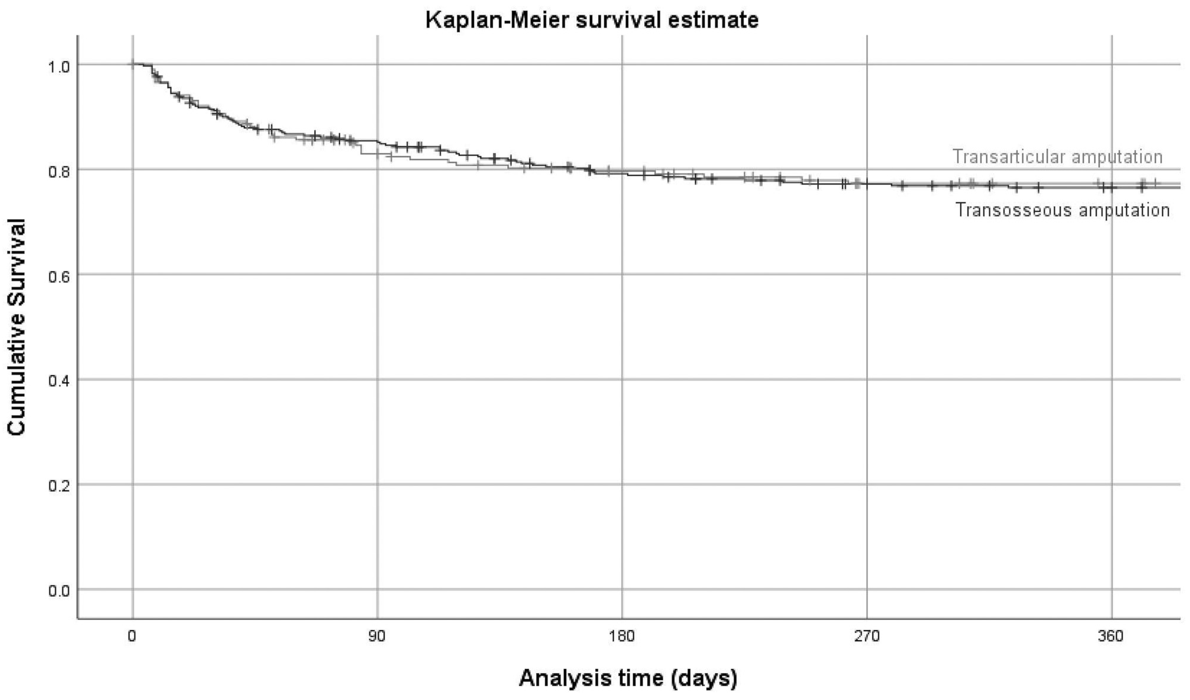
Table 2). Likewise, the Kaplan-Meyer curves showed similar survivals for both types over 1 year (log-rank test;
P = .85). The survival curves were nearly identical (
Figure 1). Revision amputations (30.2%) demonstrated significantly more clinical failures than primary amputations (20.8%;
P = 045).
Multivariate Adjustments
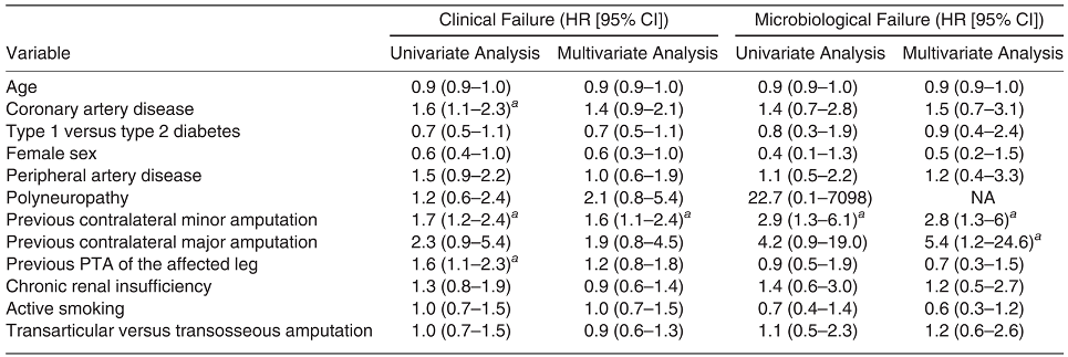
In the multivariate Cox regression analyses (
Table 3), the amputation type was not related to short -or longterm clinical failure (
Figure 1). Previous contralateral minor amputation was the only risk factor significantly associated with clinical failure (hazard ratio [HR], 1.6; 95% confidence interval [CI], 1.1–2.3). The variation in microbiological results among the included individual DFO cases was too heterogenous to be clinically categorized in the “surgical” multivariate final model.
Revision Surgery Within 1 Year
Overall, 182 episodes (33.5%) needed revision surgery within 1 year. Mean ± SD time to revision surgery was 3.5 ± 3.1 months; range, 0–11.9 months). Nine revisions were soft-tissue revision due to soft-tissue infection (resolvable with soft-tissue surgery alone), and the remaining 173 revisions were reamputations. Indications for surgery were osteomyelitis in 130 episodes (71.4%), gangrene in 20 (11.0%), soft-tissue infection in 12 (6.6%), chronic wounds needing closure in 11 (6.0%), surgical correction of malpositions in five (2.7%), tissue necrosis in three (1.6%), and acute arterial occlusion in one (0.5%).
Furthermore, 19 of 351 included extremities (5.4%) needed major amputation. Mean ± SD time to major amputation was 4.6 ± 3.3 months; range, 0.4–11.7 months). A separate multivariate Cox regression analysis revealed that presence of peripheral artery disease (HR, 3.2; 95% CI, 1.3–8.3) and presence of type 2 diabetes (HR, 3.1; 95% CI, 1.5–6.1) were related to major amputation, whereas type of amputation (transosseous versus transarticular) was not (HR, 1.2; 95% CI, 0.6–2.4).
Discussion
The most important finding of this study is that there is no difference in risk of failure between patients undergoing transarticular versus transosseous amputation in DFO irrespective of whether performed at the forefoot, midfoot, or hindfoot level.
In this large, single-center, retrospective study comparing the clinical and microbiological outcomes of DFO treated with amputation, either transarticular or transosseous, we found the risk of failure to be 22.5% in both groups within 1 year. This revision risk is similar to that previously reported for similar procedures in the literature [
3,
4,
5,
6,
7,
8,
36]. We also found that for the two amputation types, the Kaplan-Meier curves and the time to failure were not significantly different, and they were similar for amputations at the hindfoot, midfoot, and toe levels. Because we found no benefit of one amputation type over the other during the observed period and no differences concerning the anatomical region where the amputation was performed, these results reaffirm established amputation level selection criteria such as radiologic extent of osteomyelitis, extent of soft-tissue destruction, perfusion status, and previous minor amputations.
Retaining the native shape of the remaining proximal bone (ie, the metatarsal head) after transarticular level amputation likely alters the topical plantar pressure less than transosseous level amputations, which are known to predispose to secondary relevant plantar pressure elevations [
37]. In polyneuropathic patients, the plantar pressure is already elevated due to chronic biomechanical alterations of the foot architecture [
38,
39]. It is unlikely that the commonly practiced technique of rounding bone edges in bone amputations can restore the physiologic bone shape and, therefore, the physiologic plantar pressure proportions [
37]. Theoretically, these combined reasons for plantar pressure elevation might be detrimental, leading to reulceration and, thereby, to a higher risk of revision amputation in transosseous amputations in neuropathic patients. However, based on our clinical experience and review of the literature, we are unconvinced by this theory. In our daily experience, adequate orthopedic shoes, frequent controls, and continued patient education will usually prevent major amputations, including in overtly neuropathic patients.
This study identified previous contralateral minor amputation as potentially significant association with ultimate clinical failure after minor amputation for DFO. Wanivenhaus et al [
3] reported that the presence of peripheral artery disease, diabetic nephropathy, and peripheral neuropathy were each independent risks for requiring revision after (diabetic) foot amputation. The present findings did not confirm their results. Other research groups have identified different variables associated with the need for revision after minor amputations. One study reported that risk factors for revision amputation in DFO included male sex, long duration of diabetes, presence of a wound infection, peripheral neuropathy, and a history of smoking [
40]. Ahn et al [
41] identified renal dialysis as a risk for revision after transmetatarsal amputations. Elsherif et al [
42] investigated patients’ demographic characteristics, history of revascularization procedures, and amputation levels but did not detect any variables that influenced the risk of revision after forefoot amputation. However, none of these studies compared transarticular versus transosseous amputation types, and all reported on much smaller sample sizes.
The present study has several limitations. First, it used a retrospective design and the enrolled population had a large case mix. We also lacked information on the exact preoperative or postoperative equine foot deformity and degree of ankle dorsiflexion in the database. Both variables could influence the risk of clinical failure [
43,
44,
45]. This was also a singlecenter study, limiting the generalizability of the results. We also considered that patients with clinical failure after DFO might have been seen for revision surgery elsewhere, but because of our specialized setting and emphasis on follow-up, we think this potential bias was minimal.
Conclusions
In this large and carefully designed retrospective comparative study targeting the question of the risk of clinical and microbiological failure in transarticular versus transosseous DFO amputations, both groups yielded the same revision risk of 22.5% within 1 year, which occurred at a similar time after surgery and independently of the anatomical location of the amputation. Because we found no compelling arguments for or against either of these two techniques, we think that the choice should be at the discretion of the treating surgeon and the individual patient.